. 2002 Dec 1;22(23):10094-105.
doi: 10.1523/JNEUROSCI.22-23-10094.2002.

Delayed rectifier K+ currents, IK, are encoded by Kv2 alpha-subunits and regulate tonic firing in mammalian sympathetic neurons

Affiliations

Delayed rectifier K+ currents, IK, are encoded by Kv2 alpha-subunits and regulate tonic firing in mammalian sympathetic neurons

Sacha A Malin et al. J Neurosci. .

Abstract

Previous studies have revealed the presence of four kinetically distinct voltage-gated K+ currents, I(Af), I(As), I(K), and I(SS), in rat superior cervical ganglion (SCG) neurons and demonstrated that I(K) and I(SS) are expressed in all cells, whereas I(Af) and I(As) are differentially distributed. Previous studies have also revealed the presence of distinct components of I(Af) encoded by alpha-subunits of the Kv1 and Kv4 subfamilies. In the experiments described here, pore mutants of Kv2.1 (Kv2.1W365C/Y380T) and Kv2.2 (Kv2.2W373C/Y388T) that function as Kv2 subfamily-specific dominant negatives (Kv2.1DN and Kv2.2DN) were generated to probe the functional role(s) of Kv2 alpha-subunits. Expression of Kv2.1DN or Kv2.2DN in human embryonic kidney-293 cells selectively attenuates Kv2.1- or Kv2.2-encoded K+ currents, respectively. Using the Biolistics Gene Gun, cDNA constructs encoding either Kv2.1DN or Kv2.2DN [and enhanced green fluorescent protein (EGFP)] were introduced into SCG neurons. Whole-cell recordings from EGFP-positive Kv2.1DN or Kv2.2DN-expressing cells revealed selective decreases in I(K). Coexpression of Kv2.1DN and Kv2.2DN eliminates I(K) in most (75%) SCG cells and, in the remaining (25%) cells, I(K) density is reduced. Together with biochemical data revealing that Kv2.1 and Kv2.2 alpha-subunits do not associate in rat SCGs, these results suggest that Kv2.1 and Kv2.2 form distinct populations of I(K) channels, and that Kv2 alpha-subunits underlie (most of) I(K) in SCG neurons. Similar to wild-type cells, phasic, adapting, and tonic firing patterns are evident in SCG cells expressing Kv2.1DN or Kv2.2DN, although action potential durations in tonic cells are prolonged. Expression of Kv2.2DN also results in membrane depolarization, suggesting that Kv2.1- and Kv2.2-encoded I(K) channels play distinct roles in regulating the excitability of SCG neurons.


Figures
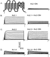
Fig. 1.
Fig. 1.
Kv2.1W365C/Y380T (Kv2.1DN) specifically attenuates Kv2.1-encoded currents in HEK cells. Whole-cell voltage-gated outward K+ currents were recorded from HEK-293 cells expressing Kv2.1DN alone (A), Kv2.1 or Kv2.1 plus Kv2.1DN (B), Kv2.2 or Kv2.2 plus Kv2.1DN (C), Kv1.4 or Kv1.4 plus Kv2.1DN (D), and EGFP. Cells were transfected with cDNA constructs (1:1) encoding these subunits, and currents were obtained from EGFP-positive cells as described in Materials and Methods. Schematic of Kv2.1DN is shown, with altered residues. A, In cells expressing Kv2.1DN alone, outward K+currents are indistinguishable from those in wild-type HEK-293 cells. B, In HEK-293 cells coexpressing Kv2.1DN and Kv2.1, current densities are reduced markedly compared with those recorded from cells expressing Kv2.1 alone (compare leftand right). C, In contrast, coexpression of Kv2.1DN with Kv2.2 reveals outward currents indistinguishable from those measured in cells expressing Kv2.2 alone. D, Outward K+ currents in cells expressing Kv2.1DN and Kv1.4 are indistinguishable from those expressing Kv1.4 alone.
Fig. 2.
Fig. 2.
Kv2.2W373C/Y388T (Kv2.2DN) specifically attenuates Kv2.2-encoded currents in HEK cells. Whole-cell voltage-gated outward K+ currents were recorded from HEK-293 cells expressing Kv2.2DN (A), Kv2.2 or Kv2.2 plus Kv2.2DN (B), Kv2.1 or Kv2.1 plus Kv2.2DN (C), and EGFP, as described in the legend to Figure 1. A, Schematic of Kv2.2DN is shown, with the altered residues indicated. In cells expressing Kv2.2DN alone, outward K+ currents are similar to those in wild-type cells. B, When Kv2.2DN is coexpressed with Kv2.2, however, Kv2.2-encoded outward K+ currents are reduced compared with cells expressing Kv2.2 alone (compareleft and right). C, In contrast, Kv2.2DN does not affect Kv2.1-encoded currents.
Fig. 3.
Fig. 3.
Expression of Kv2.1DN or Kv2.2DN reducesIK density in SCG neurons. Whole-cell voltage-gated outward K+ currents were recorded from isolated SCG neurons in response to 6 sec depolarizing voltage steps to test potentials between −10 and +50 mV from a holding potential of −90 mV. Experiments were conducted as described in Materials and Methods, with 1 μm TTX and 100 μmCdCl2 in the bath solution to block voltage-gated inward Na+ and Ca2+ currents, respectively. The records shown to the left,middle, and right were recorded from wild-type, Kv2.1DN-expressing, and Kv2.2DN-expressing cells, respectively. There are distinct and stereotyped differences in the waveforms of the currents in wild-type I, type II, and type III SCG cells (Malin and Nerbonne, 2000). The numbersgiven above the records in each column reflect the percentages of cells studied under each experimental condition that display the type I, type II, or type III outward K+ current phenotype. Although the percentages of type I, type II, and type III Kv2.1DN- or Kv2.2DN-expressing cells are not different from those in wild-type SCG cells, expression of either Kv2.1DN (middle) or Kv2.2DN (right) decreases the density of the slowly decaying current, IK, in type I cells. Expression of Kv2.1DN also decreases IKdensity in type II cells (middle).
Fig. 4.
Fig. 4.
Coexpression of Kv2.1DN and Kv2.2DN eliminatesIK in most SCG neurons. Isolated SCG neurons were transfected with both Kv2.1DN and Kv2.2DN (and EGFP) using the Biolistics Gene Gun (as described in Materials and Methods), and outward K+ currents were recorded from EGFP-positive cells as described in the legend to Figure 3. Two distinct current waveforms were evident in these recordings: most (75%) cells were found to express only IAf andISS and therefore are type I cells lackingIK; the remaining cells (25%) express IAf,IAs,IK, andISS and are classified as type II cells with reduced IK density (Table 2). The densities of IAf,IAs, andISS in Kv2.1DN plus Kv2.2DN-expressing type I and type II cells are indistinguishable from those measured in wild-type I and II cells (Table 2).
Fig. 5.
Fig. 5.
Kv2.1 and Kv2.2 are expressed in rat SCGs but do not appear to associate. A, Lysates of control and transfected HEK-293 cells and of isolated rat SCGs were fractionated in SDS-PAGE gels and immunoblotted (IB) with the monoclonal anti-Kv2.1 (left) and the polyclonal anti-Kv2.2 (right) antibodies. B, C, Lysates prepared from transfected HEK-293 cells (B) and rat SCG neurons (C) were immunoprecipitated (IP) with either the monoclonal anti-Kv2.1 or the polyclonal anti-Kv2.2 antibody, fractionated, and immunoblotted with the same antibodies. Although both the anti-Kv2.1 and anti-Kv2.2 antibodies reliably immunoprecipitate the proteins against which each of these antibodies were generated, the Kv2.1 and Kv2.2 α-subunits do not coimmunoprecipitate from lysates prepared from Kv2.1- and Kv2.2-transfected HEK-293 cells or rat SCGs. After immunoprecipitations with the anti-Kv2.1 antibody, the Kv2.2 protein is evident in the supernatants (lanes S) but not in the pellet (lanes P). Similarly, the Kv2.1 protein is found in the supernatant (S) after immunoprecipitation with the anti-Kv2.2 antibody. Closed arrows indicate Kv2.1; open arrows indicate Kv3.2.
Fig. 6.
Fig. 6.
Activation of Kv2-encoded K+currents during action potentials in SCG neurons. To explore directly the activation of Kv2.x-encoded K+ currents during action potential waveforms in SCG neurons, cells were held at the typical resting membrane potential of SCG neurons (∼48 mV; see Table3), and outward K+ currents activated by typical phasic, adapting, and tonic action potential waveforms were recorded. The voltage-clamp paradigms are illustrated at thebottom. Representative outward current waveforms in Kv2.1-expressing HEK-293 cells driven by the phasic adapting and tonic action potential waveforms are illustrated at the top. Representative outward K+ current waveforms in isolated SCG neurons (activated using the action potential voltage-clamp paradigms shown below the record) are presented at the bottom. Action potential clamp recordings from wild-type (solid line), Kv2.1DN-expressing (short dashed line), and Kv2.2DN-expressing (long dashed line) SCG cells are superimposed for comparison purposes.
Fig. 7.
Fig. 7.
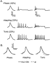
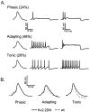
Phasic, adapting, and tonic firing patterns in SCG neurons expressing Kv2.1DN. SCG neurons were transfected with Kv2.1DN (and EGFP), and action potentials and repetitive firing patterns were recorded in response to brief or prolonged depolarizing current injections, as described in Materials and Methods. A, Current-clamp recordings from three representative Kv2.1DN-expressing cells are shown. In each cell, single action potentials were elicited by 1.5 msec current injections (left), and repetitive firing patterns were recorded in response to 100 pA (middle) or 200 pA (right) 500 msec current injections. Based on the response(s) to the 500 msec current injections, cells were classified as phasic (top), adapting (middle), or tonic (bottom) (Table 4). Although Kv2.1DN expression does not affect the distribution of firing patterns in SCG neurons, action potentials in tonic cells expressing Kv2.1DN are prolonged (B). Action potential durations in phasic and adapting cells (B), in contrast, are unaffected by Kv2.1DN expression (Table 4). wt, Wild type.
Fig. 8.
Fig. 8.
Expression of Kv2.2DN in SCG neurons increases the number of adapting cells. In SCG neurons transfected with Kv2.2DN (and EGFP), action potentials and repetitive firing patterns were recorded, and cells were classified as phasic (top), adapting (middle), or tonic (bottom), as described in the legend to Figure 7. Similar to the results obtained with Kv2.1DN, expression of Kv2.2DN prolongs action potential durations in tonic cells (B). In addition, Kv2.2DN expression alters the distribution of firing patterns: the percentage of phasic cells is reduced (by ∼40%), and the percentage of adapting cells is increased (Table 4). wt, Wild type.
Fig. 9.
Fig. 9.
Coexpression of Kv2.1DN and Kv2.2DN markedly reduces tonic firing. Isolated SCG neurons were transfected with Kv2.1DN, Kv2.2DN, and EGFP. Action potentials and repetitive firing patterns were recorded, and cells were classified as phasic (top), adapting (middle), or tonic (bottom), as described in the legend to Figure 7. In contrast to the results obtained with expression of Kv2.1DN or Kv2.1DN alone (Figs. 7, 8), expression of both dominant negative constructs markedly decreases the percentage of tonic firing cells (Table4).
Fig. 10.
Fig. 10.
Expression of Kv2.1 in SCG neurons increases tonic firing. Action potentials and repetitive firing patterns, recorded as described in the legend to Figure 7, were obtained from isolated SCG neurons 24 hr after transfection with wild-type Kv2.1 and EGFP. As in wild-type cells, the phasic (top), adapting (middle), and tonic (bottom) firing patterns were seen in recordings from cells transfected with Kv2.1. However, the percentage of tonic cells is higher and the percentages of adapting and phasic cells are lower in Kv2.1-expressing cells than in either wild-type, Kv2.1DN-expressing (Fig. 7), or Kv2.2DN-expressing (Fig. 8) SCG cells (Table 4).

Similar articles

Cited by

References

    1. An WF, Bowlby MR, Betty M, Cao J, Ling H-P, Mendoza G, Hinson JW, Mattson KI, Strassle BW, Trimmer JS, Rhodes KJ. Modulation of A-type potassium channels by a family of calcium sensors. Nature. 2000;403:553–556. - PubMed
    1. Baranauskas G, Tkatch T, Surmeier JD. Delayed rectifier currents in rat globus pallidus neurons are attributable to Kv2.1 and Kv3.1/3.2 K+ channels. J Neurosci. 1999;19:6394–6404. - PMC - PubMed
    1. Barry DM, Trimmer JS, Merlie JP, Nerbonne JM. Differential expression of voltage-gated K+ channel subunits in adult rat heart. Relation to functional K+ channels. Circ Res. 1995;77:361–369. - PubMed
    1. Barry DM, Xu H, Schuessler RB, Nerbonne JM. Functional knock-out of the transient outward current, long-QT syndrome, and cardiac remodeling in mice expressing a dominant-negative Kv4 α-subunit. Circ Res. 1998;83:560–567. - PubMed
    1. Blaine JT, Ribera AB. Heteromultimeric potassium channels formed by members of the Kv2 subfamily. J Neurosci. 1998;18:9585–9593. - PMC - PubMed

Publication types

MeSH terms