. 2002 Dec 1;22(23):10106-15.
doi: 10.1523/JNEUROSCI.22-23-10106.2002.

Potassium currents during the action potential of hippocampal CA3 neurons

Affiliations

Potassium currents during the action potential of hippocampal CA3 neurons

Jörg Mitterdorfer et al. J Neurosci. .

Abstract

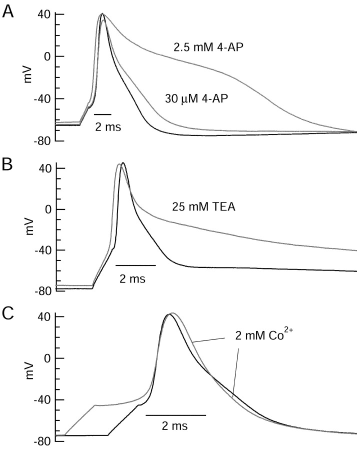
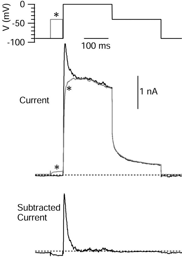
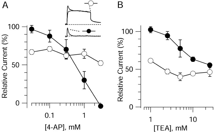
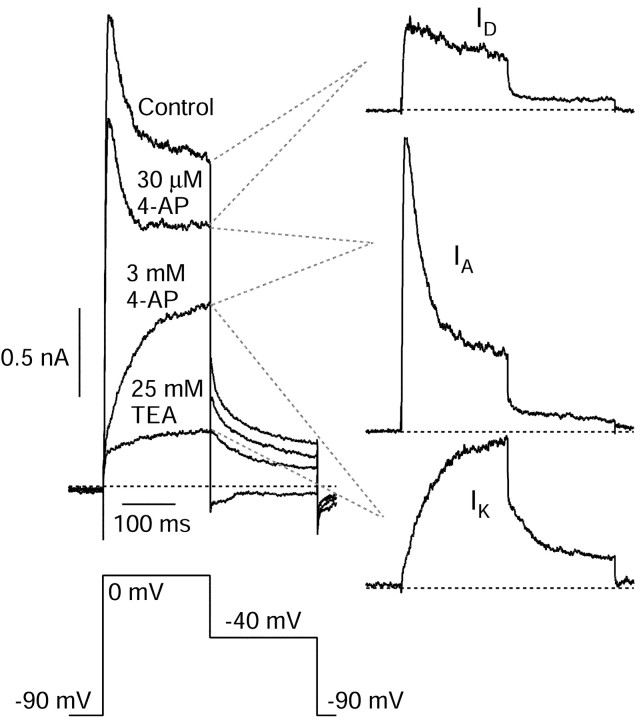
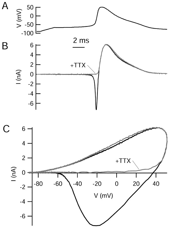
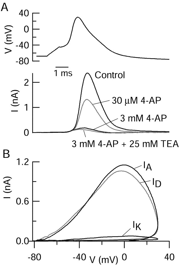
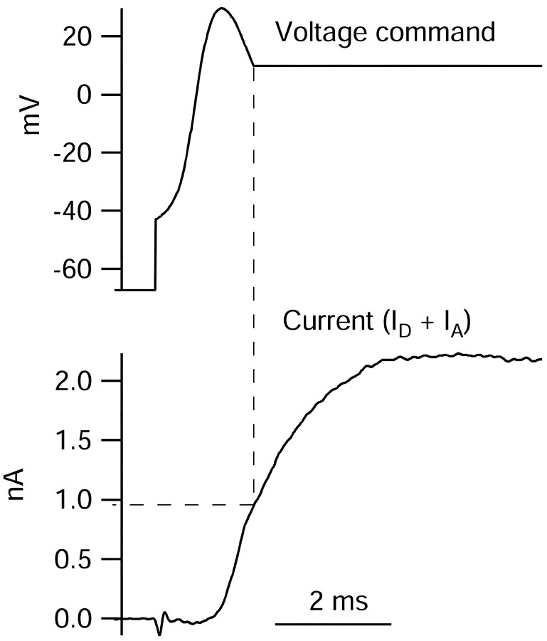
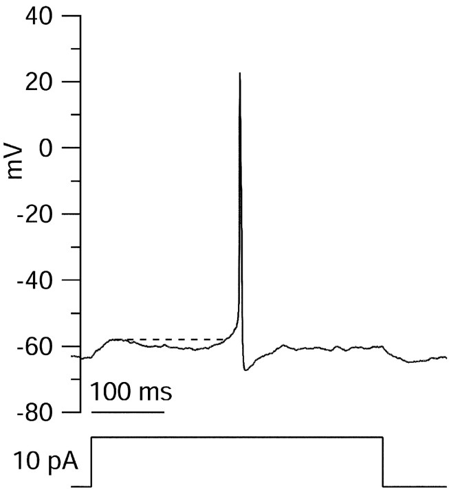
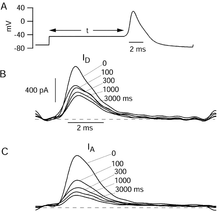
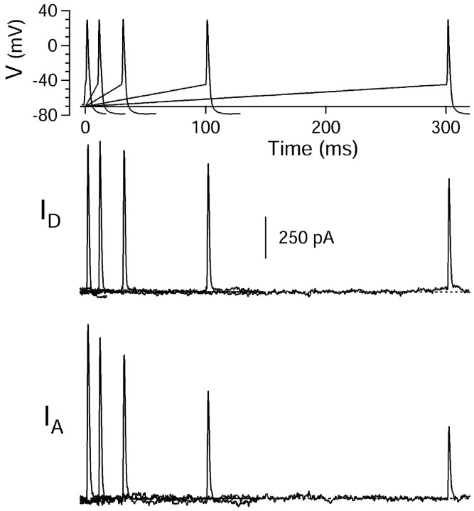
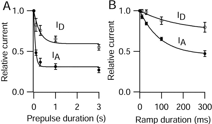
Central neurons have multiple types of voltage-dependent potassium channels, whose activation during action potentials shapes spike width and whose activation and inactivation at subthreshold voltages modulate firing frequency. We characterized the voltage-dependent potassium currents flowing during the action potentials of hippocampal CA3 pyramidal neurons and examined the susceptibility of the underlying channel types to inactivation at subthreshold voltages. Using acutely dissociated neurons that permitted rapid voltage clamp, action potentials recorded previously were used as the command voltage waveform, and individual components of potassium current were identified by pharmacological sensitivity. The overall voltage-dependent potassium current in the neurons could be split into three major components based on pharmacology and kinetics during step voltage pulses: I(D) (fast activating, slowly inactivating, and sensitive to 4-aminopyridine at 30 microm), I(A) (fast activating, fast inactivating, and sensitive to 4-aminopyridine at 3 mm), and I(K) (slowly activating, noninactivating, and sensitive to external TEA at 3-25 mm). The potassium current during the action potential was composed of approximately equal contributions of I(D) and I(A), with a negligible contribution of I(K). I(D) and I(A) had nearly identical trajectories of activation and deactivation during the action potential. Both I(A) and I(D) showed steady-state inactivation at subthreshold voltages, but maximal inactivation at such voltages was incomplete for both currents. Because of the major contribution of both I(D) and I(A) to spike repolarization, it is likely that modulation or partial inactivation at subthreshold voltages of either current can influence spike timing with minimal effect on spike width.


Figures

Fig. 1.
Fig. 1.
Action potentials in an isolated CA3 neuron. The cell was hyperpolarized by steady injection of DC (−55 pA), and action potentials were triggered by 1 msec injections of current of increasing amplitude. Bottom panel, The first derivative of the action potentials. External solution was normal Tyrode's solution with 2 mm CaCl2.
Fig. 2.
Fig. 2.
Effect of potassium channel blockers on action potential shape. A, 4-AP at 30 μm and 2.5 mm produced a dose-dependent delay in repolarization. Hyperpolarizing DC was −30 pA, and stimulus current (1 msec) was 490 pA. B, TEA at 25 mm had little effect on the initial phase of repolarization but produced a dramatic slowing of late repolarization. Hyperpolarizing DC was −40 pA, and injected current was 780 pA. C, Replacing external 2 mmcalcium with 2 mm cobalt resulted in a delay of firing and little overall change in action potential shape. Action potentials were aligned at the time of maximal upstroke to allow comparison of time course. Hyperpolarizing DC was −6 pA, and injected current was 850 pA.
Fig. 3.
Fig. 3.
Isolation of IA by prepulse inactivation. Cells were held at −90 mV, and the voltage was stepped to 0 mV with or without a prepulse to −45 mV. There was a 2 msec return to −90 mV after the prepulse. Subtraction yields fast-activating and fast-inactivating outward current. External solution was 2 mm cobalt Tyrode's solution to block voltage-dependent calcium channels and calcium-activated potassium channels. TTX (1 μm) was included to block voltage-dependent sodium channels. Asterisks indicate records with prepulse. Dotted lines indicate zero current.
Fig. 4.
Fig. 4.
Differential pharmacological sensitivity of fast-inactivating and slowly inactivating components of potassium current. Currents were elicited by the protocol in Figure 3 using pairs of test pulses to 0 mV with and without prepulses. Filled circles indicate the fast-inactivating component of current obtained by subtraction. Open circles indicate measurement of the current at the end of the step to 0 mV.Inset, top, Superimposed currents with and without prepulse. Bottom, Fast-inactivating component of current obtained by subtraction of these records. Dotted lines indicate zero current.
Fig. 5.
Fig. 5.
Strategy for pharmacological separation ofIA,ID, andIK. Left panel, Sequentially recorded traces obtained in control solution (2 mm cobalt Tyrode's solution with 1 μm TTX) and in the presence of 30 μm 4-AP, 3 mm 4-AP, and 3 mm 4-AP plus 25 mm TEA. Right panel, Subtracted currents sensitive to 30 μm4-AP (ID), 3 mm 4-AP but not 30 μm 4-AP (IA), and 25 mm TEA but not 3 mm 4-AP (IK). Horizontal dotted lines indicate zero current.
Fig. 6.
Fig. 6.
Ionic current elicited by action potential waveform clamp. A, Command waveform, consisting of a previously recorded action potential (peak, +51 mV; maximal upstroke, +435 mV/msec; maximal downstroke, −62 mV/msec). B, Ionic currents elicited by the action potential waveform in normal Tyrode's solution (containing 2 mmCaCl2) and after application of 1 μmTTX. C, Ionic currents plotted as a function of voltage during the action potential.
Fig. 7.
Fig. 7.
Pharmacological dissection of potassium current elicited by action potential waveform. A, Current elicited by action potential waveform in Tyrode's solution with calcium replaced by cobalt to eliminate voltage-activated calcium current and with 1 μm TTX to block sodium current. Action potential (peak, +30 mV; maximal upstroke, +135 mV/msec; maximal downstroke, −50 mV/msec) was recorded previously in normal Tyrode's solution. To separate IA,ID, andIK, 4-AP and TEA were applied according to the strategy summarized in Figure 5. B,IA,ID, and IKobtained by subtraction. ID was obtained as the current sensitive to 30 μm 4-AP,IA as the current sensitive to 3 mm 4-AP but not 30 μm 4-AP, andIK as the current sensitive to 25 mm TEA but not 3 mm 4-AP.
Fig. 8.
Fig. 8.
Assay of extent of activation ofIA and ID during an action potential. Total IA andID were isolated as current sensitive to 3 mm 4-AP. Voltage command consisted of a partial action potential interrupted and extended at +10 mV on the falling phase. Current reached at +10 mV during the falling phase was 0.95 nA, compared with a peak of 2.23 nA reached ∼3 msec later at the same voltage. The vertical dashed line indicates time at which falling phase of action potential reaches +10 mV; the horizontal dashed line indicates outward current at this time.
Fig. 9.
Fig. 9.
Delayed firing of an action potential near threshold. An action potential elicited near threshold with stimulation by 400 msec steps of current is shown. Note relaxation from initial depolarization, followed by a second phase of depolarization and then spike firing. Spike occurs 203 msec after start of current step. Thedashed line indicates voltage reached during initial depolarizing phase (−58 mV).
Fig. 10.
Fig. 10.
Partial inactivation ofIA and ID by prepulses to −45 mV. A, Currents were elicited by an action potential waveform preceded by a variable length prepulse to −45 mV, from a steady holding potential of −70 mV.Arrows indicate variable length of prepulse (0-3000 msec).B, ID (obtained as fraction of current sensitive to 30 μm 4-AP) elicited by the action potential after prepulses of indicated lengths.C, IA (obtained as fraction of current sensitive to 3 mm 4-AP but not 30 μm 4-AP) elicited by the action potential after prepulses of indicated lengths. External solution was 2 mmcobalt Tyrode's containing 1 μm TTX.Dashed lines indicate zero current.
Fig. 11.
Fig. 11.
Partial inactivation ofIA and ID by ramps preceding the action potential. Currents were elicited by an action potential waveform preceded by ramps (of variable lengths from 0 to 300 msec) from −70 mV to −45 mV (top panel).ID (middle panel) was obtained as the fraction of current sensitive to 30 μm4-AP elicited by the waveforms. IA was obtained as the fraction of current sensitive to 3 mm 4-AP but not 30 μm 4-AP. External solution was 2 mm cobalt Tyrode's containing 1 μmTTX.
Fig. 12.
Fig. 12.
Time course of reduction ofID and IA by prepulses or ramps to threshold potentials. A, Normalized peak current of ID (○;n = 3) or IA (●;n = 4) elicited by the action potential waveform is plotted against the duration of a step depolarization to −45 mV. Error bars show mean ± SEM. Fittedcurves are single exponential functions.ID, Time constant of 282 msec; steady-state, 59%. IA, Time constant of 75 msec; steady-state, 31%. B, Normalized peak current of ID (○; n = 7) or IA (●; n = 9) elicited by the action potential waveform is plotted against the duration of a depolarizing ramp as in Figure 11.ID, Time constant of 183 msec; steady-state, 75%. IA, Time constant of 94 msec; steady-state, 45%.

Similar articles

Cited by

References

    1. Bähring R, Boland LM, Varghese A, Gebauer M, Pongs O. Kinetic analysis of open- and closed-state inactivation transitions in human Kv4.2 A-type potassium channels. J Physiol (Lond) 2001;535:65–81. - PMC - PubMed
    1. Bergles DA. PhD thesis. Stanford University; 1995. The actions of norepinephrine on hippocampal interneurons.
    1. Bossu JL, Capogna M, Debanne D, McKinney RA, Gahwiler BH. Somatic voltage-gated potassium currents of rat hippocampal pyramidal cells in organotypic slice cultures. J Physiol (Lond) 1996;495:367–381. - PMC - PubMed
    1. Brown DA, Gahwiler BH, Griffith WH, Halliwell JV. Membrane currents in hippocampal neurons. Prog Brain Res. 1990;83:141–160. - PubMed
    1. Castro PA, Cooper EC, Lowenstein DH, Baraban SC. Hippocampal heterotopia lack functional Kv4.2 potassium channels in the methylazoxymethanol model of cortical malformations and epilepsy. J Neurosci. 2001;21:6626–6634. - PMC - PubMed

Publication types

MeSH terms