. 2002 Dec 1;22(23):10163-71.
doi: 10.1523/JNEUROSCI.22-23-10163.2002.

Small conductance Ca2+-activated K+ channels modulate synaptic plasticity and memory encoding

Affiliations

Small conductance Ca2+-activated K+ channels modulate synaptic plasticity and memory encoding

Robert W Stackman et al. J Neurosci. .

Abstract

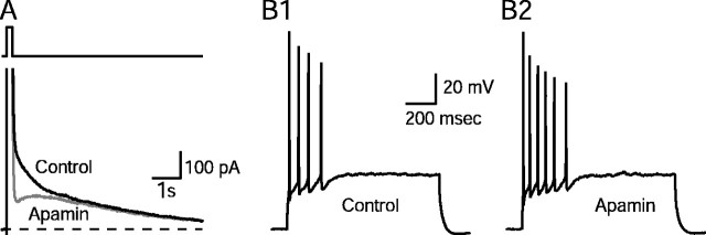
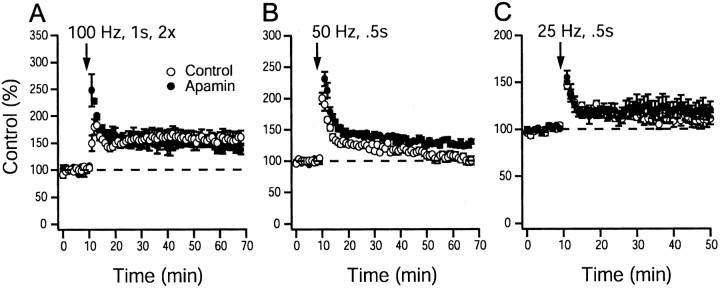
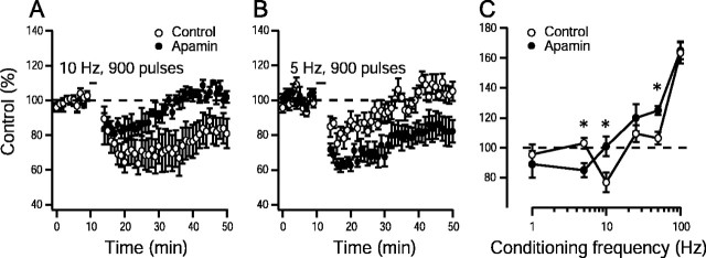
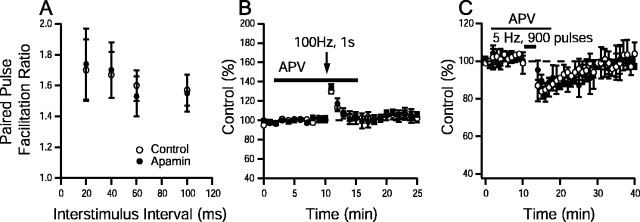
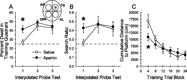
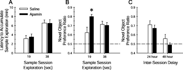
Activity-dependent changes in neuronal excitability and synaptic strength are thought to underlie memory encoding. In hippocampal CA1 neurons, small conductance Ca2+-activated K+ (SK) channels contribute to the afterhyperpolarization, affecting neuronal excitability. In the present study, we examined the effect of apamin-sensitive SK channels on the induction of hippocampal synaptic plasticity in response to a range of stimulation frequencies. In addition, the role of apamin-sensitive SK channels on hippocampal-dependent memory encoding and retention was also tested. The results show that blocking SK channels with apamin increased the excitability of hippocampal neurons and facilitated the induction of synaptic plasticity by shifting the modification threshold to lower frequencies. This facilitation was NMDA receptor (NMDAR) dependent and appeared to be postsynaptic. Mice treated with apamin demonstrated accelerated hippocampal-dependent spatial and nonspatial memory encoding. They required fewer trials to learn the location of a hidden platform in the Morris water maze and less time to encode object memory in an object-recognition task compared with saline-treated mice. Apamin did not influence long-term retention of spatial or nonspatial memory. These data support a role for SK channels in the modulation of hippocampal synaptic plasticity and hippocampal-dependent memory encoding.


Figures

Fig. 1.
Fig. 1.
Blockade of the apamin-sensitive afterhyperpolarization (mAHP) increases excitability.A, IAHPs were evoked in the whole-cell configuration by a 200 msec depolarizing pulse to +20 mV followed by a return to the −55 mV holding potential.IAHPs were obtained in the presence and absence of apamin (100 nm). After application of apamin, the medium-duration component (ImAHP) of the tail current was selectively inhibited. Dashed lineindicates zero current. B, Apamin increased the number of action potentials. B1, Response of a pyramidal neuron to a 1 sec depolarizing current pulse. B2, Response of the same neuron to the same depolarizing current pulse in the presence of apamin (control cells fired an average ± SEM of 4.7 ± 1.2 action potentials/depolarizing pulse, which increased to 6.7 ± 1.7 with apamin; n = 5; p = 0.04; paired Student's t test).
Fig. 2.
Fig. 2.
Apamin block of SK channel activity enhances plasticity induced by high-frequency stimulation.A, A 100 Hz, 1 sec tetanus in control and apamin (100 nm)-treated slices (164 ± 7%,n = 9 slices per 6 animals for controls; 165 ± 6%, n = 10 slices per 6 animals for apamin;p > 0.05; unpaired Student's ttest). B, A 50 Hz, 0.5 sec stimulation protocol in control and apamin (100 nm)-treated slices (106 ± 4%, n = 12 slices per 8 animals for control slices, 125 ± 3%, n = 13 slices per 8 animals for apamin-treated slices; p < 0.05; unpaired Student's t test). C, A 25 Hz, 0.5 sec stimulation protocol in control and apamin (100 nm)-treated slices (109 ± 9%, n= 8 slices per 6 animals for controls; 120 ± 6%,n = 8 slices per 6 animals for apamin;p > 0.05; unpaired Student's ttest). Control and apamin-treated slices were interleaved. Synaptic strength was measured as the initial slope of the recorded field EPSP. Dashed line indicates baseline response inA–C.
Fig. 3.
Fig. 3.
Apamin block of SK channel activity shifts the synaptic modification threshold to lower frequencies. Induction of synaptic plasticity by 10 Hz, 900 pulse stimulation in control slices (77 ± 6%; n = 10 slices per 6 animals) and apamin (100 nm)-treated slices (101 ± 7%; n = 9 slices per 5 animals;p < 0.05; unpaired Student's ttest) (A) and 5 Hz, 900 pulse stimulation protocol in control slices (103 ± 4%; n = 8 slices per 5 animals) and apamin (100 nm)-treated slices (85 ± 5%; n = 9 slices per 5 animals;p < 0.05; unpaired Student's ttest) (B). Dashed line indicates baseline response. C, Frequency–response relationship for the induction of LTP and LTD in controls and experiments from slices in which apamin (100 nm) was applied. The mean effect of 900 pulses of conditioning stimulation delivered at various frequencies to the Shaffer collaterals on the synaptic response measured 40–50 min after conditioning is shown. ∗p < 0.05 versus respective control data point; Student's t test. Dashed line indicates the transition between LTD and LTP.
Fig. 4.
Fig. 4.
SK channels do not have presynaptic effects in CA1. A, Paired-pulse facilitation (PPF), measured as the ratio of the second response to the first, was plotted as a function of interstimulus interval for controls and in the presence of apamin (n > 8 for all interstimulus intervals). No significant differences were detected (p > 0.05; paired Student's t test). B, Time course of post-tetanic potentiation elicited by 100 Hz, 1 sec tetanus in control and apamin-treated slices. Post-tetanic potentiation (peak enhancement in controls, 132 ± 6% of baseline,n = 9 slices per 4 animals; peak enhancement in apamin, 134 ± 5% of baseline, n = 9 slices per 4 animals) was not different between groups (p > 0.05; unpaired Student'st test). C, Time course of short-term depression elicited by 5 Hz, 900 pulse stimulation in control and apamin-treated slices (80 ± 7% of baseline,n = 6 slices per 3 animals; peak depression in apamin, 82 ± 5% of baseline, n = 6 slices per 3 animals). No significant differences were detected between groups (p > 0.05; paired Student'st test). Synaptic strength was measured as the initial slope of the recorded field EPSP. Solid line in Band C indicates the duration of d-APV application.
Fig. 5.
Fig. 5.
Apamin block of SK channels facilitates the encoding of spatial memory. A, A modified Morris water maze task was used to examine the effects of apamin on encoding of spatial memory. Mice were trained for four trials per day for 6 d, and 30 sec probe tests were presented immediately after the fourth, 12th, and 20th trial. Mean ± SEM percentage of time spent dwelling (Percent Dwell) in the training quadrant during the interpolated probe tests revealed that mice treated with 0.4 mg/kg apamin (n = 10) spent significantly more time in the training quadrant during the first probe test than saline-treated (n = 9) control mice (∗p < 0.009 vs saline-treated mice on probe test 1; planned comparison Student's t test). Thedashed line at 25% represents chance performance.AL, Adjacent left; AR, adjacent right; OPP, opposite; TQ, training quadrant. B, Mean ± SEM search ratio reflects the accuracy with which mice search in the correct location within the training quadrant of the pool. Search ratio is computed as the number of times the animal crosses into the zone (see circular regions of inset diagram) encompassing the platform (shaded zone) divided by the total number of crossings into all four zones. The dashed line at 0.25 represents chance performance during the probe tests or the lack of spatial bias for any particular pool location. Apamin-treated mice exhibited a significantly higher search ratio than saline-treated mice during the first probe test (∗p < 0.02 vs saline-treated mice on probe test 1; planned comparison Student'st test). Measures of the percentage of time spent dwelling in the training quadrant or search ratio from the second or third probe tests were equivalent between the two groups, indicating that there were no group differences in platform search behavior after more training. C, Mean ± SEM cumulative distance to platform measures of saline- and apamin-treated mice plotted in blocks of four training trials. This measure indicates the proximity of the mice to the platform during each training trial. Consistent with the data from probe test 1, apamin-treated mice swam in closer proximity to the platform during the first four trial block of training than saline-treated mice (∗p < 0.04; post hoc Tukey multiple comparisons test).
Fig. 6.
Fig. 6.
Apamin block of SK channel activity facilitates the encoding of nonspatial object memory but does not influence the retention of object memory. Object-recognition memory was quantified by computing the novel object preference ratio, the amount of time spent exploring the novel object during the test session divided by the total time spent exploring both the familiar and novel object. A, The object recognition task was modified to test the influence of apamin on object memory encoding. As described in Materials and Methods, during the sample session, saline- and apamin-treated mice were restricted to either 19 sec (minimal training) or 38 sec (extensive training) of sample object exploration. The amount of time required to accumulate either 19 or 38 sec of sample object exploration did not differ between apamin- and saline-treated mice (p values >0.05; unpaired Student's t test). B, Restricting the amount of object exploration during the sample session to 19 sec weakens the degree of preference exhibited by the mouse during a test session 24 hr later. This is illustrated by the lower novel object preference ratio of the saline-treated mice (n = 9) limited to 19 sec of sample object exploration. However, apamin (0.4 mg/kg)-treated mice (n = 10) that were limited to only 19 sec of sample object exploration exhibited a significantly greater novel object preference during the 24 hr test session (∗p < 0.04 vs saline-treated mice permitted 19 sec of object exploration; planned comparison Student'st test). When apamin (0.4 mg/kg)-treated (n = 10) and saline-treated (n= 9) mice were permitted 38 sec of sample object exploration, there was no difference in novel object preference ratio during the 24 hr test session. Each dashed line at 0.5 represents chance performance or a lack of discrimination between the novel and familiar object. C, Object memory retention decays over a similar time course in apamin- and saline-treated mice. Both apamin- and saline-treated mice exhibited similar strong preference for the novel object during a 24 hr retention test; mean ± SEM novel object preference ratios were not significantly different. Four days later, the same mice received a second sample session with two new objects. When tested for retention 48 hr later, both apamin- and saline-treated mice failed to show a strong preference for the novel object over the familiar object. These data indicate that apamin does not affect the retention of object memory.

References

    1. Alford S, Frenguelli BG, Schofield JG, Collingridge GL. Characterization of Ca2+ signals induced in hippocampal CA1 neurones by the synaptic activation of NMDA receptors. J Physiol (Lond) 1993;469:693–716. - PMC - PubMed
    1. Andreasen M, Lambert JD. The excitability of CA1 pyramidal cell dendrites is modulated by a local Ca2+-dependent K+-conductance. Brain Res. 1995;698:193–203. - PubMed
    1. Artola A, Singer W. Long-term depression of excitatory synaptic transmission and its relationship to long-term potentiation. Trends Neurosci. 1993;16:480–487. - PubMed
    1. Bear MF. Mechanism for a sliding synaptic modification threshold. Neuron. 1995;15:1–4. - PubMed
    1. Behnisch T, Reymann KG. Inhibition of apamin-sensitive calcium dependent potassium channels facilitate the induction of long-term potentiation in the CA1 region of rat hippocampus in vitro. Neurosci Lett. 1998;253:91–94. - PubMed

Publication types

MeSH terms

Substances

LinkOut - more resources