. 2002 Dec 1;22(23):10172-81.
doi: 10.1523/JNEUROSCI.22-23-10172.2002.

Changes in conformation and subcellular distribution of alpha4beta2 nicotinic acetylcholine receptors revealed by chronic nicotine treatment and expression of subunit chimeras

Affiliations

Changes in conformation and subcellular distribution of alpha4beta2 nicotinic acetylcholine receptors revealed by chronic nicotine treatment and expression of subunit chimeras

Patricia C Harkness et al. J Neurosci. .

Abstract

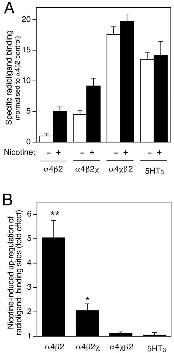
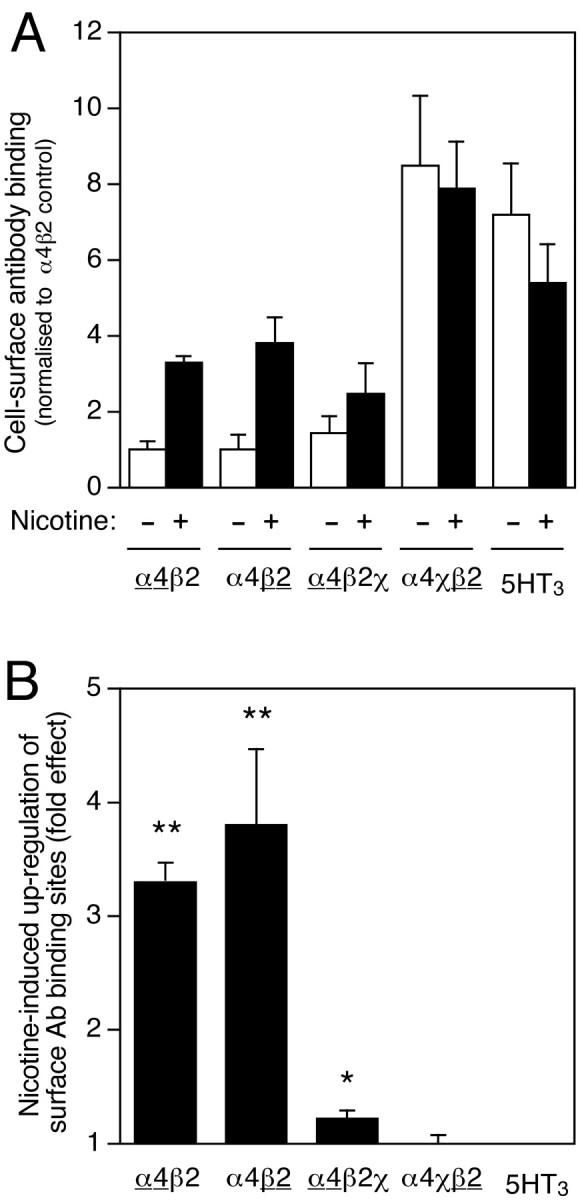
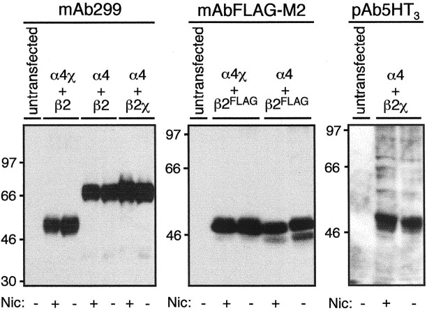
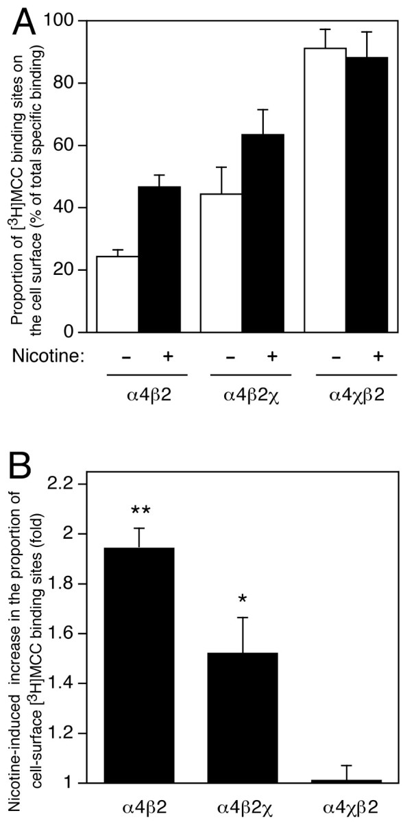
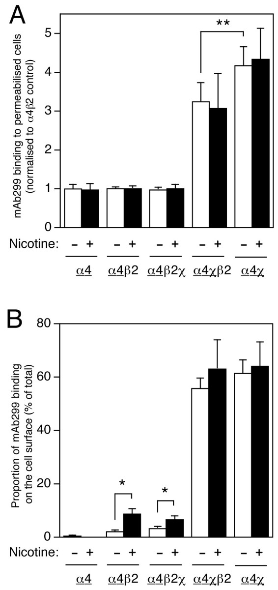
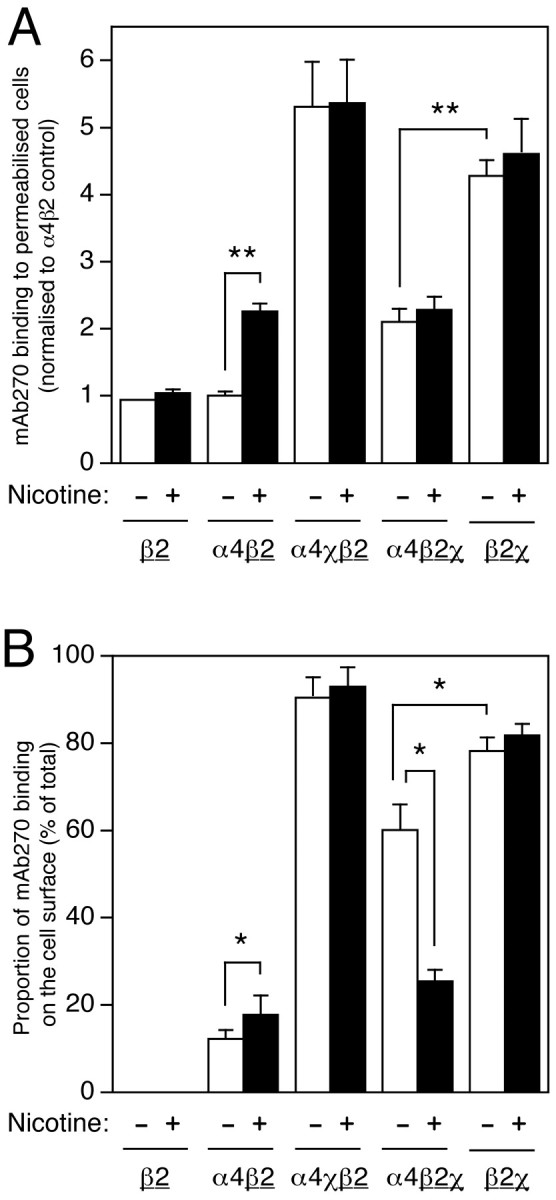
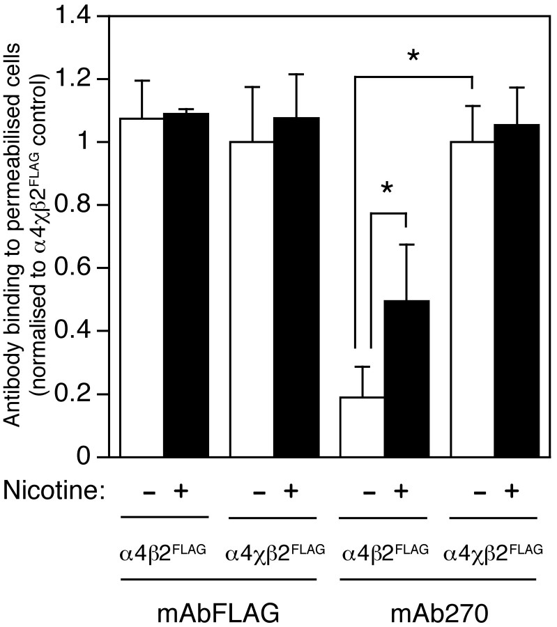
Chronic exposure to nicotine, as occurs during tobacco smoking, is one of several factors that have been reported to cause an upregulation of neuronal nicotinic acetylcholine receptors (nAChRs). Here, the influence of both chronic exposure to nicotine (10 microm, 24 hr) and the coexpression of subunit chimeras has been examined in cultured cell lines expressing recombinant alpha4beta2 nAChRs, a major nicotinic receptor subtype expressed in the mammalian brain. Evidence is presented which demonstrates that both chronic exposure to nicotine and the coexpression of subunit chimeras upregulates levels of receptor expressed on the cell surface. Immunoblotting data indicate that neither chronic nicotine treatment nor coexpressed subunit partners greatly affect the level of total subunit protein. This finding, together with radioligand and antibody binding studies conducted on both intact and permeabilized cells, reveals that receptor upregulation corresponds to an increase in the proportion of total receptor expressed on the cell surface. It is also apparent that nicotine-induced nAChR upregulation is very strongly dependent on subunit composition and subunit domains. An important aspect of this study is that direct evidence has been obtained indicating that both chronic exposure to nicotine and coexpressed subunit partners can influence subunit conformation. The influence of chronic nicotine treatment on subunit folding may help to explain the phenomenon of nicotine-induced receptor upregulation. The finding that subunit conformation can be influenced by coassembled subunit partners is in agreement with models of receptor assembly which propose that subunit folding continues after initial subunit-subunit interactions.


Figures

Fig. 1.
Fig. 1.
Influence of chronic nicotine treatment and subunit composition on radioligand binding to transfected TSA201 cells.A, Specific radioligand binding to cell homogenates from transfected cells was determined using a saturating concentration of [3H]epibatidine (2 nm, for α4β2, α4β2χ, and α4χβ2) or [3H]GR-65630 (10 nm, for 5HT3A). For all subunit combinations (α4β2, α4β2χ, α4χβ2, and 5HT3A), radioligand binding was determined in cells that had been grown in the absence (white bars) or presence (black bars) of nicotine (10 μm, 24 hr). Data represent means ± SEM of four independent experiments that have been normalized to the level of binding obtained with α4β2 in the absence of nicotine (0.15 ± 0.04 pmol/mg protein; n = 8). B, Data have been replotted to show the extent of nicotine-induced upregulation of radioligand binding for each subunit combination. Significant differences from control, determined by two-tailed Student'st test, are indicated (*p < 0.05; **p < 0.001).
Fig. 2.
Fig. 2.
Influence of chronic nicotine treatment and subunit composition on cell-surface antibody binding. A, Surface antibody binding to intact cell monolayers was determined with mAb270 (anti-β2), mAb299 (anti-α4), or pAb120 (anti-5HT3A) using an enzyme-linked antibody binding assay (see Materials and Methods). Surface levels of α4χβ2 were assayed by mAb270 binding, and levels of α4β2χ were assayed by mAb299 binding, thus ensuring that only assembled cell-surface complexes were detected (neither α4 nor β2 is expressed on the cell surface when expressed individually). Surface levels of α4β2 were assayed separately with both mAb270 and mAb299. For all subunit combinations (α4β2, α4β2χ, α4χβ2, and 5HT3A), antibody binding was determined in cells that had been grown in the absence (white bars) or presence (black bars) of nicotine (10 μm, 24 hr). The background signal from mock-transfected coverslips has been subtracted. Data are the means ± SEM of four independent experiments and have been normalized to the level of surface antibody binding determined with α4β2 in the absence of nicotine.B, Data have been replotted to show the extent of nicotine-induced upregulation of antibody binding for each subunit combination. Where two subunits are coexpressed, the subunit assayed by mAb binding is underlined. Statistical significance of the results, determined by two-tailed Student's t test, is indicated (*p = 0.05; **p < 0.01).
Fig. 3.
Fig. 3.
Influence of chronic nicotine treatment and subunit composition on total subunit protein levels determined by immunoblotting. Total cellular protein (150 μg) from TSA201 cells transfected with various subunit combinations and grown in the presence or absence of nicotine (10 μm, 24 hr) was separated by SDS-PAGE and immunoblotted with mAb299, which recognizes α4 and α4χ, mAbFLAG-M2, which recognizes epitope-tagged β2FLAG, or pAb5HT3, which recognizes β2χ. Specific immunoreactive bands (absent from mock-transfected control cells) were detected for α4 (∼70 kDa), α4χ (∼55 kDa), β2FLAG (∼55 kDa), and β2χ (∼55 kDa). The positions of protein molecular weight markers are shown.
Fig. 4.
Fig. 4.
Influence of chronic nicotine treatment and subunit composition on the proportion of radioligand binding sites expressed on the cell surface. A, Specific binding of the membrane-impermeant nicotinic radioligand [3H]MCC was determined for intact and homogenized cells in the absence (white bars) and presence (black bars) of nicotine (10 μm, 24 hr). Data are presented as the proportion of binding sites on the cell surface (as a percentage of total specific binding). Data are means ± SEM of four to six independent experiments using a saturating concentration of ligand (25 nm).B, Data have been replotted to show the extent of nicotine-induced upregulation of cell-surface radioligand binding for each subunit combination. Statistical significance of the results, determined by two-tailed Student's t test, is indicated (*p = 0.067; **p < 0.01).
Fig. 5.
Fig. 5.
Influence of chronic nicotine treatment and subunit composition on subcellular distribution of α4 and α4χ subunits. Transfected TSA201 cells, grown on coverslips, were labeled with mAb299 (specific for α4 and α4χ subunits) either after membrane permeabilization or as intact cell monolayers. Data are presented as total antibody binding to permeabilized cells (A) and as the proportion of antibody binding sites on the cell surface (B). The level of mAb299 binding was determined in an enzyme-linked assay (see Materials and Methods). For all subunit combinations, mAb299 binding was determined in cells that had been grown in the absence (white bars) or presence (black bars) of nicotine (10 μm, 24 hr). Data are the means of five to nine independent experiments, each performed in duplicate, and have been normalized to the level of mAb299 binding determined for permeabilized cells transfected with α4β2 in the absence of nicotine. The background signal from mock-transfected coverslips has been subtracted. B, The proportion of mAb299 binding detected on the cell surface for each subunit combination has been determined from parallel experiments performed on permeabilized and intact cell monolayers. Because α4χ can form homomeric (as well as heteromeric) cell-surface complexes (Cooper et al., 1999), mAb299 would be expected to detect a heterogeneous population of cell-surface complexes in cells cotransfected with α4χ + β2. To emphasize which subunit is being assayed by mAb299 binding (α4 or α4χ, rather than β2 or β2x), the appropriate subunit is underlined. Significant differences, determined by two-tailed Student's t test, are indicated (*p < 0.05; **p < 0.02).
Fig. 6.
Fig. 6.
Influence of chronic nicotine treatment and subunit composition on subcellular distribution of β2 and β2χ subunits. Transfected TSA201 cells, grown on coverslips, were labeled with mAb270 (specific for β2 and β2χ subunits) either after membrane permeabilization or as intact cell monolayers. Data are presented as total antibody binding to permeabilized cells (A) and as the proportion of antibody binding sites on the cell surface (B). The level of mAb270 binding was determined in an enzyme-linked assay (see Materials and Methods). For all subunit combinations, mAb270 binding was determined in cells that had been grown in the absence (white bars) or presence (black bars) of nicotine (10 μm, 24 hr). Data are the means of 5–11 independent experiments, each performed in duplicate, and have been normalized to the level of mAb270 binding determined with permeabilized cells transfected with α4β2 in the absence of nicotine. The background signal from mock-transfected coverslips has been subtracted. B, The proportion of mAb270 binding detected on the cell surface for each subunit combination has been determined from parallel experiments performed on permeabilized and intact cell monolayers. Because β2χ can form homomeric (as well as heteromeric) cell-surface complexes (Cooper et al., 1999), mAb270 would be expected to detect a heterogeneous population of cell-surface complexes in cells cotransfected with α4 + β2χ. To emphasize which subunit is being assayed by mAb270 binding (β2 or β2χ, rather than α4 or α4χ), the appropriate subunit is underlined. Significant differences, determined by two-tailed Student'st test, are indicated (*p < 0.05; **p < 0.01).
Fig. 7.
Fig. 7.
Influence of chronic nicotine treatment and subunit composition on levels of β2FLAG detected in permeabilized cells. Transfected TSA201 cells, grown on coverslips, were labeled with either mAb270 or mAbFLAG-M2 (both of which recognize β2FLAG). The level of antibody binding was determined after membrane permeabilization in an enzyme-linked assay using an HRP-linked secondary antibody (see Materials and Methods). For all subunit combinations, antibody binding was determined in cells that had been grown in the absence (white bars) or presence (black bars) of nicotine (10 μm, 24 hr). Data are the means of two to three independent experiments, each performed in duplicate, and have been normalized to the level of binding determined with cells transfected with α4χβ2FLAG in the absence of nicotine. The background signal from mock-transfected coverslips has been subtracted. Significant differences, determined by two-tailed Student'st test, are indicated (*p < 0.05).

Similar articles

Cited by

References

    1. Bencherif M, Fowler K, Lukas R, Lippiello PM. Mechanisms of up-regulation of neuronal nicotinic acetylcholine receptors in clonal cell lines and primary cultures of fetal rat brain. J Pharmacol Exp Ther. 1995;275:987–994. - PubMed
    1. Benwell MEM, Balfour DJK, Anderson JM. Evidence that tobacco smoking increases the density of (−)-[3H]nicotine binding sites in human brain. J Neurochem. 1988;50:1243–1247. - PubMed
    1. Buisson B, Gopalakrishnan M, Arneric SP, Sullivan JP, Bertrand D. Human α4β2 neuronal nicotinic acetylcholine receptor in HEK 293 cells: a patch-clamp study. J Neurosci. 1996;16:7880–7891. - PMC - PubMed
    1. Chen D, Dang H, Patrick JW. Contributions of N-linked glycosylation to the expression of a functional α7-nicotinic receptor in Xenopus oocytes. J Neurochem. 1998;70:349–357. - PubMed
    1. Cooper ST, Millar NS. Host cell-specific folding and assembly of the neuronal nicotinic acetylcholine receptor α7 subunit. J Neurochem. 1997;68:2140–2151. - PubMed

Publication types

MeSH terms

LinkOut - more resources