. 2002 Dec 1;22(23):10209-16.
doi: 10.1523/JNEUROSCI.22-23-10209.2002.

DRPEER: a motif in the extracellular vestibule conferring high Ca2+ flux rates in NMDA receptor channels

Affiliations

DRPEER: a motif in the extracellular vestibule conferring high Ca2+ flux rates in NMDA receptor channels

Junryo Watanabe et al. J Neurosci. .

Abstract

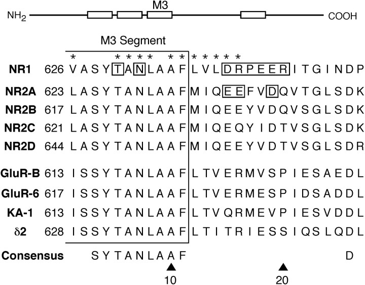
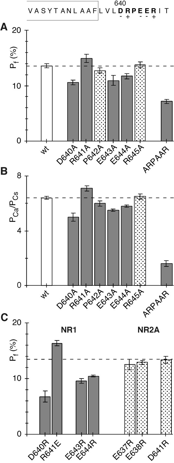
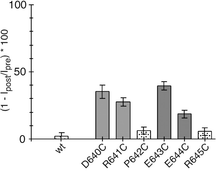
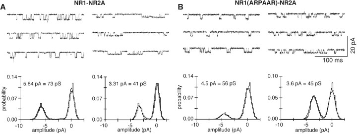
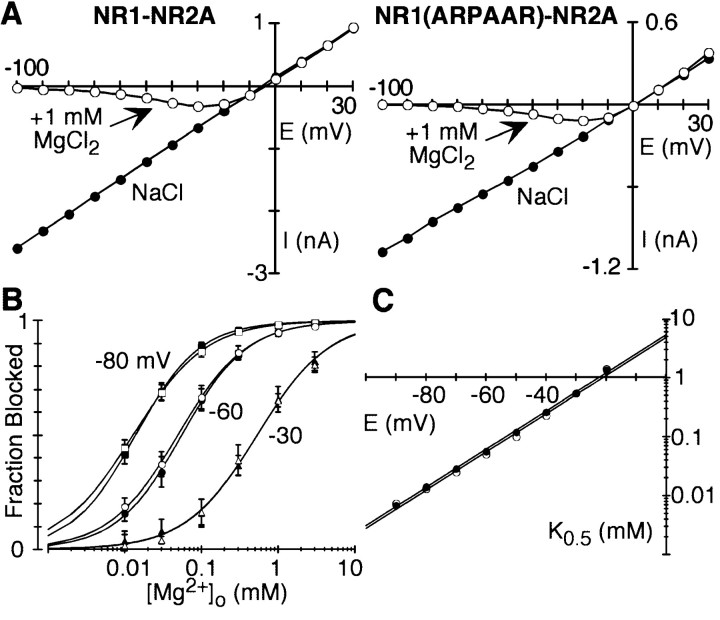
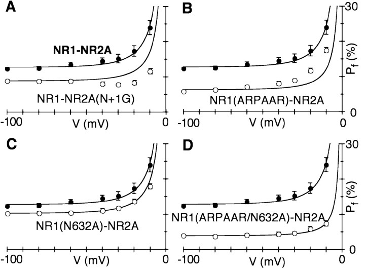
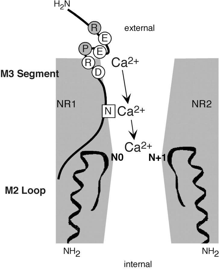
The high flux rate of Ca2+ through NMDA receptor (NMDAR) channels is critical for their biological function and may depend on a Ca2+ binding site in the extracellular vestibule. We screened substitutions of hydrophilic residues exposed in the vestibule and identified a cluster of charged residues and a proline, the DRPEER motif, positioned C terminal to M3, that is unique to the NR1 subunit. Charge neutralization or conversion of residues in DRPEER altered fractional Ca2+ currents in a manner consistent with its forming a binding site for Ca2+. Similarly, in a mutant channel in which all of the negative charges are neutralized (ARPAAR), the block by extracellular Ca2+ of single-channel current amplitudes is attenuated. In these same channels, the block by extracellular Mg2+ is unaffected. DRPEER is located extracellularly, and its contribution to Ca2+ influx is distinct from that of the narrow constriction. We conclude that key residues in DRPEER, acting as an external binding site for Ca2+, along with a conserved asparagine in the M3 segment proper, contribute to the high fractional Ca2+ currents in these channels under physiological conditions. Therefore, these domains represent critical molecular determinants of NMDAR function in synaptic physiology.


Figures

Fig. 1.
Fig. 1.
Sequence alignment of the C-terminal half and residues C terminal to the M3 segment in GluR subtypes. In this schematic drawing of GluR subunits, the four hydrophobic segments (M1–M4) are indicated as open boxes. Theenlarged region shows a sequence alignment of amino acid residues on the C-terminal end and C terminal to M3. For clarity, only a subset of the receptor subtypes is shown (the region is highly conserved within subunits of the same subtype). The sequence numbers on the left are for the mature protein. For NR1, the asterisks indicate positions exposed to the water interface (Beck et al., 1999). The boxed positionsfor NR1 and NR2A were tested for their effects on Ca2+ permeability. The lower consensus sequence is found in all GluR subunits.
Fig. 2.
Fig. 2.
Fractional Ca2+ currents in wild-type and mutant NMDAR channels. A, MeanPf values in wild-type (wt) and mutant NR1–NR2A channels containing alanine substitutions of the DRPEER motif. Cells were bathed in 143.5 mm NaCl and 1.8 mm CaCl2. The holding potential (V) was −60 mV to the reversal potential (Jatzke et al., 2002). Pf values significantly different from those in wild type are shown asgray bars. B, Mean Ca2+ permeability ratios (PCa/PCs) in wt and mutant NR1–NR2A channels containing alanine substitutions of the DRPEER motif.PCa/PCsvalues were derived from changes in reversal potentials ongoing from a Cs+-based reference solution to a solution in which CsCl was replaced by 1.8 mm Ca2+ and 140 mm NMDG (see Materials and Methods).PCa/PCsvalues significantly different from those in wild type are shown asgray bars. C, MeanPf values in mutant channels containing oppositely charged substitutions of the DRPEER motif or of homologous positions in NR2A (see Fig. 1). Results are recorded and displayed as in A, except that the wild-type value is shown as adashed line.
Fig. 3.
Fig. 3.
Accessibility of substituted cysteines in DRPEER to MTSEA. The mean percentage of change (n >5) in current amplitudes measured before (Ipre) and after (Ipost) exposure to MTSEA (2 mm, 60 sec application) (see Materials and Methods) is shown. MTS reagents were applied in the presence of glutamate. Statistically significant positions are shown as gray bars. wt, Wild type.
Fig. 4.
Fig. 4.
Block by Ca2+ in wild-type and mutant NMDAR channels. A, Single-channel currents at −80 mV in the absence (1.5 mm EGTA) (left) or presence (right) of 1 mm Ca2+. Traces are from an outside-out patch isolated from a Xenopus oocyte expressing wild-type NR1–NR2A channels. Bottom, All-point amplitude histogram in the absence (left) or presence (right) of 1 mmCa2+. Continuous curves are maximum likelihood fits of Gaussian distributions. B, Same as A, except the oocyte was expressing NR1(ARPAAR)–NR2A.
Fig. 5.
Fig. 5.
Extracellular Mg2+ block in wild-type and mutant NMDAR channels. A, Peak current–voltage relationship for glutamate-activated currents in wild-type or NR1(ARPAAR)–NR2A channels with NaCl externally either in the absence (●) or in the presence (○) of 1 mmMg2+. The control recording is an average of currents recorded before and after the Mg2+exposure. B, Mean fraction blocked, 1 −IB/I0(n >5 for each concentration), in the presence (IB) or absence (I0) of different Mg2+ concentrations at three potentials for wild-type (solid symbols) or ARPAAR (open symbols) channels. Continuous curves are fitted Langmuir isotherms [1/(1 +K0.5(E)/[Mg2+]o)], where K0.5(E) is the half block at any one potential and [Mg2+]o is the external Mg2+ concentration. C,K0.5 as a function of membrane potential, with the straight line indicating a linear equation fit from −90 to −20 mV, from which the half-block at 0 mV [K0.5 (0 mV)] and the voltage dependence of the block (δ) were derived (Wollmuth et al., 1998a).
Fig. 6.
Fig. 6.
Voltage dependence of fractional Ca2+ currents. Mean Pfvalues measured over a wide voltage range in wild-type (●) and mutant (○) channels containing substitutions of different domains are shown. Potentials are expressed relative to the reversal potential. Thesolid lines in each plot are predictedPf values based on thePCa/PNaderived from the Pf measurement at −80 mV, using concentrations of monovalents and Ca2+corrected for activity coefficients (Jatzke et al., 2002). The derivedPCa/PNavalues are as follows: NR1–NR2A, 3.55 ± 0.06 (n = 12); NR1–NR2A(N+1G), 2.40 ± 0.08 (n = 6); NR1(ARPAAR)–NR2A, 1.68 ± 0.05 (n = 7); NR1(N632A)–NR2A, 2.85 ± 0.09 (n = 6); and NR1(ARPAAR/N632A)–NR2A, 0.96 ± 0.05 (n = 6).
Fig. 7.
Fig. 7.
Multidomain model of Ca2+influx in NMDAR channels. A schematic of the determinants of Ca2+ influx in NMDAR channels is shown. The M2 loop and M3 are indicated as thick lines. The narrow constriction is positioned at the approximate tip of the M2 loop and at the approximate center of the pore (Villarroel et al., 1995; Zarei and Dani, 1995). It is also formed by asparagines occupying nonhomologous positions, the NR2 N+1 site, and the NR1 N site (Wollmuth et al., 1996). Residues in DRPEER that are apparently exposed to the water interface are shown as open circles.

References

    1. Ascher P, Nowak L. The role of divalent cations in the N-methyl-d-aspartate responses of mouse central neurones in culture. J Physiol (Lond) 1988;399:247–266. - PMC - PubMed
    1. Beck C, Wollmuth LP, Seeburg PH, Sakmann B, Kuner T. NMDAR channel segments forming the extracellular vestibule inferred from the accessibility of substituted cysteines. Neuron. 1999;22:559–570. - PubMed
    1. Burnashev N. Calcium permeability of glutamate-gated channels in the central nervous system. Curr Opin Neurobiol. 1996;6:311–317. - PubMed
    1. Burnashev N, Schoepfer R, Monyer H, Ruppersberg JP, Günther W, Seeburg PH, Sakmann B. Control by asparagine residues of calcium permeability and magnesium blockade in the NMDA receptor. Science. 1992;257:1415–1419. - PubMed
    1. Burnashev N, Zhou Z, Neher E, Sakmann B. Fractional calcium currents through recombinant GluR channels of the NMDA, AMPA and kainate receptor subtypes. J Physiol (Lond) 1995;485:403–418. - PMC - PubMed

Publication types

MeSH terms

LinkOut - more resources