. 2002 Dec 1;22(23):10302-12.
doi: 10.1523/JNEUROSCI.22-23-10302.2002.

Multicistronic lentiviral vector-mediated striatal gene transfer of aromatic L-amino acid decarboxylase, tyrosine hydroxylase, and GTP cyclohydrolase I induces sustained transgene expression, dopamine production, and functional improvement in a rat model of Parkinson's disease

Affiliations

Multicistronic lentiviral vector-mediated striatal gene transfer of aromatic L-amino acid decarboxylase, tyrosine hydroxylase, and GTP cyclohydrolase I induces sustained transgene expression, dopamine production, and functional improvement in a rat model of Parkinson's disease

Mimoun Azzouz et al. J Neurosci. .

Abstract

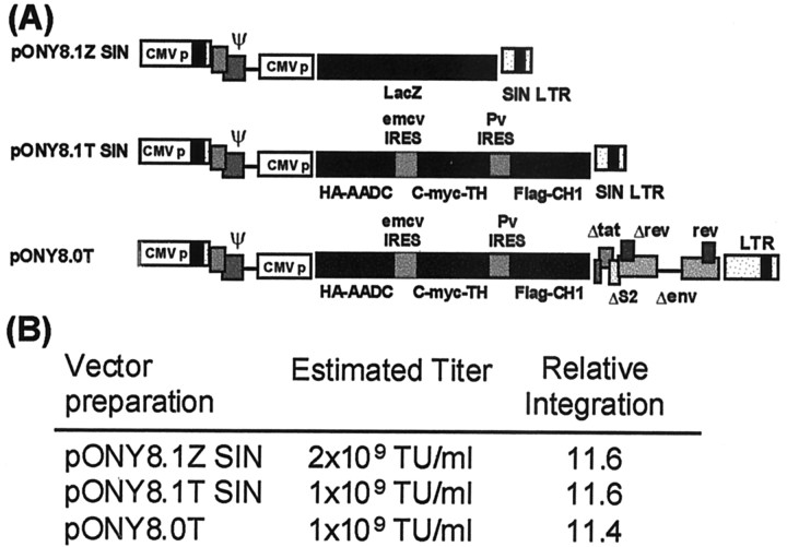
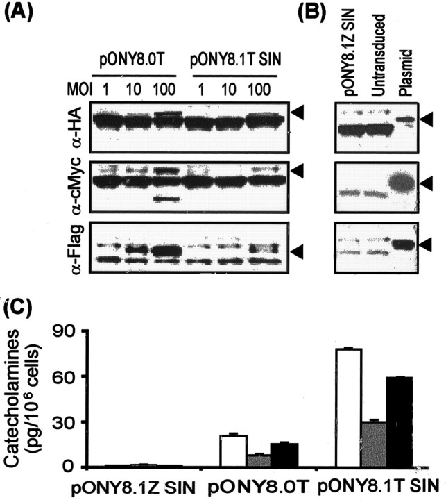
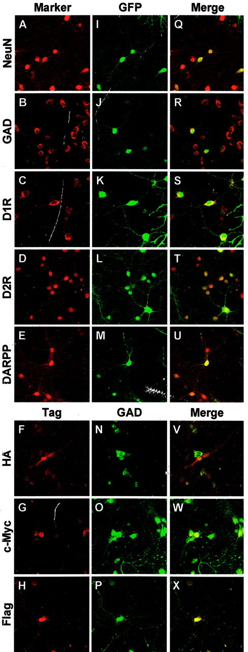
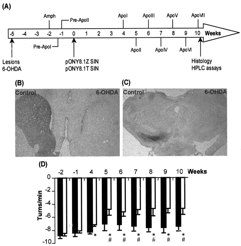
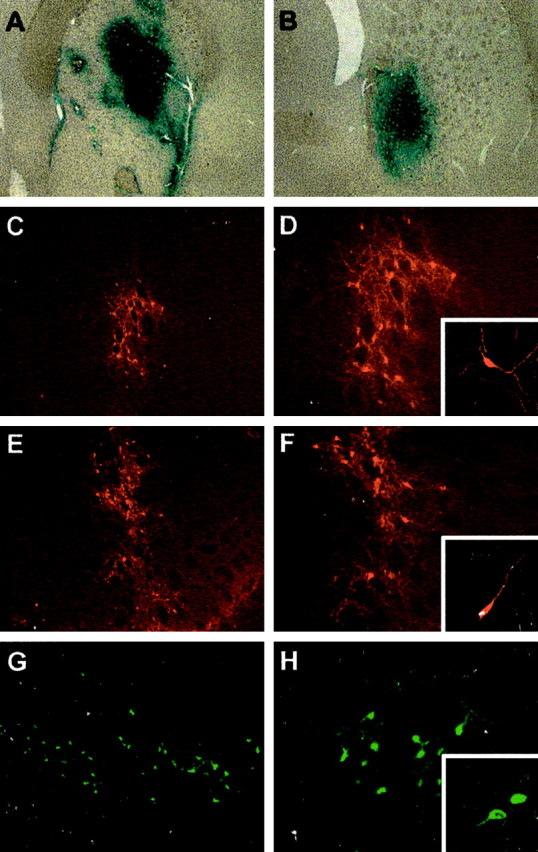
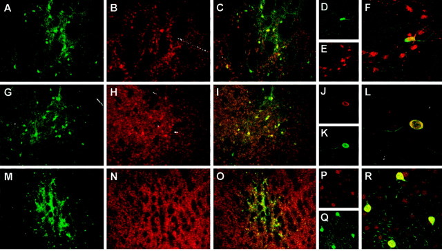
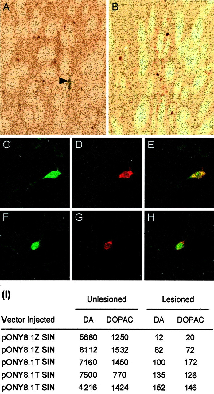
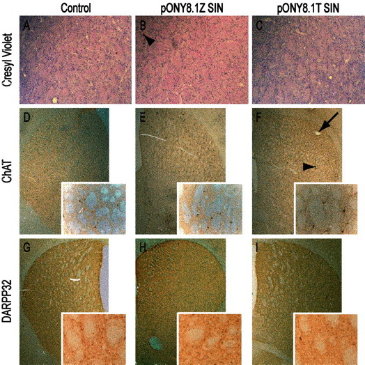
Parkinson's disease (PD) is a neurodegenerative disorder characterized by the selective loss of dopaminergic neurons in the substantia nigra. This loss leads to complete dopamine depletion in the striatum and severe motor impairment. It has been demonstrated previously that a lentiviral vector system based on equine infectious anemia virus (EIAV) gives rise to highly efficient and sustained transduction of neurons in the rat brain. Therefore, a dopamine replacement strategy using EIAV has been investigated as a treatment in the 6-hydroxydopamine (6-OHDA) animal model of PD. A self-inactivating EIAV minimal lentiviral vector that expresses tyrosine hydroxylase (TH), aromatic amino acid dopa decarboxylase (AADC), and GTP cyclohydrolase 1 (CH1) in a single transcription unit has been generated. In cultured striatal neurons transduced with this vector, TH, AADC, and CH1 proteins can all be detected. After stereotactic delivery into the dopamine-denervated striatum of the 6-OHDA-lesioned rat, sustained expression of each enzyme and effective production of catecholamines were detected, resulting in significant reduction of apomorphine-induced motor asymmetry compared with control animals (p < 0.003). Expression of each enzyme in the striatum was observed for up to 5 months after injection. These data indicate that the delivery of three catecholaminergic synthetic enzymes by a single lentiviral vector can achieve functional improvement and thus open the potential for the use of this vector for gene therapy of late-stage PD patients.


Figures

Fig. 1.
Fig. 1.
EIAV vectors used in this study. A, Genetic configuration of pONY8.1Z SIN, pONY8.1T SIN, and pONY8.0T vector genomes. B, Titers and integration efficiency of EIAV vectors were estimated by quantitative real-time RT-PCR (Martin-Rendon et al., 2002). Biological titers were calculated by comparison with known standards. For relative integration efficiencies, D17 cells were transduced at an MOI of 10 based on the calculated biological titers, and total DNA was isolated from the transduced cells 7–10 d after transduction. The relative integration efficiency of the different EIAV genomes in the host cells relative to β-actin (ΔCt values) was equivalent for the three vector genomes.
Fig. 2.
Fig. 2.
Expression of catecholamine biosynthetic enzymes in heterologous cell lines transduced with EIAV vectors.A, Western blot on total cell extracts prepared from D17 cells 8 d after transduction. Cells were transduced with either pONY8.0T or pONY8.1T SIN at MOIs of 1, 10, and 100 as indicated.B, Control blots showing extracts from cells transduced with pONY8.1Z SIN at an MOI of 100, untransduced cells, and HEK293T cells transfected with the appropriate monocistronic plasmid pNE2 (HA-AADC), pNE4a (C-myc-TH), or pNE6 (Flag-CH1). The blots were probed with mouse monoclonal antibodies against the tagged proteins at a 1:1000 dilution. Arrowheads indicate the apparent molecular weights of the tagged proteins: 52 kDa for HA-AADC, 42 kDa for C-myc-TH, and 26 kDa for Flag-CH1. C, HEK293T cells were transduced with pONY8.1Z SIN, pONY8.0T, or pONY8.1T SIN at an MOI of 100, and cell lysates were prepared 10 d after transduction. Catecholamines were extracted from the cell lysates, separated by HPLC, and detected electrochemically. Significant production (picograms per 106 cells) of l-dopa (white bars), DOPAC (gray bars), and dopamine (black bars) was detected in cells transduced with either pONY8.0T or pONY8.1T SIN but not cells transduced with pONY8.1Z SIN.
Fig. 3.
Fig. 3.
Confocal microscopic images demonstrating transduction of cultured striatal neurons with EIAV vectors. Striatal neurons (DIV 6) were transduced with pONY8.1G SIN at an MOI of 10 and, after an additional 6 d in culture, were processed for immunocytochemistry. Cells were fixed and stained with antibodies against NeuN (A), GAD (B), D1R (C), D2R (D), or DARPP (E) and with the appropriate Texas Red- and Cy3-coupled secondary antibody. GFP expression was observed in the same cells (I–M), and colocalization of GFP with the cell specific markers can clearly be seen in the merged images (Q–U). For HPLC and catecholamine release experiments, striatal neurons were transduced with pONY8.1Z SIN or pONY8.1T SIN (DIV 12). At DIV 19, neurons were tested for catecholamine release and were processed for immunocytochemistry. Cells transduced with pONY8.1T SIN were stained with antibodies against each of the three specific tag epitopes, HA-AADC, C-myc-TH, and Flag-CH1 (F–H, respectively, all Texas Red) and were colocalized with the anti-GAD antibody (N–P, FITC). Merged images are shown (V–X) that demonstrate that each of the three genes encoded by pONY8.1T SIN are expressed in the GABAergic medium spiny striatal neurons in culture.
Fig. 4.
Fig. 4.
In vivo gene transfer of EIAV vectors in 6-OHDA-treated rats. A, Time course for surgery and behavioral testing from lesion (−5 weeks) to histology and biochemical analysis (>10 weeks). Medial forebrain lesions were performed 5 weeks before viral vector injections. 6-OHDA (4 μl at 4 μg/μl) was injected into one site and induced a severe loss of TH immunoreactivity in the ipsilateral substantia nigra compacta (B) and striatal terminals (C). Three weeks after the 6-OHDA lesion, rats were prescreened for amphetamine (Amph) and apomorphine-induced behavioral motor asymmetry (Pre-ApoI, Pre-ApoII, 0.05 mg/kg, s.c.). Before viral injection into the striatum (4 weeks after 6-OHDA lesion), all groups showed clear apomorphine-induced contralateral rotation that was equivalent across the groups (D). Viral vector injections were performed in three sites into the striatum (2 μl/site), and apomorphine-induced rotational behavior was measured weekly from 4 and 10 weeks after viral administration. Rats that were injected with pONY8.1T SIN show substantial reduction (−48%) in rotational asymmetry that is significantly different from the pONY8.1Z SIN-treated group (p < 0.003). Rotations are expressed as net turns (ipsilateral to contralateral) per minute. The number of turns per minute remained relatively stable in the treated group between 4 and 10 weeks after viral administration.Asterisks indicate a significant difference from the pONY8.1Z SIN-treated control animals at each time point. Thepound sign denotes a significant difference in the number of rotations from the baseline value recorded before viral injection (Pre-ApoI, Pre-ApoII), Bonferroni test, (p < 0.003).
Fig. 5.
Fig. 5.
Transduction efficiency of pONY8.1Z SIN (A, B) and pONY8.1T SIN (C–H) in 6-OHDA-lesioned striatum 10 weeks after viral injections. Extensive reporter gene expression was observed in the striatum from the first (A) to the third (B) sites of pONY8.1Z SIN injection, and transduction was comparable with the data obtained with pONY8.0Z (Mazarakis et al., 2001). Expression of HA-AADC (C, D), C-myc-TH (E, F), and Flag-CH1 (G, H) in pONY8.1T SIN-injected striatum was confirmed by immunocytochemistry, and robust transduction was observed with each of the tags. For each tag, D, F, H are higher magnifications of C, E, G, respectively, and the insets show confocal images of transduced cells with neuronal morphology. Magnification: A, B, 10×; C, E, G, 25×; D, F, H, 50×, insets, 120×.
Fig. 6.
Fig. 6.
Long-term (5 months) expression and neuronal specificity after transduction with pONY8.1T SIN viral vectors. Extensive gene transfer at the site of injection in the caudate putamen was observed by immunocytochemistry 5 months after pONY8.1T SIN viral delivery. Each of the three tagged enzymes, HA-AADC (A), C-myc-TH (G), and Flag-CH1 (M), was detected and colocalized with NeuN staining (B, H, N) as shown in themerged images (C, I, O). Confocal analysis of the transduced cell types in the rat pONY8.1T-SIN-injected striatum (D–F, J–L, P–R) showed that the transduced cells were neurons, as demonstrated with HA (D), C-myc (J), Flag (P), and NeuN staining (E, K, Q) in the same sections. Colocalization of tags and NeuN expression can be seen inyellow in the merged images (F, L, R). Magnification: A–C, G–I, M–O, 25×;D–F, P–R, J–L, 120×.
Fig. 7.
Fig. 7.
Specific viral vector-mediated expression of AADC and TH in the denervated striatum 5 months after pONY8.1T SIN delivery. Photomicrographs demonstrate AADC (A) and TH (B) expression, and confocal microscopic images reveal the colocalization of expression of AADC (C, F) with either Flag (D) or C-myc (G) antibodies. Composite confocal images show colocalization of AADC and Flag (E) or C-myc (H) in yellow. Thearrowhead in A shows clear evidence of the presence of the needle tract with minor tissue reaction and the absence of damage to the striatum. Magnification: A,B, 25×; C–H, 120×. In similarly treated animals, in vivo synthesis of catecholamines in the striata of 6-OHDA-lesioned rats injected with either pONY8.1Z SIN or pONY8.1T SIN is demonstrated. I, Levels of dopamine and DOPAC (picograms per milligram of wet tissue) in lesioned and unlesioned striata measured by HPLC with electrochemical detection.
Fig. 8.
Fig. 8.
Evaluation of cytotoxicity in striatal neurons in vivo after EIAV vector transduction and increased dopamine turnover. Photomicrographs demonstrate cresyl violet staining (A–C) and anti-ChAT (D–F, insets) and anti-DARPP32 (G–I, insets) immunostaining on striatal sections from control (A, D, G) and pONY8.1Z SIN-injected (B, E, H) and pONY8.1T SIN-injected striata (C,F, I). No damage or reduction of the number of striatal neurons is observed. Insetsshow normal morphology of ChAT- or DARPP32-positive neurons in pONY8.1T SIN-injected brain compared with pONY8.1Z SIN and control striata. Arrowheads in B andF indicate needle tracts, and the arrowin F indicates a needle mark made during brain sectioning for recognition of the right striatum. Magnification:A–C, 25×; D–I, 10×;insets, 50×.

Similar articles

Cited by

References

    1. Abercrombie M. Estimation of nuclear population from microtome sections. Anat Rec. 1946;94:239–247. - PubMed
    1. Ariano MA, Sortwell CE, Ray M, Altemus KL, Sibley DR, Levine MS. Agonist-induced morphologic decrease in cellular D1A dopamine receptor staining. Synapse. 1997;27:313–321. - PubMed
    1. Bankiewicz KS, Eberling JL, Kohutnicka M, Jagust W, Pivirotto P, Bringas J, Cunningham J, Budinger TF, Harvey-White J. Convection-enhanced delivery of AAV vector in parkinsonian monkeys; in vivo detection of gene expression and restoration of dopaminergic function using pro-drug approach. Exp Neurol. 2000;164:2–14. - PubMed
    1. Barker RA, Dunnett SB. Functional integration of neural grafts in Parkinson's disease. Nat Neurosci. 1999;2:1047–1048. - PubMed
    1. Barzilai A, Melamed E, Shirvan A. Is there a rationale for neuroprotection against dopamine toxicity in Parkinson's disease? Cell Mol Neurobiol. 2001;21:215–235. - PMC - PubMed

MeSH terms

LinkOut - more resources