. 2002 Dec 1;22(23):10346-56.
doi: 10.1523/JNEUROSCI.22-23-10346.2002.

The netrin 1 receptors Unc5h3 and Dcc are necessary at multiple choice points for the guidance of corticospinal tract axons

Affiliations

The netrin 1 receptors Unc5h3 and Dcc are necessary at multiple choice points for the guidance of corticospinal tract axons

Jacqueline H Finger et al. J Neurosci. .

Abstract

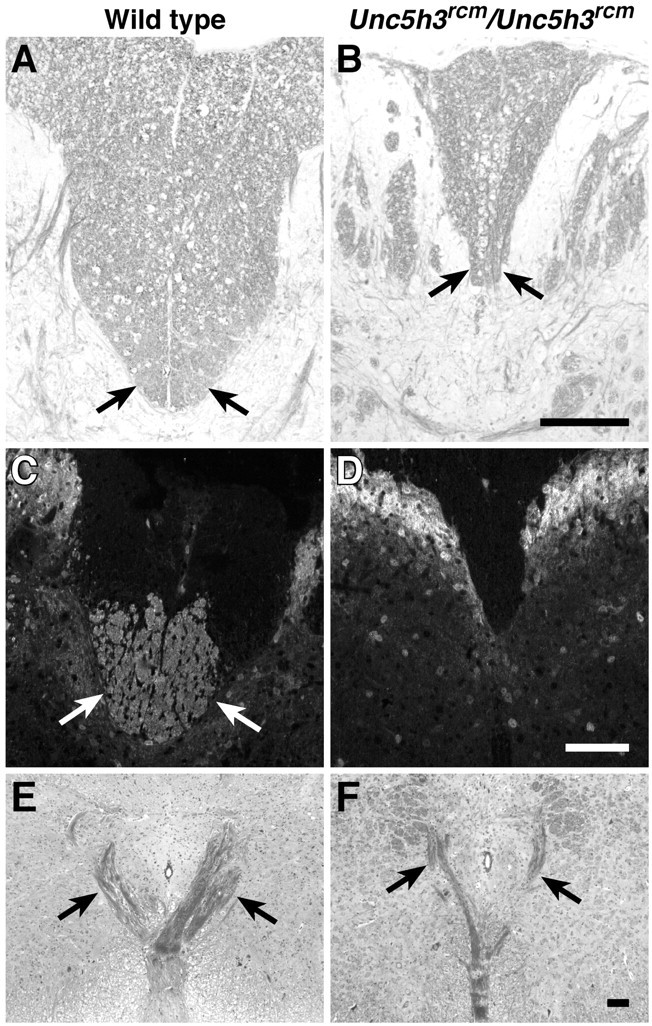
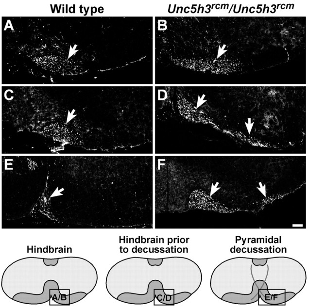
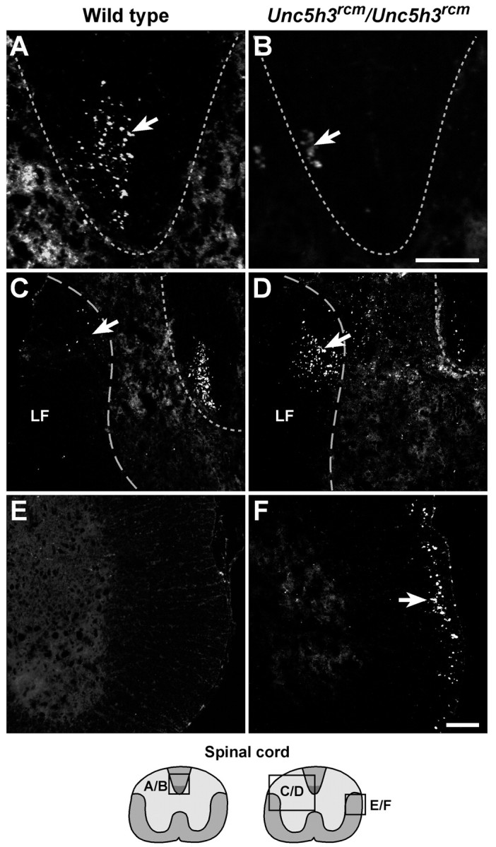
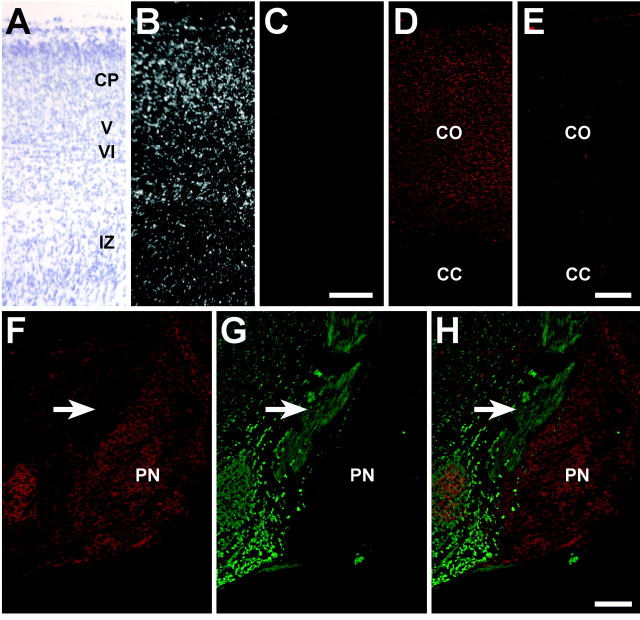
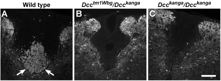
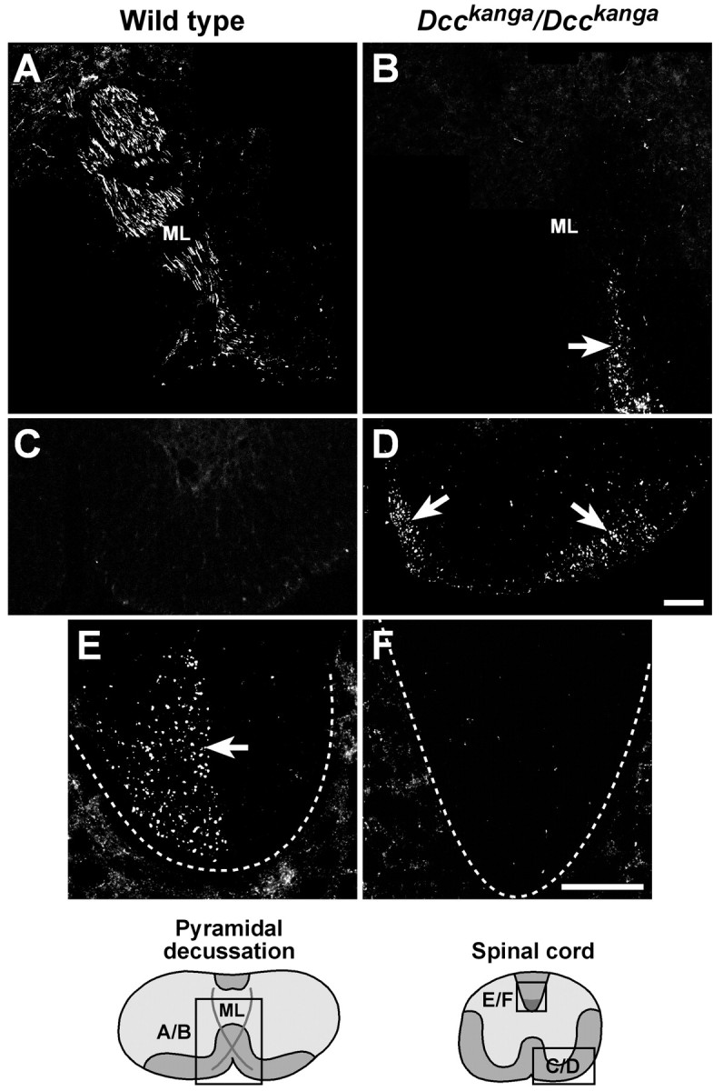
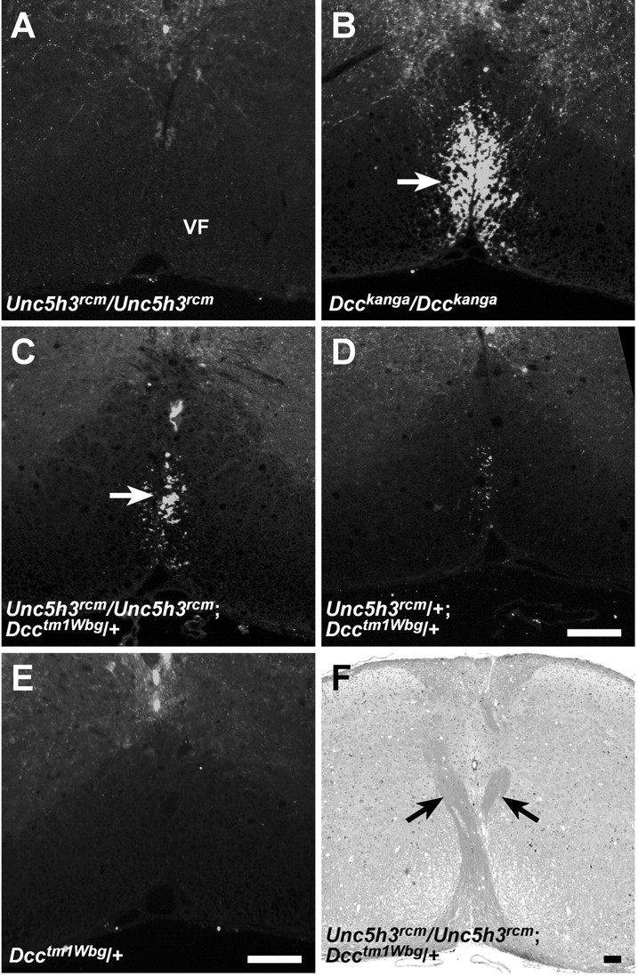
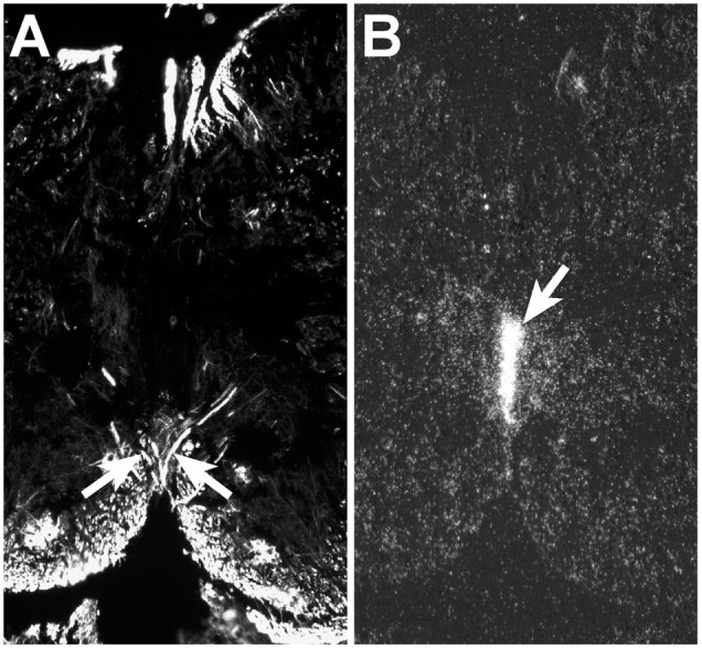
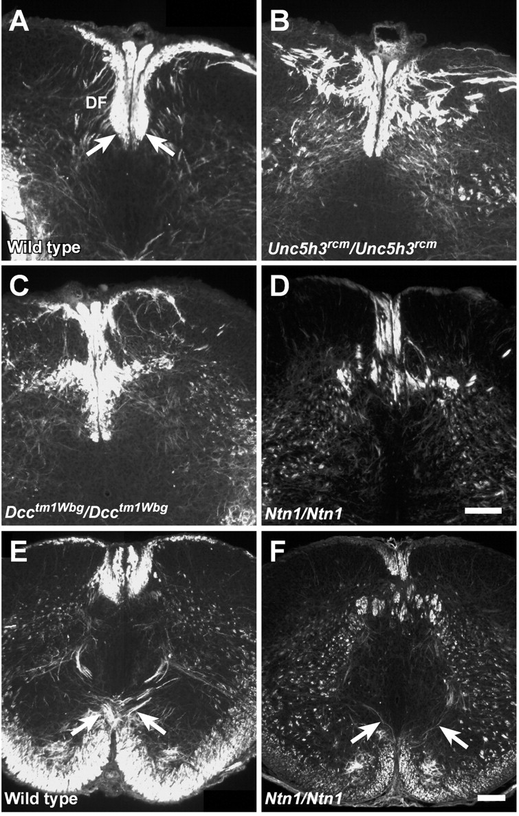
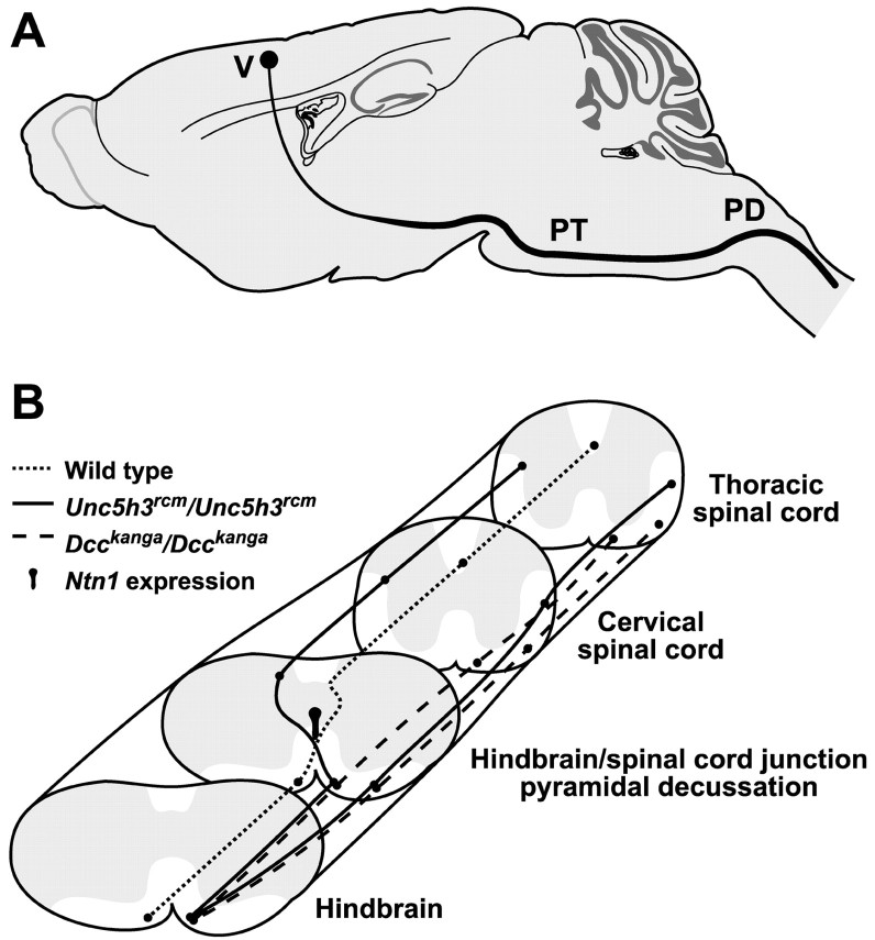
Migrating axons require the correct presentation of guidance molecules, often at multiple choice points, to find their target. Netrin 1, a bifunctional cue involved in both attracting and repelling axons, is involved in many cell migration and axon pathfinding processes in the CNS. The netrin 1 receptor DCC and its Caenorhabditis elegans homolog UNC-40 have been implicated in directing the guidance of axons toward netrin sources, whereas the C. elegans UNC-6 receptor, UNC-5 is necessary for migrations away from UNC-6. However, a role of vertebrate UNC-5 homologs in axonal migration has not been demonstrated. We demonstrate that the Unc5h3 gene product, shown previously to regulate cerebellar granule cell migrations, also controls the guidance of the corticospinal tract, the major tract responsible for coordination of limb movements. Furthermore, we show that corticospinal tract fibers respond differently to loss of UNC5H3. In addition, we observe corticospinal tract defects in mice homozygous for a spontaneous mutation that truncates the Dcc transcript. Postnatal day 0 netrin 1 mutant mice also demonstrate corticospinal tract abnormalities. Last, interactions between the Dcc and Unc5h3 mutations were observed in gene dosage experiments. This is the first evidence of an involvement in axon guidance for any member of the vertebrate unc-5 family and confirms that both the cellular and axonal guidance functions of C. elegans unc-5 have been conserved in vertebrates.


Figures

Fig. 1.
Fig. 1.
CST abnormalities inUnc5h3rcm/Unc5h3rcmmice. The dorsal funiculus (arrows) is shown in transverse sections of cervical spinal cord from wild-type (A, C) andUnc5h3rcm/Unc5h3rcm(B, D) mice stained with LFB and counterstained with cresyl violet (A, B) or antibody against CaM kinase II (C, D). The ipsilateral ventral pyramidal tract crosses the midline dorsally, forming the pyramidal decussation (E, wild type; F,Unc5h3rcm/Unc5h3rcm). Note the thinner decussation of pyramidal tract fibers (arrows) inUnc5h3rcm/Unc5h3rcmbrain, and the CST is missing from the dorsal funiculus of the mutant spinal cord. Scale bars, 100 μm.
Fig. 2.
Fig. 2.
The migration of the CST through theUnc5h3rcm/Unc5h3rcmhindbrain. BDA injected into the motor cortex of wild-type (A, C, E) andUnc5h3rcm/Unc5h3rcm(B, D, F) animals was visualized with streptavidin-Cy3. The trajectories of the ipsilateral pyramidal tract (arrow) appear similar in both wild-type (A) andUnc5h3rcm/Unc5h3rcm(B) mice through the hindbrain before the pyramidal decussation. Just before the pyramidal decussation (C, D), the pyramidal tract broadens (arrows) inUnc5h3rcm/Unc5h3rcmmice, whereas the labeled fibers in the wild-type brain remain bundled (arrow). At the pyramidal decussation, the mutant tract splits (arrows) into medial and lateral fiber bundles (F). The relative level of sections is shown in the accompanying diagrams at the bottom. Scale bars, 100 μm.
Fig. 3.
Fig. 3.
Aberrant CST axon migration inUnc5h3rcm/Unc5h3rcmspinal cord. BDA injected into the motor cortex of wild-type (A, C, E) andUnc5h3rcm/Unc5h3rcm(B, D, F) animals was visualized with streptavidin-Cy3. Labeled contralateral CST fibers (arrow) are present in the wild-type (A) dorsal funiculus (dotted line), whereas only a few labeled fibers are seen in the dorsal funiculus ofUnc5h3rcm/Unc5h3rcmanimals (B, arrow). In contrast to the wild-type spinal cord, many labeled axons are visible in the contralateral lateral funiculus (C, D, elongated dotted line, arrows) and the dorsal gray matter at the thoracic level of the spinal cord ofUnc5h3rcm/Unc5h3rcmmice. The ipsilateral lateral funiculus of the wild-type cervical spinal cord (E) does not contain labeled CST fibers, whereas many labeled ipsilateral CST fibers move from the ventral pyramidal tract into the ipsilateral lateral funiculus of theUnc5h3rcm/Unc5h3rcmspinal cord (F, arrow). The relative level of sections is shown in the accompanying diagrams at thebottom. LF, Contralateral lateral funiculus. Scale bars, 100 μm.
Fig. 4.
Fig. 4.
Unc5h3 and DCC expression at P0.In situ hybridization was performed withUnc5h3-specific antisense and sense probes on sagittal sections of P0 wild-type forebrain (A–C). At P0, the neurons of the cortical plate and presumptive layers V and VI express the Unc5h3 transcript (B). A corresponding bright-field photograph of the sections is shown inA, and the Unc5h3-specific sense control showing no signal is shown in C. Immunofluorescence with monoclonal anti-DCC antibody and secondary and tertiary antibody control (without primary antibody) on sagittal sections of P0 wild-type forebrain (D, E) shows that DCC is expressed throughout the cortex (CO). DCC immunofluorescence on sagittal sections of P0 wild-type hindbrain (F, H, overlay) revealed very few DCC-positive fibers. The presumptive CST axons (arrow) were detected by immunofluorescence with antibody to the 160 kDa neurofilament on neighboring sections (G, H, overlay). CC, Corpus callosum;CO, cortex; CP, cortical plate;IZ, intermediate zone; PN, pontine nuclei; V, layer V; VI, layer VI. Scale bars, 100 μm.
Fig. 5.
Fig. 5.
kanga, a spontaneous mutant allele of Dcc. Luxol fast blue- and cresyl violet-stained frontal sections through the forebrain of wild-type (A),Dcckanga/Dcckanga(B), andDcctm1Wbg/Dcckanga(C) mice are shown. Note the absence of the corpus callosum (CC) and hippocampal commissures (HC) in theDcckanga/DcckangaandDcctm1Wbg/Dcckangaforebrains. D, Southern blot ofEcoRI-digested genomic DNA from wild-type,Dcckanga/+, andDcckanga/Dcckangamice probed with cDNA corresponding to a portion of theDcc coding region (bp 3149–4266). Note the 5.0 and 6.7 kb RFLPs between wild-type andDcckanga heterozygotes or homozygotes.E, RT-PCR of Dcc transcripts inDcckanga mutants. DcccDNA between exon 26 through the last exon of coding region (exon 29) does not amplify fromDcckanga/Dcckanga(Ex 26–29). However, primers corresponding to exons 26 and 27 or exons 26 and 28 (Ex 26–27, Ex 26–28, respectively) do amplify products from mutant cDNA. F, PCR analysis of exon 29 using primers from the surrounding introns (410 bp) demonstrates that exon 29 is deleted inDcckanga/Dcckangagenomic DNA. An unrelated fragment from chromosome 3 (320 bp) was amplified as an internal control for the PCR. Scale bar, 100 μm.
Fig. 6.
Fig. 6.
The CST is absent from the dorsal funiculus of the spinal cord inDcckanga/ Dcckangamice. The dorsal funiculus is shown in transverse sections of cervical spinal cord from wild-type (A),Dcctm1Wbg/Dcckanga(B), andDcckanga/Dcckanga(C) mice stained with antibody against CaM kinase II. CaM kinase II-positive fibers are present in the wild-type CST (arrows) but absent from the dorsal funiculus of mutant mice. Scale bar, 100 μm.
Fig. 7.
Fig. 7.
The CST broadens and splits in the hindbrain ofDcckanga/Dcckangamice. BDA injected into the motor cortex of wild-type (A, C, E) andDcckanga/Dcckanga(B, D, F) animals was visualized with streptavidin-Cy3. No differences in the placement of pyramidal tract fibers (arrow) in the hindbrain before the pyramidal decussation were noted between wild-type and mutant mice (A, B). Just before the expected level of the pyramidal decussation inDcckanga/Dcckangamice, labeled fibers broaden (arrows), whereas the labeled fibers (arrow) in the wild-type hindbrain remain bundled (C, D). At the pyramidal decussation, theDcckanga/Dcckangatract splits (arrows) into medial and lateral fiber bundles (F). The relative level of sections is shown in the diagrams at the bottom. Scale bars, 100 μm.
Fig. 8.
Fig. 8.
The CST migrates incorrectly in the spinal cord ofDcckanga/Dcckangamice. BDA injected into the motor cortex of wild-type (A, C, E) andDcckanga/Dcckanga(B, D, F) animals was visualized with streptavidin-Cy3. Labeled fibers cross the midline (ML) dorsally in the wild-type pyramidal decussation (A) but do not decussate inDcckanga/Dcckangamice (B, arrow). Two bundles of ipsilateral fibers were observed in theDcckanga/Dcckangaventral cervical spinal cord (arrows) but not the wild-type spinal cord (C, D). The dorsal funiculus (dotted lines) in the wild type (arrow) but not mutant spinal cord contains labeled contralateral CST axons (E, F). The relative level of sections is shown in the diagrams at the bottom. Scale bars, 100 μm.
Fig. 9.
Fig. 9.
CST fibers are present in the ventral funiculus ofUnc5h3rcm/Unc5h3rcm;Dcctm1Wbg/+ adult mice. Transverse sections of adult cervical spinal cord fromUnc5h3rcm/Unc5h3rcm(A),Dcckanga/Dcckanga(B),Unc5h3rcm/Unc5h3rcm;Dcctm1Wbg/+ (C),Unc5h3rcm/+;Dcctm1Wbg/+ (D), andDcctm1Wbg/+ (E) mice were stained with anti-CaM kinase II antibody. Note that immunopositive fibers are present in the ventral funiculus (VF; arrow) ofDcckanga/Dcckanga,Unc5h3rcm/Unc5h3rcm;Dcctm1Wbg/+, andUnc5h3rcm/+;Dcctm1Wbg/+ but notUnc5h3rcm/Unc5h3rcmand Dcctm1Wbg/+ mice. Decussating CST fibers (arrows) are still present inUnc5h3rcm/Unc5h3rcm;Dcctm1Wbg/+ mice (F). Scale bars, 100 μm.
Fig. 10.
Fig. 10.
Ntn1 is expressed adjacent to the midline crossover point of CST fibers at the pyramidal decussation in P0 mice. The pyramidal decussation of wild-type mice at P0 was visualized by anti-2H3 (160 kDa neurofilament isoform) antibody (A, arrows). In situ hybridization with an Ntn1-specific probe is shown on an adjacent section (B). At P0, Ntn1 expression is observed at the midline immediately below the central canal extending ventrally (arrow) toward the pyramidal decussation. Scale bar, 100 μm.
Fig. 11.
Fig. 11.
The dorsal funiculus is abnormal inUnc5h3rcm/Unc5h3rcm,Dcctm1Wbg/Dcctm1Wbg, and Ntn1/Ntn1 newborn mice. Transverse sections of P0 upper cervical spinal cord from wild type (A),Unc5h3rcm/Unc5h3rcm(B),Dcctm1Wbg/Dcctm1Wbg(C), and Ntn1/Ntn1(D) mice were immunostained with anti-2H3 antibody. Note the abnormal shape of the ventral portion of the dorsal funiculus (df; A, arrows) is accompanied by many neurofilament-positive misplaced axons throughout the dorsal funicular region of the mutant spinal cords. The pyramidal decussation (arrows) was visualized with anti-2H3 antibody in wild-type mice (E) but was much reduced in Ntn1/Ntn1 newborn mice (F). Scale bars, 100 μm.
Fig. 12.
Fig. 12.
CST pathways inUnc5h3rcm/Unc5h3rcmandDcckanga/Dcckangamice. A, Normal path of the CST through the brain. Axons from layer V neurons (V) in the cerebral cortex migrate through the internal capsule to the ventral aspect of the brain, where they proceed as parallel ipsilateral fiber bundles on either side of midline. In the hindbrain, these fibers form the pyramidal tract (PT), which crosses midline at the junction of the hindbrain and spinal cord (PD) before entering the dorsal spinal cord. B, CST abnormalities inUnc5h3rcm/Unc5h3rcmandDcckanga/Dcckangamice. Normally, the pyramidal tract decussates at the hindbrain–spinal cord junction and continues down the spinal cord in the ventral-most portion of the contralateral dorsal funiculus (dotted line). InUnc5h3rcm/Unc5h3rcmmice, some CST axons in the ipsilateral pyramidal tract separate from the main fiber bundle (solid lines). Although the more medial fiber bundle decussates, it does not move into the dorsal funiculus but instead enters and continues in the contralateral lateral funiculus and dorsal gray matter of the spinal cord. The lateral fiber bundle moves further laterally into the ipsilateral lateral funiculus of the spinal cord. Many CST axons in the ipsilateral pyramidal tract ofDcckanga/Dcckangamice also separate with neither fiber bundle decussating (elongated dotted lines). The medial fiber bundle continues in the ipsilateral ventral funiculus of the spinal cord. As seen inUnc5h3rcm/Unc5h3rcmmice, the lateral fiber bundle moves further laterally into the ipsilateral lateral funiculus of the spinal cord. Ntn1expression is present dorsal to the crossover point of the pyramidal decussation.

References

    1. Ackerman SL, Kozak LP, Przyborski SA, Rund LA, Boyer BB, Knowles BB. The mouse rostral cerebellar malformation gene encodes an UNC-5-like protein. Nature. 1997;386:838–842. - PubMed
    1. Chan SS, Zheng H, Su MW, Wilk R, Killeen MT, Hedgecock EM, Culotti JG. UNC-40, a C. elegans homolog of DCC (deleted in colorectal cancer), is required in motile cells responding to UNC-6 netrin cues. Cell. 1996;87:187–195. - PubMed
    1. Cho KR, Oliner JD, Simons JW, Hedrick L, Fearon ER, Preisinger AC, Hedge P, Silverman GA, Vogelstein B. The DCC gene: structural analysis and mutations in colorectal carcinomas. Genomics. 1994;19:525–531. - PubMed
    1. Cohen NR, Taylor JS, Scott LB, Guillery RW, Soriano P, Furley AJ. Errors in corticospinal axon guidance in mice lacking the neural cell adhesion molecule L1. Curr Biol. 1998;8:26–33. - PubMed
    1. Colavita A, Culotti JG. Suppressors of ectopic UNC-5 growth cone steering identify eight genes involved in axon guidance in Caenorhabditis elegans. Dev Biol. 1998;194:72–85. - PubMed

Publication types

MeSH terms