. 2002 Dec 1;22(23):10377-87.
doi: 10.1523/JNEUROSCI.22-23-10377.2002.

Glia induce dendritic growth in cultured sympathetic neurons by modulating the balance between bone morphogenetic proteins (BMPs) and BMP antagonists

Affiliations

Glia induce dendritic growth in cultured sympathetic neurons by modulating the balance between bone morphogenetic proteins (BMPs) and BMP antagonists

Pamela J Lein et al. J Neurosci. .

Abstract

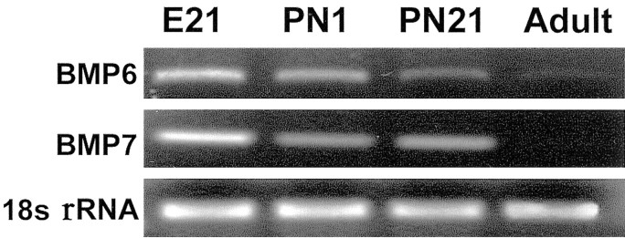
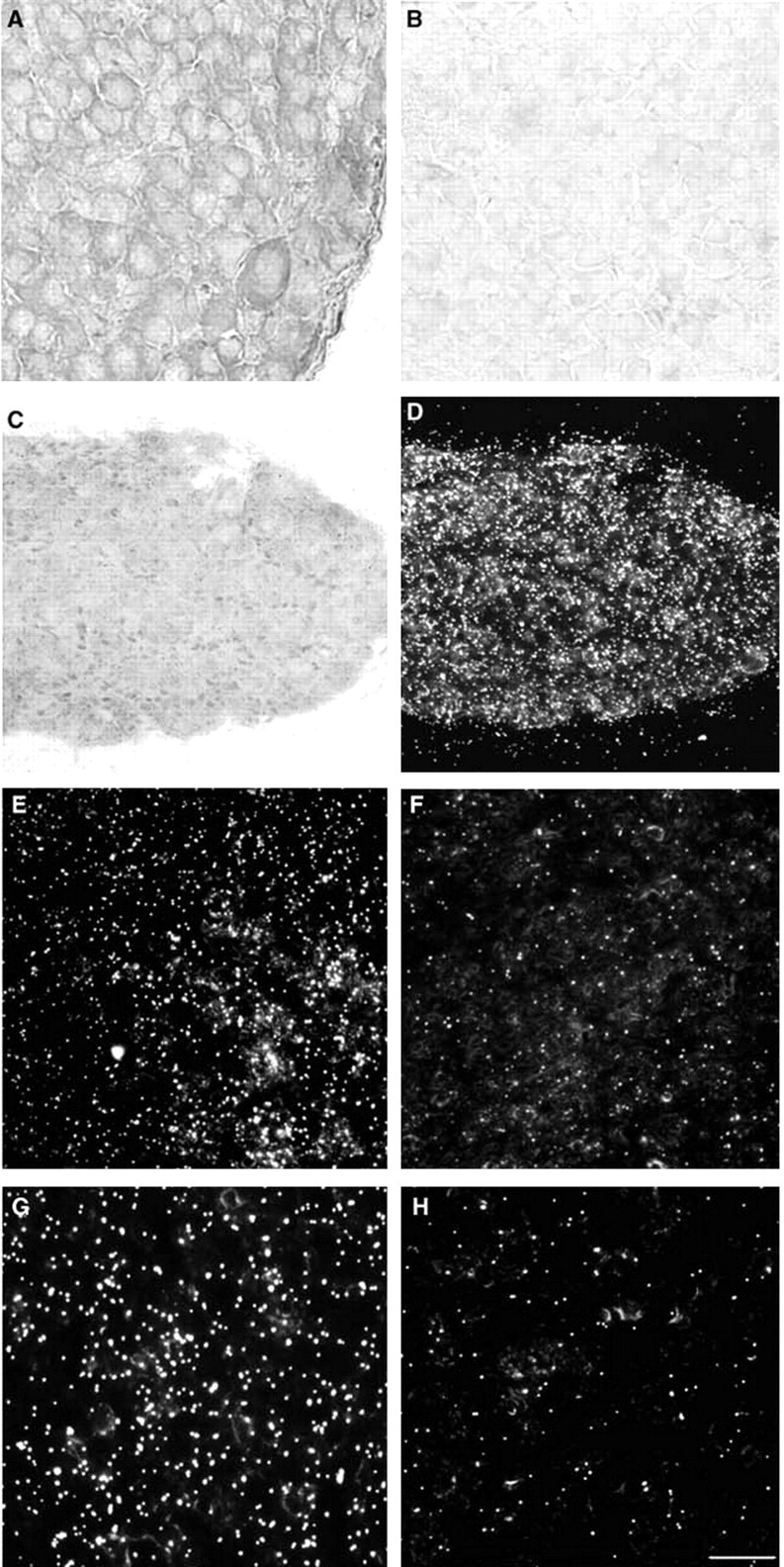
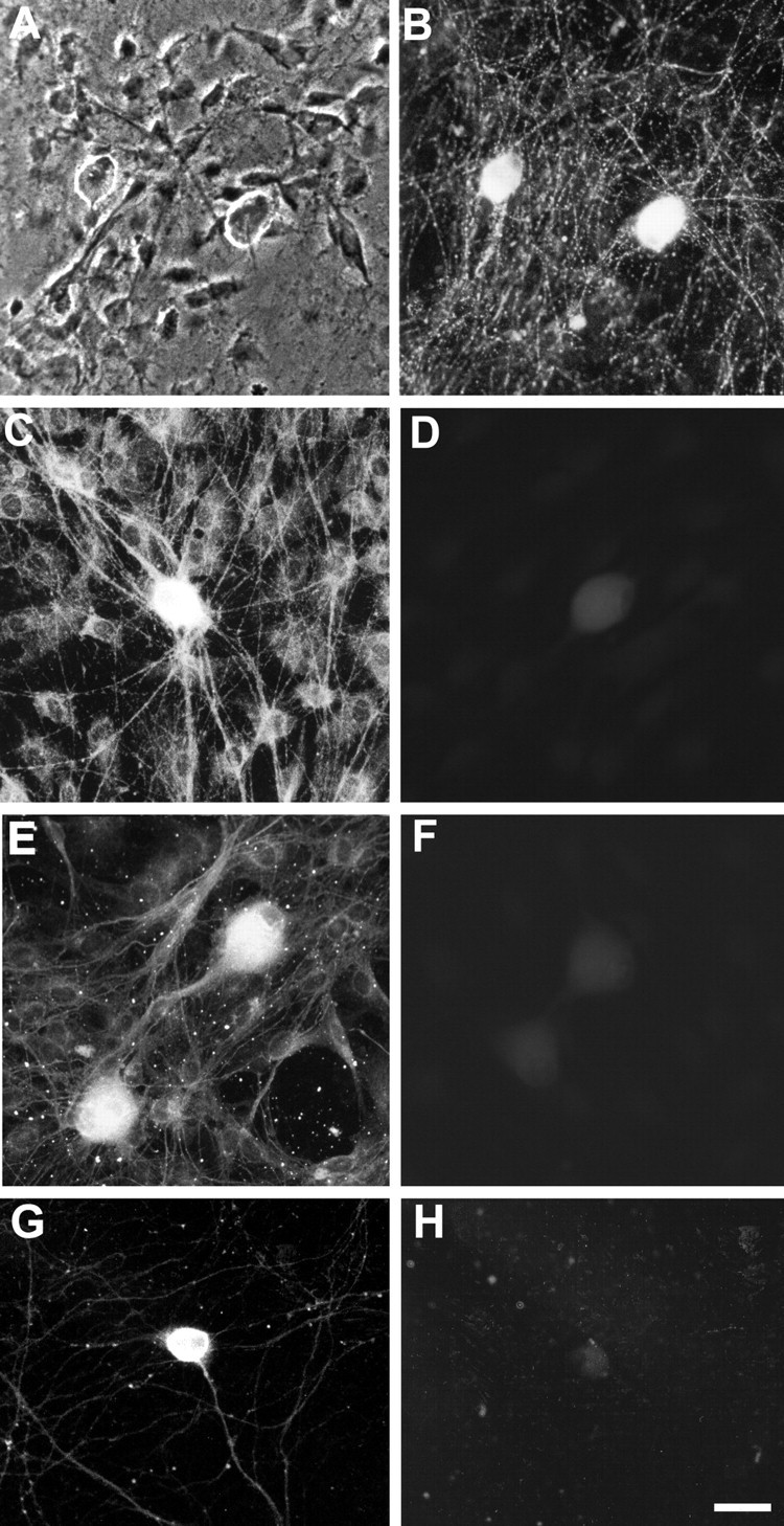
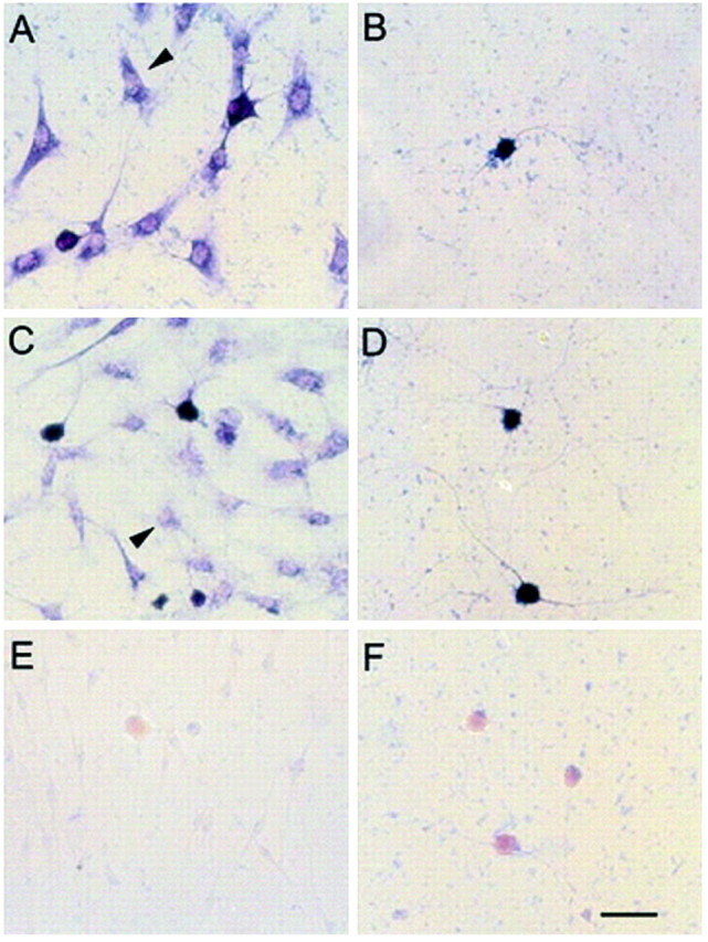
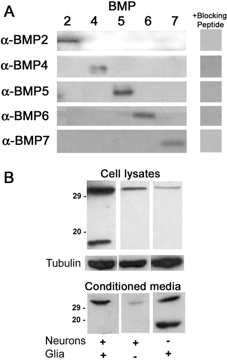
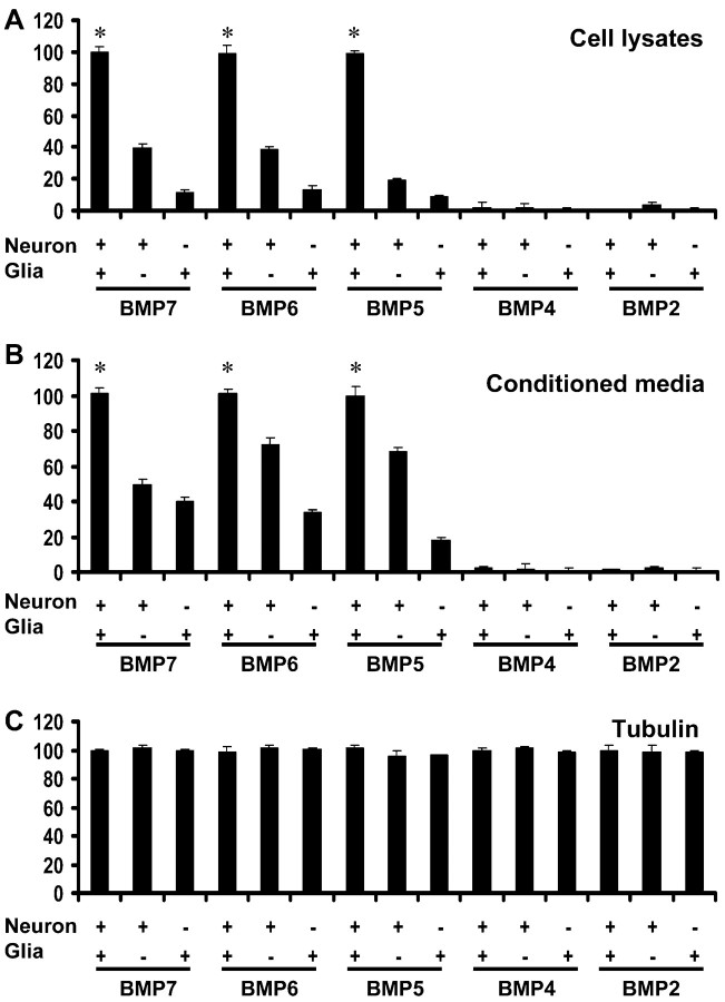
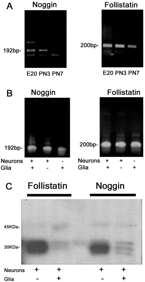
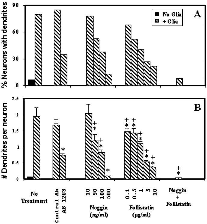
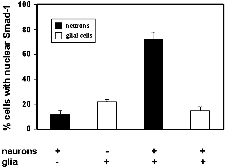
Dendritic growth in cultured sympathetic neurons requires specific trophic interactions. Previous studies have demonstrated that either coculture with glia or exposure to recombinant bone morphogenetic proteins (BMPs) is both necessary and sufficient to induce dendrite formation. These observations led us to test the hypothesis that BMPs mediate glial-induced dendritic growth. In situ hybridization and immunocytochemical studies indicate that the spatiotemporal expression of BMP5, -6, and -7 in rat superior cervical ganglia (SCG) is consistent with their proposed role in dendritogenesis. In vitro, both SCG glia and neurons were found to express BMP mRNA and protein when grown in the presence or absence of the other cell type. However, addition of ganglionic glia to cultured sympathetic neurons causes a marked increase in BMP proteins coincident with a significant decrease in follistatin and noggin. Functional assays indicate that glial-induced dendritic growth is significantly reduced by BMP7 antibodies and completely inhibited by exogenous noggin and follistatin. These data suggest that glia influence the rapid perinatal expansion of the dendritic arbor in sympathetic neurons by increasing BMP activity via modulation of the balance between BMPs and their antagonists.


Figures

Fig. 1.
Fig. 1.
BMP mRNA is expressed in SCG at times corresponding to initial and maximal dendritic growth. Transcripts forBMP6 and BMP7 are detected by RT-PCR in total RNA extracted from rat SCG at E21. PCR products for both BMPs are still present at PN1 but decline over development such that expression is not evident in equal amounts of total RNA isolated from adult SCG. In contrast, levels of 18S rRNA are not altered as a function of developmental stage.
Fig. 2.
Fig. 2.
BMP protein and transcript are present in sympathetic ganglia of perinatal rat pups during the time of rapid growth and expansion of the dendritic arbor. A,B, Bright-field micrographs of HRP reaction product in frozen sections from SCG of a PN1 rat pup. A, Sections immunostained with mAb 12G3 raised against BMP7 exhibit significant HRP reaction product throughout the neuronal cytoplasm and in extracellular spaces; in contrast, labeling is not observed in neuronal nuclei.B, Labeling is not observed in control sections treated only with the secondary antibody. C, D, Bright-field (C) and dark-field (D) micrographs (200×) of a SCG from a PN7 rat pup hybridized with 35S-labeled riboprobes and counterstained with cresyl violet. Sections hybridized with BMP7 antisense probe (C, D) show significant labeling throughout most of the ganglion. Higher magnification (400×) dark-field images of SCG from a PN7 rat hybridized with35S-labeled BMP7 antisense (E) or BMP6 antisense (G) probes exhibit diffuse distribution of grains. The grain density in sections labeled with BMP7 or BMP6 antisense probes (3.2 × 10⁶ ± 0.14 × 10⁶ grains/cm2 and 3.3 × 10⁶ ± 0.05 × 10⁶ grains/cm2, respectively) is significantly greater than the grain density observed in sections hybridized with BMP7 sense probe (F) (0.6 × 10⁶ ± 0.06 × 10⁶grains/cm2) or BMP6 sense probe (H) (0.73 × 10⁶ ± 0.06 × 10⁶grains/cm2). Scale bar: 200× magnification, 50 μm; 400× magnification, 25 μm.
Fig. 3.
Fig. 3.
BMP immunoreactivity is associated with both ganglionic glial cells and sympathetic neurons in coculture.A, B, Phase-contrast (A) and fluorescence (B) micrographs of cocultures reacted with mAb 12G3 against BMP7 before fixation or permeabilization. Neuronal soma exhibit diffuse cell surface immunoreactivity, and neuronal processes are clearly delineated by a punctate staining pattern. C, In cocultures immunostained with mAb 12G3 after fixation and permeabilization, BMP7 immunoreactivity is evident throughout the cytoplasm of both glial cells and neurons. In neurons, the processes as well as the soma are brightly stained. D, Preincubation of mAb 12G3 with recombinant BMP7 significantly decreases BMP7 immunoreactivity in permeabilized cocultures. E, Both neurons and glial cells exhibit BMP6 immunoreactivity throughout their cytoplasm, which is significantly reduced by preincubating BMP6 antiserum with specific blocking peptide (F). Immunocytochemical localization of BMP7 in neurons cultured in the absence of glial cells (G, H) indicates significant immunoreactivity in the soma and processes of neurons permeabilized before reaction with BMP7 mAb (G), but very little if any surface staining for BMP7 in neurons reacted with BMP7 mAb before permeabilization (H). Scale bar, 50 μm.
Fig. 4.
Fig. 4.
Cultured ganglionic glial cells and sympathetic neurons express BMP6 and –7 mRNA. Sympathetic neurons were grown for 4–5 d in the presence (A, C,E) or absence (B, D,F) of ganglionic glial cells before hybridization with digoxigenin-labeled antisense riboprobes for BMP7 (A, B) or BMP6 (C,D). Glial cells (arrowhead) express mRNA for BMP7 (A) and BMP6 (C) throughout their cytoplasm. Neurons cocultured with ganglionic glial cells also exhibit intense somatic labeling for BMP7 (A) and BMP6 (C) transcripts. A similar distribution of BMP7 (B) and BMP6 (D) mRNA is observed in neurons grown in the absence of glia. Negligible labeling is observed in either neurons or glial cells hybridized with BMP7 sense riboprobe (E,F). Scale bar, 50 μm.
Fig. 5.
Fig. 5.
Western blot analyses of BMPs in cocultures of sympathetic neurons and ganglionic glial cells. A, The specificity of the antibodies used for Western blot analysis was evaluated by reacting blots of recombinant BMP2, -4, -5, -6, or -7 (50 ng) with antibodies raised against each of these BMPs. Each BMP antibody (0.5 μg/ml) reacted only with the BMP against which it was raised, and this interaction was inhibited by preincubation of the antibody with specific blocking peptide (2.5 μg/ml).B, Cell lysates and conditioned medium from neuron and glial cocultures (far left lane), purified neuronal cultures (middle lane), or purified cultures of ganglionic glial cells (far right lane) were probed using antibody selective for BMP7. Equal amounts of protein were loaded into all wells, and blots of cell lysates were also probed for α-tubulin. The levels of both the monomeric (∼17 kDa) and dimeric (∼30 kDa) forms of BMP7 varied between the different culture conditions.
Fig. 6.
Fig. 6.
BMPs are present in significantly greater concentrations in cocultures of sympathetic neurons and ganglionic glial cells relative to cultures of neurons only or glial cells only.A, B, Densitometric analyses of Western blots of cell lysates (A) and conditioned medium (B) from cocultures of sympathetic neurons and ganglionic glia, cultures of neurons only, or cultures of ganglionic glial cells only. Equal amounts of proteins were loaded into all wells, probed with antibodies selective for BMP7, -6, -5, -4, or -2 (0.5 μg/ml), and quantified by densitometry. Bars represent levels of total (combined dimeric and monomeric) BMP. C, Blots of cell lysates were also probed for α-tubulin. Data are presented as the mean ± SEM (n = 3 per condition). *p < 0.01 versus corresponding value for neuron–glia cocultures.
Fig. 7.
Fig. 7.
Expression of the BMP antagonists noggin and follistatin in SCG cells. A, Noggin and follistatin mRNA were detected by RT-PCR in total RNA extracted from rat SCG at E20 and PN3, which correspond to times of initial dendrite extension (E20) and rapid expansion of the dendritic arbor (PN3) in these neurons. At later developmental times when the dendritic arbor is undergoing maximal expansion (PN7), transcript levels for both noggin and follistatin decrease. B, RT-PCR analyses of total RNA from cultured SCG cells indicate that both neurons and glial cells express noggin and follistatin mRNA, and these levels are not altered significantly by coculture. C, Protein immunoprecipitated with BMP antibodies from neuron–glia cocultures or neuron monocultures probed with antibodies specific for noggin or follistatin. Coculture of neurons with glial cells significantly decreases levels of BMPs complexed to noggin and follistatin relative to levels present in neuronal monocultures.
Fig. 8.
Fig. 8.
Reagents that block BMP function inhibit glial-induced dendritic growth. Sympathetic neurons were cocultured with ganglionic glial cells in the absence or presence of mAb 12G3 raised against BMP7 (15 μg/ml) or an isotype-matched control antibody (15 μg/ml), or varying concentrations of the BMP antagonists noggin and follistatin. After 12–14 d of exposure to these reagents, cultures were immunostained for MAP-2, and dendritic growth was quantified as the percentage of neurons with dendrites (A) and the number of dendrites per neuron (B). Data are represented as the mean ± SEM (n ≥ 70 per condition). *p < 0.001 versus neuron–glia cocultures grown in the absence of BMP function-blocking reagents; + p < 0.001 versus neurons cultured in the absence of glia.
Fig. 9.
Fig. 9.
Coculture of sympathetic neurons with ganglionic glial cells induces Smad-1 nuclear translocation in neurons but not in glial cells. After 5 d in vitro, a subset of neuronal cultures was reseeded with highly purified populations of ganglionic glial cells. Some of the glial cell suspension was plated into dishes without neurons. Three days after plating the glial cells, neuronal monocultures, glial monocultures, and neuron–glia cocultures were immunostained for Smad-1, and the percentage of neurons and glial cells with nuclear Smad-1 localization was quantified. In neurons and glial cells grown in monoculture, Smad-1 was predominantly localized to the cytoplasm. Coculture of neurons with glia did not alter the distribution of Smad-1 in glial cells but did significantly increase the percentage of neurons with nuclear Smad-1 localization. Data are represented as the mean ± SEM (n = 20 microscopic fields at 200× magnification; 5 fields each from 4 different cultures from 2 separate dissections).

Similar articles

Cited by

References

    1. Ai X, Cappuzzello J, Hall AK. Activin and bone morphogenetic proteins induce calcitonin gene-related peptide in embryonic sensory neurons in vitro. Mol Cell Neurosci. 1999;14:506–518. - PubMed
    1. Barres BA. A new role for glia: generation of neurons! Cell. 1999;97:667–670. - PubMed
    1. Beck HN, Drahushuk K, Jacoby DB, Higgins D, Lein PJ. Bone morphogenetic protein-5 (BMP-5) promotes dendritic growth in cultured sympathetic neurons. BMC Neurosci. 2001;2:12. - PMC - PubMed
    1. Bruckenstein DA, Higgins D. Morphological differentiation of embryonic rat sympathetic neurons in tissue culture. I. Conditions under which neurons form axons but not dendrites. Dev Biol. 1988;128:324–336. - PubMed
    1. Calvet S, Doherty P, Prochiantz A. Identification of a signaling pathway activated specifically in the somatodendritic compartment by a heparan sulfate that regulates dendrite growth. J Neurosci. 1998;18:9751–9765. - PMC - PubMed

Publication types

MeSH terms