. 2002 Dec 1;22(23):10399-407.
doi: 10.1523/JNEUROSCI.22-23-10399.2002.

Cellular mechanisms regulating activity-dependent release of native brain-derived neurotrophic factor from hippocampal neurons

Affiliations

Cellular mechanisms regulating activity-dependent release of native brain-derived neurotrophic factor from hippocampal neurons

Agnieszka Balkowiec et al. J Neurosci. .

Abstract

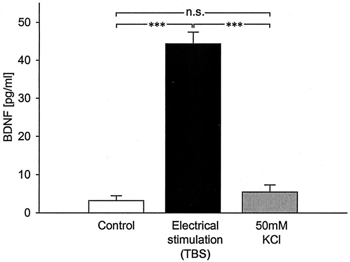
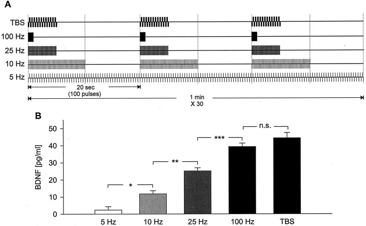
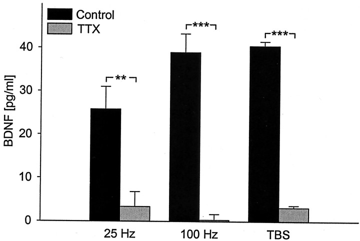
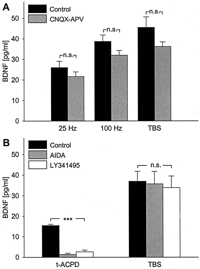
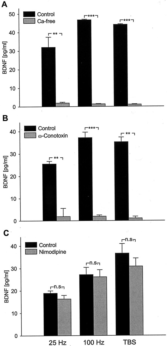
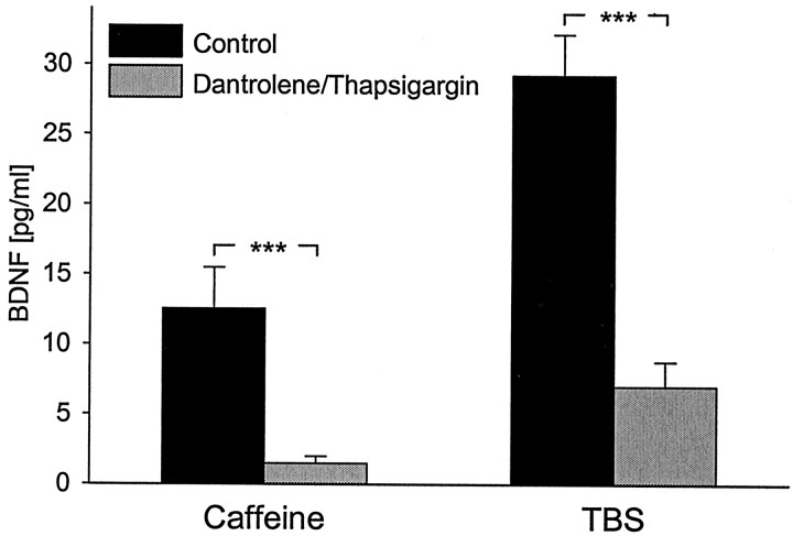
Brain-derived neurotrophic factor (BDNF) plays a critical role in activity-dependent modifications of neuronal connectivity and synaptic strength, including establishment of hippocampal long-term potentiation (LTP). To shed light on mechanisms underlying BDNF-dependent synaptic plasticity, the present study was undertaken to characterize release of native BDNF from newborn rat hippocampal neurons in response to physiologically relevant patterns of electrical field stimulation in culture, including tonic stimulation at 5 Hz, bursting stimulation at 25 and 100 Hz, and theta-burst stimulation (TBS). Release was measured using the ELISA in situ technique, developed in our laboratory to quantify secretion of native BDNF without the need to first overexpress the protein to nonphysiological levels. Each stimulation protocol resulted in a significant increase in BDNF release that was tetrodotoxin sensitive and occurred in the absence of glutamate receptor activation. However, 100 Hz tetanus and TBS, stimulus patterns that are most effective in inducing hippocampal LTP, were significantly more effective in releasing native BDNF than lower-frequency stimulation. For all stimulation protocols tested, removal of extracellular calcium, or blockade of N-type calcium channels, prevented BDNF release. Similarly, depletion of intracellular calcium stores with thapsigargin and treatment with dantrolene, an inhibitor of calcium release from caffeine-ryanodine-sensitive stores, markedly inhibited activity-dependent BDNF release. Our results indicate that BDNF release can encode temporal features of hippocampal neuronal activity. The dual requirement for calcium influx through N-type calcium channels and calcium mobilization from intracellular stores strongly implicates a role for calcium-induced calcium release in activity-dependent BDNF secretion.


Figures

Fig. 1.
Fig. 1.
Short-term patterned electrical stimulation, but not short-term continuous KCl depolarization, significantly increases release of native BDNF from hippocampal neurons. Mean levels of BDNF measured in sister cultures of newborn hippocampal neurons after 3 d in vitro plus 30 min of the following: (1) control conditions (no stimulation; n = 48), (2) TBS (25 bursts of 4 pulses at 100 Hz, delivered at 5 Hz, every 20 sec;n = 30), or (3) continuous depolarization with 50 mm KCl (n = 39). ***p < 0.001; n.s., not significant.
Fig. 2.
Fig. 2.
High-frequency patterns of electrical stimulation that are known to induce LTP are significantly more effective at releasing native BDNF from hippocampal neurons than low-frequency stimulation. A, Schematic representation of the stimulation pattern applied to each group of cultures.B, Mean levels of BDNF released in sister cultures of newborn hippocampal neurons during 30 min of electrical field stimulation with 100 biphasic rectangular pulses of 4 msec, delivered at 5 Hz (n = 16), 10 Hz (n = 14), 25 Hz (n = 40), 100 Hz (n= 42), or TBS (n = 30), with interburst intervals, respectively, of 0, 10, 16, 19, and 15 sec, as shown inA. *p < 0.05; **p < 0.01; ***p < 0.001;n.s., not significant.
Fig. 3.
Fig. 3.
Activity-dependent release of native BDNF depends on action potentials. Mean levels of BDNF released in sister cultures of newborn hippocampal neurons during 30 min of electrical field stimulation [100 biphasic rectangular pulses of 4 msec, delivered at 25 Hz (n = 12), 100 Hz (n = 10), and TBS (n = 4), once every 20 sec] in the absence (black bars) or presence (gray bars) of 1.5 μm TTX, an inhibitor of voltage-dependent sodium channels. **p < 0.01; ***p < 0.001.
Fig. 4.
Fig. 4.
Release of native BDNF evoked by patterned electrical stimulation does not require glutamatergic synaptic activity. A, Mean levels of BDNF released in sister cultures of newborn hippocampal neurons during 30 min of electrical field stimulation [100 biphasic rectangular pulses of 4 msec, delivered at 25 Hz (n = 8), 100 Hz (n = 8), and TBS (n = 10), once every 20 sec] in the absence (black bars) or presence of CNQX (20 μm) and APV (100 μm), antagonists of, respectively, non-NMDA and NMDA glutamate receptors (CNQX-APV; gray bars). B, Mean levels of BDNF released in sister cultures during 30 min of either exposure to 100 μmtrans-[1S,3R]-ACPD, an agonist of mGluR I and II (t-ACPD; n= 12), or electrical field stimulation at the theta-burst pattern (TBS; n = 12) in the absence (black bars) or presence of metabotropic glutamate receptor antagonists AIDA (500 μm; gray bars) and LY341495 (100 μm; white bars); ***p < 0.001; n.s., not significant.
Fig. 5.
Fig. 5.
Release of native BDNF evoked by patterned electrical stimulation requires calcium influx through N-type voltage-activated calcium channels. Mean levels of BDNF released in sister cultures of newborn hippocampal neurons during 30 min of electrical field stimulation (100 biphasic rectangular pulses of 4 msec, delivered at 25 Hz, 100 Hz, and TBS, once every 20 sec):A, in the presence (Control; black bars) or absence (Ca-free; gray bars) of extracellular calcium (25 Hz, n = 6; 100 Hz, n = 4; TBS, n = 4);B, in the absence (Control; black bars) or presence of 1 μm ω-Conotoxin GVIA, an N-type channel antagonist (ω-Conotoxin; gray bars) (25 Hz, n = 4; 100 Hz,n = 10; TBS, n = 4);C, in the absence (Control; black bars) or presence of 2 μm nimodipine, an L-type channel antagonist (Nimodipine; gray bars) (25 Hz, n = 4; 100 Hz,n = 4; TBS, n = 6). **p < 0.01; ***p < 0.001;n.s., not significant.
Fig. 6.
Fig. 6.
Release of native BDNF in response to patterned electrical stimulation requires calcium mobilization from ryanodine-sensitive stores. Mean levels of BDNF released in sister cultures of newborn hippocampal neurons during 30 min exposure to either 30 μm caffeine or electrical field stimulation at the theta-burst pattern (TBS) in the absence (black bars) or presence of dantrolene (50 μm), a selective antagonist of ryanodine receptor channels. Cultures were pretreated with thapsigargin (10 μm), which selectively inhibits the endoplasmic reticulum Ca2+-ATPase (gray bars);n = 8. ***p < 0.001.

References

    1. Akaneya Y, Tsumoto T, Kinoshita S, Hatanaka H. Brain-derived neurotrophic factor enhances long-term potentiation in rat visual cortex. J Neurosci. 1997;17:6707–6716. - PMC - PubMed
    1. Altar CA, Cai N, Bliven T, Juhasz M, Conner JM, Acheson AL, Lindsay RM, Wiegand SJ. Anterograde transport of brain-derived neurotrophic factor and its role in the brain. Nature. 1997;389:856–860. - PubMed
    1. Androutsellis-Theotokis A, McCormack WJ, Bradford HF, Stern GM, Pliego-Rivero FB. The depolarisation-induced release of [125I]BDNF from brain tissue. Brain Res. 1996;743:40–48. - PubMed
    1. Balkowiec A, Katz DM. Activity-dependent release of endogenous brain-derived neurotrophic factor from primary sensory neurons detected by ELISA in situ. J Neurosci. 2000;20:7417–7423. - PMC - PubMed
    1. Balkowiec A, Katz DM. Cellular mechanisms underlying activity-dependent release of endogenous BDNF from hippocampal neurons. Soc Neurosci Abstr. 2001;27:801.14.

Publication types

MeSH terms

LinkOut - more resources