

Projection to the inferior colliculus from the basal nucleus of the amygdala
- PMID: 12451144
- PMCID: PMC6758740
- DOI: 10.1523/JNEUROSCI.22-23-10449.2002
Projection to the inferior colliculus from the basal nucleus of the amygdala
Abstract
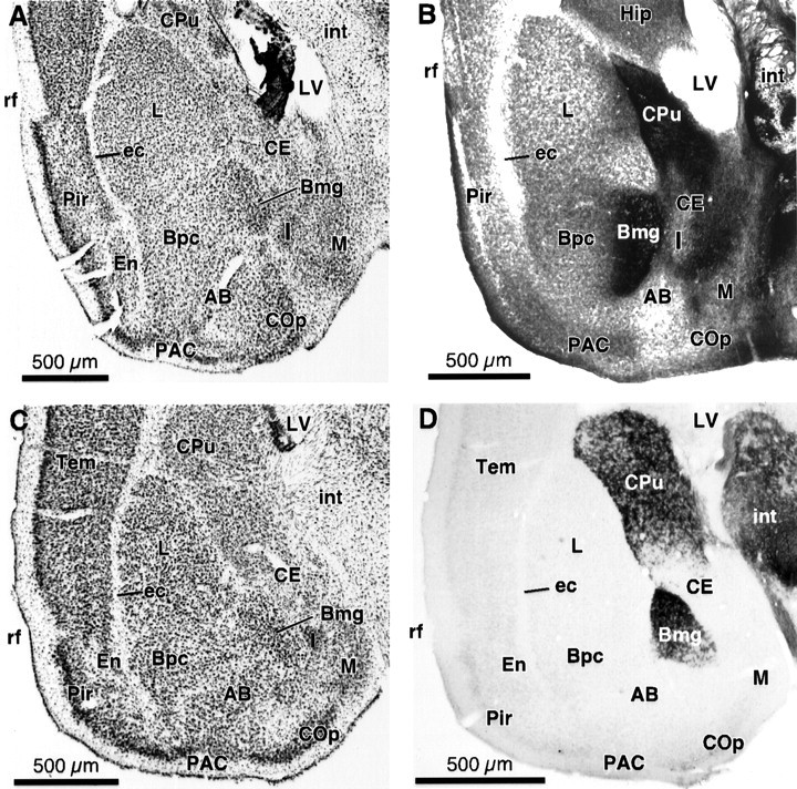
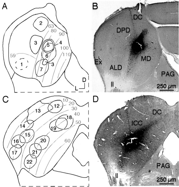
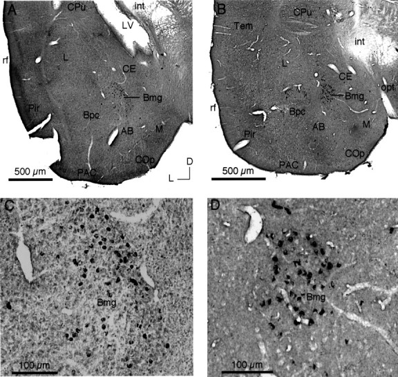
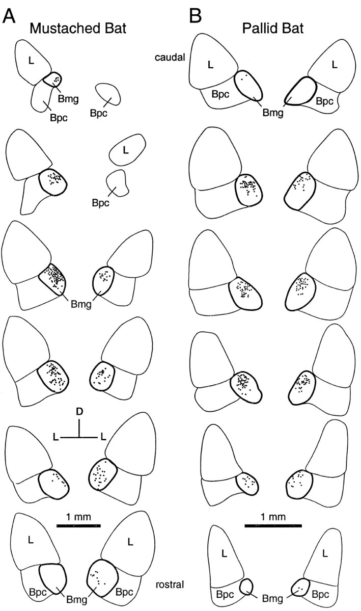
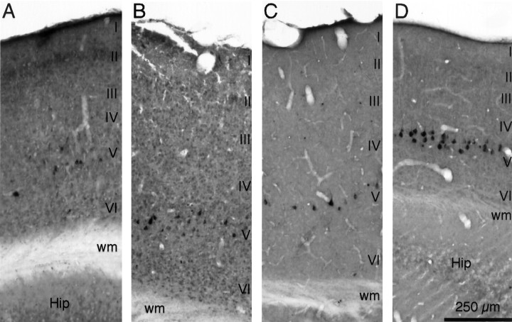
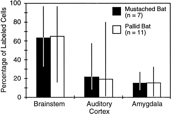
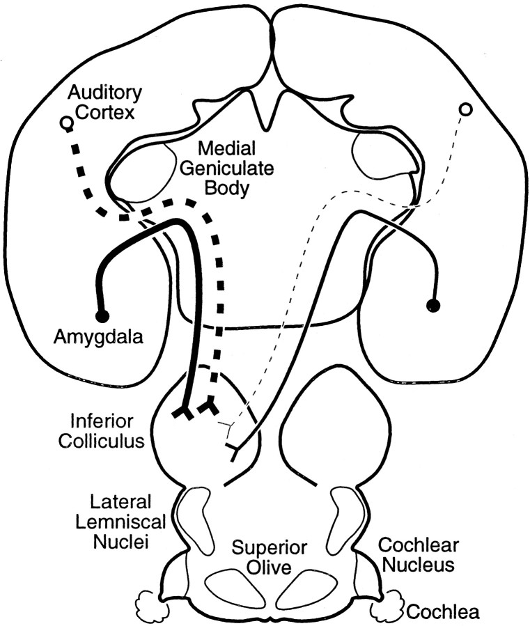
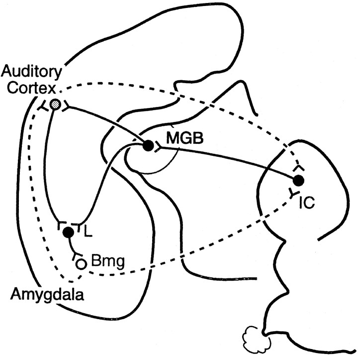
This report describes a projection from the amygdala, a forebrain center mediating emotional expression, to the inferior colliculus (IC), the midbrain integration center of the ascending auditory system. In the IC of mustached bats (Pteronotus parnellii) and pallid bats (Antrozous pallidus), we placed deposits of retrograde tracers at physiologically defined sites and then searched for retrogradely labeled somata in the forebrain. Labeling was most sensitive in experiments using cholera toxin B-subunit as tracer. We consistently observed retrograde labeling in a single amygdalar subdivision, the magnocellular subdivision of the basal nucleus (Bmg). The Bmg is distinctive across mammals, containing the largest cells in the amygdala and the most intense acetylcholinesterase staining. Labeled amygdalar cells occurred ipsilateral and contralateral to IC deposits, but ipsilateral labeling was greater, averaging 72%. Amygdalar labeling was observed after tracer deposits throughout the IC, including its central nucleus (ICC). In comparison, labeling in the auditory cortex (layer V) was heavily ipsilateral (averaging 92%). Cortical labeling depended on the location of IC deposits: dorsomedial deposits resulted in the most labeled cells, whereas ventrolateral deposits labeled few or no cortical cells. Cortical labeling occurred after several deposits in the ICC. Across experiments, the average number of labeled cells in the amygdala was similar to that in the auditory cortex, indicating that the amygdalocollicular projection is significant. The results demonstrate a direct, widespread projection from the basal amygdala to the IC. They also suggest the presence of a rapid thalamoamygdalocollicular feedback circuit that may impose emotional content onto processing of sensory stimuli at a relatively low level of an ascending sensory pathway.
Figures

Similar articles
-
Descending projections from the auditory cortex to the inferior colliculus in the gerbil, Meriones unguiculatus.J Comp Neurol. 2005 May 30;486(2):101-16. doi: 10.1002/cne.20542. J Comp Neurol. 2005. PMID: 15844210
-
Inputs to combination-sensitive neurons in the medial geniculate body of the mustached bat: the missing fundamental.J Neurosci. 1995 Jun;15(6):4693-711. doi: 10.1523/JNEUROSCI.15-06-04693.1995. J Neurosci. 1995. PMID: 7540682 Free PMC article.
-
Functional organization of mustached bat inferior colliculus: II. Connections of the FM2 region.J Comp Neurol. 1989 Jun 1;284(1):85-107. doi: 10.1002/cne.902840107. J Comp Neurol. 1989. PMID: 2754032
-
Roles of inhibition for transforming binaural properties in the brainstem auditory system.Hear Res. 2002 Jun;168(1-2):60-78. doi: 10.1016/s0378-5955(02)00362-3. Hear Res. 2002. PMID: 12117510 Review.
-
Descending projections to the auditory midbrain: evolutionary considerations.J Comp Physiol A Neuroethol Sens Neural Behav Physiol. 2023 Jan;209(1):131-143. doi: 10.1007/s00359-022-01588-5. Epub 2022 Nov 3. J Comp Physiol A Neuroethol Sens Neural Behav Physiol. 2023. PMID: 36323876 Free PMC article. Review.
Cited by
-
Intracellular recordings reveal integrative function of the basolateral amygdala in acoustic communication.J Neurophysiol. 2023 Jun 1;129(6):1334-1343. doi: 10.1152/jn.00103.2023. Epub 2023 Apr 26. J Neurophysiol. 2023. PMID: 37098994 Free PMC article.
-
Neural Correlates of Perceptual Plasticity in the Auditory Midbrain and Thalamus.J Neurosci. 2025 Mar 5;45(10):e0691242024. doi: 10.1523/JNEUROSCI.0691-24.2024. J Neurosci. 2025. PMID: 39753303
-
Rhythmic Relating: Bidirectional Support for Social Timing in Autism Therapies.Front Psychol. 2022 May 16;13:793258. doi: 10.3389/fpsyg.2022.793258. eCollection 2022. Front Psychol. 2022. PMID: 35693509 Free PMC article.
-
A new concept for noninvasive tinnitus treatment utilizing multimodal pathways.Annu Int Conf IEEE Eng Med Biol Soc. 2013;2013:3122-5. doi: 10.1109/EMBC.2013.6610202. Annu Int Conf IEEE Eng Med Biol Soc. 2013. PMID: 24110389 Free PMC article.
-
Coding the meaning of sounds: contextual modulation of auditory responses in the basolateral amygdala.J Neurosci. 2013 Oct 30;33(44):17538-48. doi: 10.1523/JNEUROSCI.2205-13.2013. J Neurosci. 2013. PMID: 24174686 Free PMC article.
References
-
- Aggleton JP, Burton MJ, Passingham RE. Cortical and subcortical afferents to the amygdala of the rhesus monkey (Macaca mulatta). Brain Res. 1980;190:347–368. - PubMed
-
- Alheid GF, de Olmos JS, Beltramino CA. Amygdala and extended amygdala. In: Paxinos G, editor. The rat nervous system. Academic; San Diego: 1995. pp. 495–578.
-
- Amaral DG, Price JL. Amygdalo-cortical projections in the monkey (Macaca fascicularis). J Comp Neurol. 1984;230:465–496. - PubMed
-
- Amorapanth P, LeDoux JE, Nader K. Different lateral amygdala outputs mediate reactions and actions elicited by a fear-arousing stimulus. Nat Neurosci. 2000;3:74–79. - PubMed
Publication types
MeSH terms
Substances
Grants and funding
LinkOut - more resources
Full Text Sources
Other Literature Sources