

Induction of IL-15 by TCR/CD3 aggregation depends on IFN-gamma and protects against apoptosis of immature thymocytes in vivo
- PMID: 12452826
- PMCID: PMC1906542
- DOI: 10.1046/j.1365-2249.2002.02013.x
Induction of IL-15 by TCR/CD3 aggregation depends on IFN-gamma and protects against apoptosis of immature thymocytes in vivo
Abstract
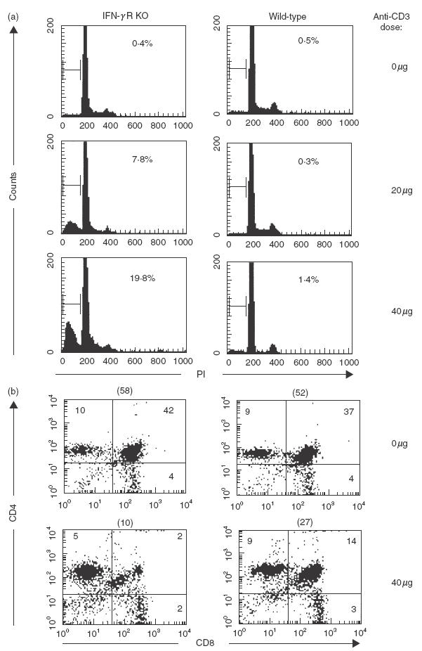
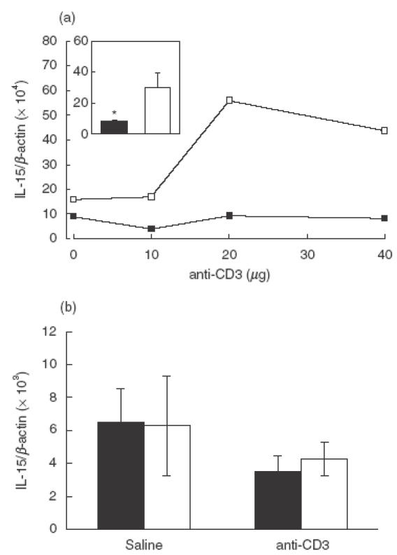
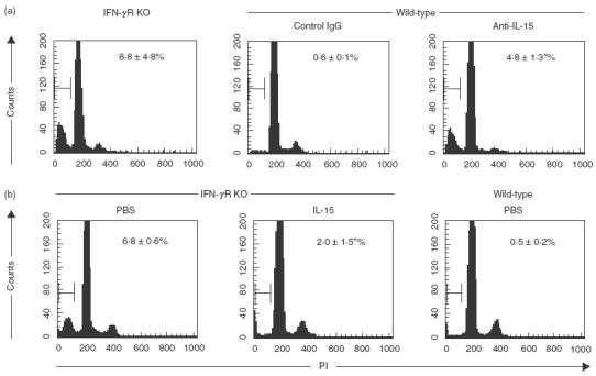
TCR/CD3 aggregation by injection of anti-CD3 Ab produces T cell activation, release of cytokines such as IFN-gamma, and apoptosis in the cortical region of the thymus. We show that anti-CD3 Ab induces IL-15 mRNA in spleens of wild-type but not IFN-gamma receptor-knock-out (IFN-gammaR KO) mice. The loss of IL-15 mRNA induction in IFN-gammaR KO mice was associated with increased thymocyte apoptosis. Pretreatment of wild-type mice with neutralizing anti-IL-15 Ab increased the anti-CD3-triggered thymocyte apoptosis, thus mimicking the sensitive phenotype of IFN-gammaR KO mice. Inversely, anti-CD3-induced apoptosis in IFN-gammaR KO mice was suppressed by administration of recombinant IL-15. In IFN-gammaR KO mice and in wild-type mice that were treated with anti-IL-15, augmented apoptosis affected mainly CD4+CD8+ immature thymocytes. IL-15 as well as IL-15Ralpha mRNA expression in thymocytes was not increased by anti-CD3. These data demonstrate that systemic IL-15 exerts anti-apoptotic activity on immature T cells and establish a regulatory mechanism whereby TCR/CD3 engagement induces IL-15 expression via an IFN-gamma-dependent pathway. The self-amplifying nature of this IFN-gamma/IL-15 connection may constitute a regulatory pathway in central tolerance to self.
Figures
Similar articles
-
IFN-gamma receptor-deficient mice are hypersensitive to the anti-CD3-induced cytokine release syndrome and thymocyte apoptosis. Protective role of endogenous nitric oxide.J Immunol. 1995 Oct 15;155(8):3823-9. J Immunol. 1995. PMID: 7561088
-
Protection from anti-TCR/CD3-induced apoptosis in immature thymocytes by a signal through thymic shared antigen-1/stem cell antigen-2.J Exp Med. 1996 May 1;183(5):2355-60. doi: 10.1084/jem.183.5.2355. J Exp Med. 1996. PMID: 8642345 Free PMC article.
-
JNK2 is required for efficient T-cell activation and apoptosis but not for normal lymphocyte development.Curr Biol. 1999 Feb 11;9(3):116-25. doi: 10.1016/s0960-9822(99)80065-7. Curr Biol. 1999. PMID: 10021384
-
CD3-mediated apoptosis of human medullary thymocytes and activated peripheral T cells: respective roles of interleukin-1, interleukin-2, interferon-gamma and accessory cells.Eur J Immunol. 1993 Jul;23(7):1623-9. doi: 10.1002/eji.1830230734. Eur J Immunol. 1993. PMID: 8325338
-
Cell cycle control of T cell apoptosis induced by activation through the T cell antigen receptor.Adv Exp Med Biol. 1996;406:57-67. doi: 10.1007/978-1-4899-0274-0_6. Adv Exp Med Biol. 1996. PMID: 8910671 Review. No abstract available.
References
-
- Hirsch R, Gress RE, Pluznik DH, Eckhaus M, Bluestone JA. Effects of in vivo administration of CD3 monoclonal antibody on T cell function in mice. II. In vivo activation of T cells. J Immunol. 1989;142:737–43. - PubMed
-
- Alegre M, Vandenabeele P, Depierreux M, et al. Cytokine release syndrome induced by the 145–2C11 anti-CD3 monoclonal antibody in mice. prevention by high doses of methylprednisolone. J Immunol. 1991;146:1184–91. - PubMed
-
- Ferran C, Dy M, Sheehan K, et al. Cascade modulation by anti-tumor necrosis factor monoclonal antibody of interferon-γ, interleukin-3 and interleukin-6 release after triggering of the CD3/T cell receptor activation pathway. Eur J Immunol. 1991;21:2349–53. - PubMed
-
- Matthys P, Froyen G, Huang S, et al. Interferon-γ receptor-deficient mice are hypersensitive to the anti-CD3-induced cytokine release syndrome and thymocyte apoptosis: Protective role of endogenous nitric oxide. J Immunol. 1995;155:3823–9. - PubMed
-
- Shi Y, Bissonnette RP, Parfrey N, Szalay M, Kubo RT, Green DR. In vivo administration of monoclonal antibodies to the CD3 T cell receptor complex induces cell death (apoptosis) in imature thymocytes. J Immunol. 1991;146:3340–6. - PubMed
Publication types
MeSH terms
Substances
LinkOut - more resources
Full Text Sources
Molecular Biology Databases
Research Materials
