. 2002 Dec;130(3):459-66.
doi: 10.1046/j.1365-2249.2002.01998.x.

Murine glomerular mesangial cell uptake of apoptotic cells is inefficient and involves serum-mediated but complement-independent mechanisms

Affiliations

Murine glomerular mesangial cell uptake of apoptotic cells is inefficient and involves serum-mediated but complement-independent mechanisms

J Cortes-Hernandez et al. Clin Exp Immunol. 2002 Dec.

Abstract

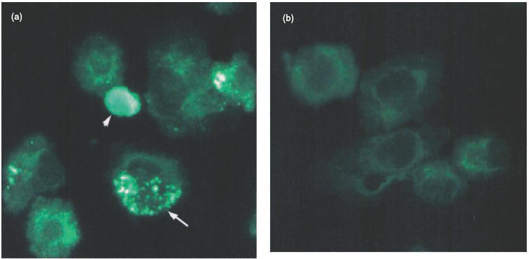
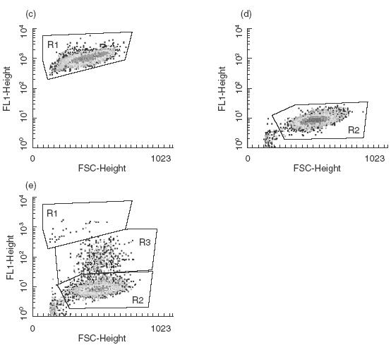
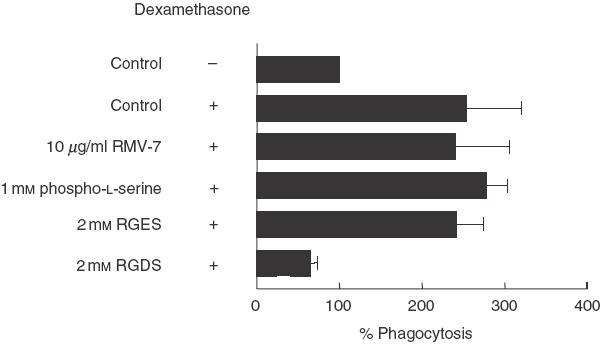
An increased number of apoptotic bodies have been detected in glomeruli of non-nephritic kidneys of C1q-deficient mice. In these mice an in vivo impaired uptake of apoptotic cells by peritoneal macrophages was also demonstrated. Here we investigated whether C1q plays a role in the in vitro clearance of apoptotic cells by glomerular mesangial cells. Phagocytosis was assessed using a novel flow cytometric assay that was validated by immunofluorescence studies. The uptake of apoptotic cells by mesangial cells, measured as percentage of mesangial cells ingesting apoptotic cells, was approximately 25%, 10% and 10% for a T cell lymphoma line (RMA), thymocytes and neutrophils, respectively. The uptake reached a plateau phase after 3 h, was specific for apoptotic cells and was mediated by serum but not by complement components C1q or C3. The phagocytosis of apoptotic cells was significantly inhibited by Arg-Gly-Asp-Ser (RGDS), a peptide capable of blocking the interaction of thrombospondin with CD36 or the vitronectin receptor. Pretreatment of the mesangial cells with dexamethasone (200 nm) but not with LPS increased the uptake markedly. These findings indicate that murine mesangial cells are capable of taking up syngeneic apoptotic cells, although much less efficiently than professional phagocytic cells. They also show that serum proteins other than complement components mediate the removal of apoptotic cells by murine mesangial cells in vitro.


Figures

Fig. 1
Fig. 1
Phagocytosis of apoptotic RMA cells by murine mesangial cells in vitro. (a) Representative photomicrographs of cytospin preparations of PKH26-labelled mesangial cells incubated with CFSE-labelled apoptotic RMA cells for 3 h in the presence of mouse serum. Note that the ingested apoptotic RMA cells gave a green granular pattern within the mesangial cells (example arrowed), while the uningested RMA cells retained their condensed morphology and appeared merely to adhere to the mesangial cells (arrowhead). (b) PKH26-stained mesangial cells alone. (c,d) FACS profiles of CFSE-labelled apoptotic RMA and unlabelled mesangial cells, respectively. (e) FACS profile of unlabelled mesangial cells incubated with CFSE-labelled apoptotic RMA cells for three hours in presence of mouse serum. Three populations of cells can be defined according to their size and fluorescence: uningested apoptotic RMA cells (R1), ingested apoptotic RMA cells (R3) and mesangial cells alone (R2).
Fig. 1
Fig. 1
Phagocytosis of apoptotic RMA cells by murine mesangial cells in vitro. (a) Representative photomicrographs of cytospin preparations of PKH26-labelled mesangial cells incubated with CFSE-labelled apoptotic RMA cells for 3 h in the presence of mouse serum. Note that the ingested apoptotic RMA cells gave a green granular pattern within the mesangial cells (example arrowed), while the uningested RMA cells retained their condensed morphology and appeared merely to adhere to the mesangial cells (arrowhead). (b) PKH26-stained mesangial cells alone. (c,d) FACS profiles of CFSE-labelled apoptotic RMA and unlabelled mesangial cells, respectively. (e) FACS profile of unlabelled mesangial cells incubated with CFSE-labelled apoptotic RMA cells for three hours in presence of mouse serum. Three populations of cells can be defined according to their size and fluorescence: uningested apoptotic RMA cells (R1), ingested apoptotic RMA cells (R3) and mesangial cells alone (R2).
Fig. 2
Fig. 2
Kinetic of apoptotic RMA cell clearance. Graph showing the percentage of mesangial cells from 129/sv and C57BL/6 mice phagocytosing apoptotic RMA cells at different time-points. The kinetic of the uptake was similar with phagocytosis reaching a plateau phase after 3 h of co-culture. Data shown are representative of three independent experiments. •, C57BL/6; ▴, 129/Sv.
Fig. 3
Fig. 3
Phagocytic uptake of murine apoptotic cells of different origin. Graph showing the percentage of mesangial cells phagocytosing apoptotic RMA cells (∼80% apoptotic) or neutrophils (∼55% apoptotic) or thymocytes (∼45% apoptotic) at different time-points. Plateau was reached after 3 h of co-culture irrespective of the origin of the cells. The higher uptake observed using the RMA cells paralleled the higher percentage of apoptosis present in the RMA cell preparations. Data shown are representative of three independent experiments., ▪, Mitomycin-induced apoptotic RMA; ▴, starvation-induced apoptotic neutrophils; •, dexamethazone-induced apoptotic thymocytes.
Fig. 4
Fig. 4
Phagocytosis of apoptotic RMA cells by mesangial cells from wild-type (n = 4) and C1q-deficient mice (n = 4). Graph showing the percentage of phagocytosis after 3 h of co-culture in the presence of 20% serum from wild-type animals or C1q- or C3-deficient mice and in the absence of serum. The results are expressed as a mean percentage of the phagocytic signal in the well containing wild-type serum for each experiment ± s.e.m. The control well was standardized to 100%. No inhibition in phagocytosis was detected using complement-deficient sera, while the absence of serum reduced the uptake to about 5–7% of the control. No difference of phagocytic uptake was observed between mesangial cells isolated from C1q-deficient or wild-type mice. ▪, Wild-type serum; formula image
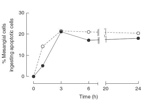
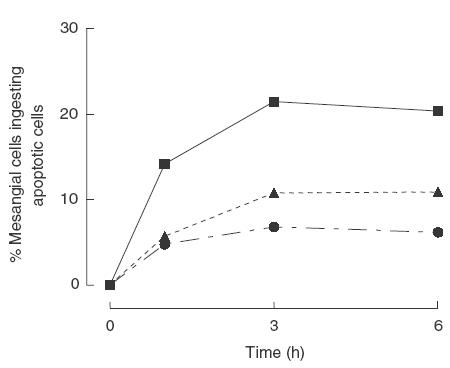
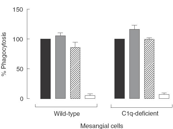
, C1q-deficient serum; formula image, C3-deficient serum; □, no serum.
Fig. 5
Fig. 5
The effect of RGDS and RGES tetrapeptides, phospho-l-serine and RMV-7 upon mesangial cell phagocytosis of apoptotic RMA cells at the 3-h time-point. The higher inhibition of phagocytosis was seen following the addition of 2 mm RGDS in the mesangial cell/apoptotic RMA interaction medium while the control tetrapeptide RGES had no effect. The results are expressed as a mean percentage of the phagocytic signal in the well containing wild-type serum for each experiment ± s.e.m. The control well was standardized to 100%. Data shown are the result of four independent experiments. *P < 0·05; **P < 0·0001.
Fig. 6
Fig. 6
The effect of RGDS and RGES tetrapeptides, phospho-l-serine and RMV-7 upon dexamethasone-treated mesangial cell phagocytosis of apoptotic RMA cells at the 3-h time-point. Inhibition of phagocytosis was seen only following the inclusion of 2 mm RGDS in the mesangial cell/apoptotic RMA interaction medium. Note that addition of dexamethasone to wild-type serum induced a significant increase in the phagocytic uptake to about 250% of the control. The results are expressed as a mean percentage of the phagocytic signal in the well containing wild-type serum for each experiment ± s.e.m. The control well was standardized to 100%. Data shown are the result of three independent experiments.

Similar articles

Cited by

References

    1. Fadok VA, Bratton DL, Henson PM. Phagocyte receptors for apoptotic cells: recognition, uptake and consequences. J Clin Invest. 2001;108:957–62. - PMC - PubMed
    1. Savill J, Dransfield I, Hogg N, et al. Vitronectin receptor-mediated phagocytosis of cells undergoing apoptosis. Nature. 1990;343:170–3. - PubMed
    1. Ren Y, Silverstein RL, Allen J, et al. CD36 gene transfer confers capacity for phagocytosis of cells undergoing apoptosis. J Exp Med. 1995;181:1857–62. - PMC - PubMed
    1. Fadok VA, Bratton DL, Rose DM, et al. A receptor for phosphatidylserine-specific clearance of apoptotic cells. Nature. 2000;405:85–90. - PubMed
    1. Devitt A, Moffatt OD, Raykundalia C, et al. Human CD14 mediates recognition and phagocytosis of apoptotic cells. Nature. 1998;392:505–9. - PubMed

Publication types