. 2002 Dec;40(12):4713-9.
doi: 10.1128/JCM.40.12.4713-4719.2002.

Development of a real-time fluorescence PCR assay for rapid detection of the diphtheria toxin gene

Affiliations

Development of a real-time fluorescence PCR assay for rapid detection of the diphtheria toxin gene

Elizabeth A Mothershed et al. J Clin Microbiol. 2002 Dec.

Abstract

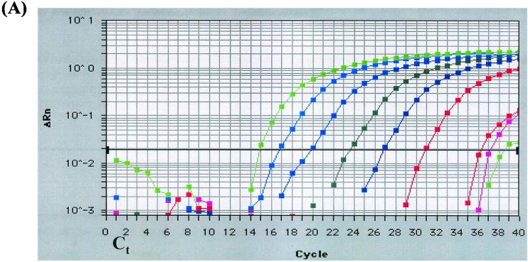
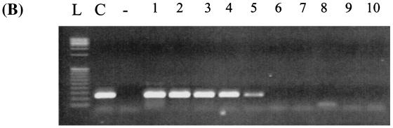
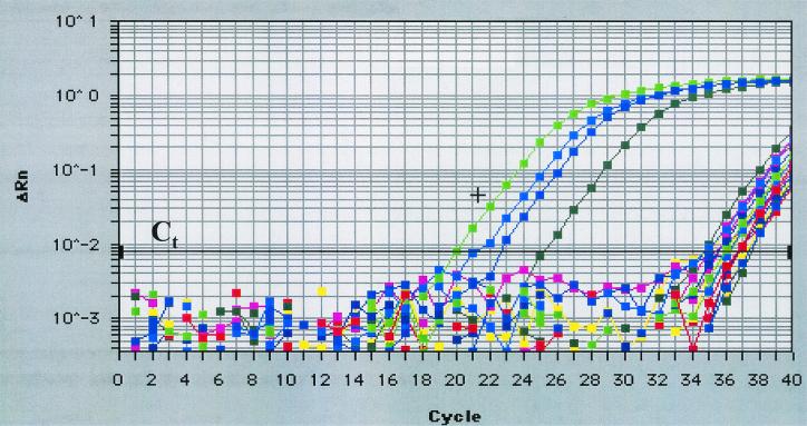
We developed and evaluated a real-time fluorescence PCR assay for detecting the A and B subunits of diphtheria toxin (tox) gene. When 23 toxigenic Corynebacterium diphtheriae strains, 9 nontoxigenic C. diphtheriae strains, and 44 strains representing the diversity of pathogens and normal respiratory flora were tested, this real-time PCR assay exhibited 100% sensitivity and specificity. It allowed for the detection of both subunits of the tox gene at 750 times greater sensitivity (2 CFU) than the standard PCR (1,500 CFU). When used directly on specimens collected from patients with clinical diphtheria, one or both subunits of the tox gene were detected in 34 of 36 specimens by using the real-time PCR assay; only 9 specimens were found to be positive by standard PCR. Reamplification by standard PCR and DNA sequencing of the amplification product confirmed all real-time PCR tox-positive reactions. This real-time PCR format is a more sensitive and rapid alternative to standard PCR for detection of the tox gene in clinical material.


Figures

FIG. 1.
FIG. 1.
Comparative lower limit of detection of the tox real-time PCR assay and standard PCR. Corynebacterium diphtheriae (NCTC 10648) was grown on sheep blood agar at 37°C for 16 h, and serial 10-fold dilutions were prepared in HI broth. DNA was extracted from undiluted culture and from 10−1 to 10−8 dilutions and used in the tox real-time PCR assay and in the standard PCR assay. (A) Real-time PCR amplification generated by the Prism 7700 sequence detector showing amplification of target DNA from nine samples (from left to right: undiluted C. diphtheriae growth in HI broth and dilutions 10−1 to 10−8). ΔRn, difference in reporter fluorescence between the sample and the no-template controls; Ct, threshold cycle (i.e., the cycle at which a statistically significant increase in fluorescence is first detected). (B) Amplification of the A subunit of the tox gene (primers tox 1 and tox 2) by standard PCR. Lanes: L, low-mass DNA ladder; C, undiluted C. diphtheriae growth in HI broth; −, no-template control; 1 to 10, dilutions 10−1 to 10−10, respectively.
FIG. 1.
FIG. 1.
Comparative lower limit of detection of the tox real-time PCR assay and standard PCR. Corynebacterium diphtheriae (NCTC 10648) was grown on sheep blood agar at 37°C for 16 h, and serial 10-fold dilutions were prepared in HI broth. DNA was extracted from undiluted culture and from 10−1 to 10−8 dilutions and used in the tox real-time PCR assay and in the standard PCR assay. (A) Real-time PCR amplification generated by the Prism 7700 sequence detector showing amplification of target DNA from nine samples (from left to right: undiluted C. diphtheriae growth in HI broth and dilutions 10−1 to 10−8). ΔRn, difference in reporter fluorescence between the sample and the no-template controls; Ct, threshold cycle (i.e., the cycle at which a statistically significant increase in fluorescence is first detected). (B) Amplification of the A subunit of the tox gene (primers tox 1 and tox 2) by standard PCR. Lanes: L, low-mass DNA ladder; C, undiluted C. diphtheriae growth in HI broth; −, no-template control; 1 to 10, dilutions 10−1 to 10−10, respectively.
FIG. 2.
FIG. 2.
Real-time amplification plot of 18 representative clinical samples. Positive control DNA from C. diphtheriae (NCTC 10648 toxigenic) and three clinical samples amplified early (20 to 25 cycles). The majority of samples amplified late (>34 cycles) in the 40-cycle program. +, Positive control DNA; Ct, cycle threshold (i.e., the cycle at which a statistically significant increase in fluorescence is first detected).

References

    1. Dittmann, S., M. Wharton, C. Vitek, M. Ciotti, A. Galazka, S. Guichard, I. Hardy, U. Kartoglu, S. Koyama, J. Kreysler, B. Martin, D. Mercer, T. Ronne, C. Roure, R. Steinglass, P. Strebel, R. Sutter, and M. Trostle. 2000. Successful control of epidemic diphtheria in the states of the Former Union of Soviet Socialist Republics: lessons learned. J. Infect. Dis. 181(Suppl. 1):S10-S22. - PubMed
    1. Efstratiou, A., and P. A. Maple. 1994. WHO manual for the laboratory diagnosis of diptheria, p. 69-71. Document ICP-EPI 038(C). World Health Organization, Geneva, Switzerland.
    1. Funke, G., and K. A. Berhard. 1999. Coryneform gram-positive rods, p. 319-345. In P. R. Murray, E. J. Baron, M. A. Pfaller, F. C. Tenover, and R. H. Yolken (ed.), Manual of clinical microbiology, 7th ed. ASM Press, Washington, D.C.
    1. Henricson, B., M. Segarra, J. Garvin, J. Burns, S. Jankins, C. Kim, T. Popovic, A. Golaz, and B. Akey. 2000. Toxigenic Corynebacterium diphtheriae associated with an equine wound infection. J. Vet. Diagn. Investig. 12:253-257. - PubMed
    1. Higgins, J. A., J. Ezzell, B. J. Hinnebusch, M. Shipley, E. A. Henchal, and M. S. Ibrahim. 1998. 5′ nuclease PCR assay to detect Yersinia pestis. J. Clin. Microbiol. 36:2284-2288. - PMC - PubMed

Publication types

LinkOut - more resources