. 2002 Dec 10;99(25):16180-5.
doi: 10.1073/pnas.212643199. Epub 2002 Nov 26.

Disulfide bond-mediated dimerization of HLA-G on the cell surface

Affiliations

Disulfide bond-mediated dimerization of HLA-G on the cell surface

Jonathan E Boyson et al. Proc Natl Acad Sci U S A. .

Abstract

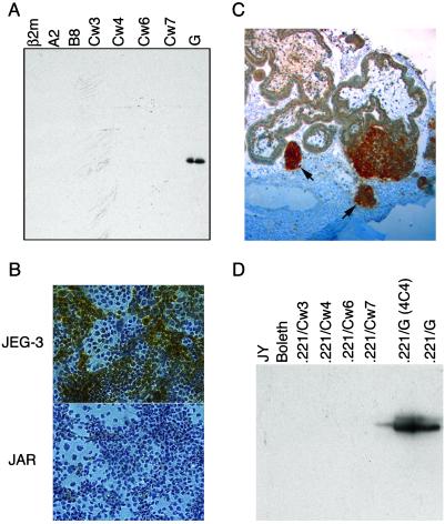
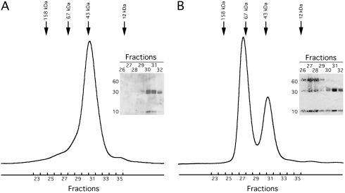
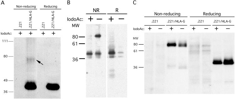
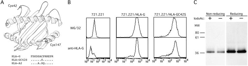
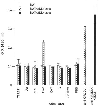
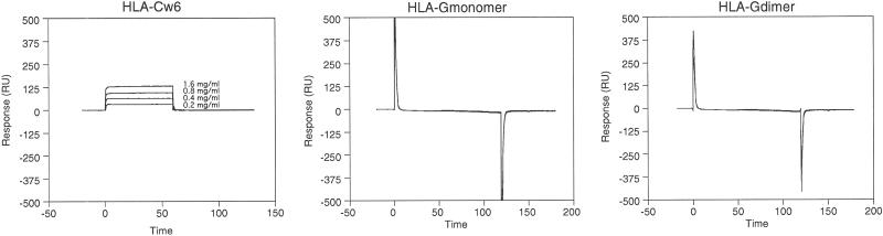
HLA-G is a nonclassical class I MHC molecule with an unknown function and with unusual characteristics that distinguish it from other class I MHC molecules. Here, we demonstrate that HLA-G forms disulfide-linked dimers that are present on the cell surface. Immunoprecipitation of HLA-G from surface biotinylated transfectants using the anti-beta2-microglobulin mAb BBM.1 revealed the presence of an approximately equal 78-kDa form of HLA-G heavy chain that was reduced by using DTT to a 39-kDa form. Mutation of Cys-42 to a serine completely abrogated dimerization of HLA-G, suggesting that the disulfide linkage formed exclusively through this residue. A possible interaction between the HLA-G monomer or dimer and the KIR2DL4 receptor was also investigated, but no interaction between these molecules could be detected through several approaches. The cell-surface expression of dimerized HLA-G molecules may have implications for HLA-Greceptor interactions and for the search for specific receptors that bind HLA-G.


Figures

Fig 1.
Fig 1.
Characterization of HLA-G-specific mAbs. (A) Western blot of bacterially produced solubilized inclusion bodies from various soluble recombinant HLA molecules detected by MEM-G/1 mAb. (B) Cytospin preparations of the HLA-G+ choriocarcinoma cell line JEG-3 and the HLA-G cell line JAR were stained with the MEM-G/1 mAb. Whereas JAR is completely negative, HLA-G staining (brown) can be seen clearly in the JEG-3 sample. Note that not all of the JEG-3 cells stain positively, which is consistent with the heterogeneous nature of this tumor line. (C) Immunohistochemistry of paraffin-embedded placental sections using the MEM-G/1 mAb. HLA-G staining (dark brown) was detected on the cell islands (denoted by arrows) that had migrated from the villi. (D) The MEM-G/11 mAb was used to immunoprecipitate HLA-G from surface-biotinylated B-lymphoblastoid cell lines and from various class I MHC transfected into the MHC-deficient 721.221 B-lymphoblastoid cell line. ECL visualization demonstrated that only the two different clones of HLA-G+ transfectants were recognized.
Fig 2.
Fig 2.
Soluble HLA-G dimerizes in vitro. (A) Chromatogram of recombinant soluble HLA-G analyzed by size-exclusion chromatography using a Superdex 200 10/30. Arrows depict MM calibration standards. (Inset) A nonreducing SDS/PAGE gel of the fractions before pooling. Because it is a soluble truncated form of HLA-G, the heavy chain has an expected MM of 32 kDa, and the refolded HLA-G monomer has an expected MM of ≈44 kDa. (B) Chromatogram of HLA-G after a 21-day incubation at 4°C run on a Superdex 200 10/30. Approximately 25% of the sample eluted in a volume corresponding to a MM of ≈43 kDa and 75% of the sample eluted in a volume corresponding to roughly twice the MM of the monomer. (Inset) A nonreducing SDS/PAGE gel of the fractions from both peaks.
Fig 3.
Fig 3.
Cell-surface dimerization of HLA-G. (A) 721.221/HLA-G transfectants and the 721.221 parental cell line were lysed in SDS/PAGE buffer containing iodoacetamide and run under nonreducing and reducing conditions. After blotting to nitrocellulose, HLA-G heavy chains were detected by using the HLA-G-specific mAb, MEM-G/1. (B) Immunoprecipitation of cell surface-biotinylated HLA-A2 molecules from 721.221/HLA-A2 transfectants. Inclusion of iodoacetamide abrogated the formation of HLA-A2 dimer artifacts, because under nonreducing conditions the ≈85-kDa MM band does not appear in samples lysed in the presence of iodoacetamide. (C) Cell-surface molecules of 721.221/HLA-G transfectants and the 721.221 parental cell line were biotinylated, and cells were lysed in the presence or absence of iodoacetamide. Class I MHC molecules were immunoprecipitated with BBM.1 and run under both nonreducing and reducing conditions. Even in the presence of iodoacetamide, HLA-G dimers were detected.
Fig 4.
Fig 4.
Identification and mutagenesis of extracellular cysteines in HLA-G. (A) A ribbon diagram of the crystal structure of HLA-A2 with Cys-42 and Cys-147 residues superimposed. Shown below is a portion of the α1 domain HLA-G sequence containing Cys-42 which was chosen for mutagenesis to a serine. Dots (.) indicate the residue identity with the HLA-G sequence. (B) Cell-surface expression of HLA-G/C42S. 721.221 B-LCLs transfected with HLA-G/C42S were stained with the conformation-specific mAb W6/32 and the HLA-G-specific mAb, MEM-G/11. After FACS-sorting, HLA-G/C42S and the wild-type HLA-G transfectants expressed similar levels of protein. (C) Mutagenesis of Cys-42 to Ser-42 completely abrogates HLA-G dimerization. HLA-G/C42S transfectants were cell surface-biotinylated and lysed in the presence or absence of iodoacetamide. Class I MHC molecules were immunoprecipitated and run under both nonreducing and reducing conditions. Even in the absence of iodoacetamide, HLA-G dimers could not be detected.
Fig 5.
Fig 5.
Cell surface-expressed KIR2DL4-ζ is not crosslinked by HLA-G. BW cells stably transfected with KIR-ζ constructs were cocultured with various class I MHC transfectants. After 72 h, supernatants were collected and IL-2 secretion was measured by ELISA. As positive controls, some wells were coated with anti-KIR2DL1-specific mAb EB6 and anti-KIR2DL4/KIR2DL5 polyclonal antibody.
Fig 6.
Fig 6.
SPR analysis of HLA-G–KIR2DL4 interaction. KIR-Ig coupled to sensor chips by means of a goat anti-human IgG antibody were used to detect interactions between HLA-Cw6 and KIR2DL1-Ig (A), HLA-G monomer and KIR2DL4- and KIR2DL1-Ig (B), and HLA-G dimer and KIR2DL4- and KIR2DL1-Ig (C). The varying concentrations of HLA-Cw6 that flowed over the surface are indicated. Neither HLA-G monomer nor HLA-G dimer bound to the KIR-Ig fusion proteins when run at a concentration of 5 mg/ml (>100 μM).

References

    1. Kovats S., Main, E. K., Librach, C., Stubblebine, M., Fisher, S. J. & DeMars, R. (1990) Science 248, 220-223. - PubMed
    1. McMaster M. T., Librach, C. L., Zhou, Y., Lim, K. H., Janatpour, M. J., DeMars, R., Kovats, S., Damsky, C. & Fisher, S. J. (1995) J. Immunol. 154, 3771-3778. - PubMed
    1. Alizadeh M., Legras, C., Semana, G., Le Bouteiller, P., Genetet, B. & Fauchet, R. (1993) Hum. Immunol. 38, 206-212. - PubMed
    1. Kirszenbaum M., Djoulah, S., Hors, J., Le Gall, I., de Oliveira, E. B., Prost, S., Dausset, J. & Carosella, E. D. (1997) Hum. Immunol. 53, 140-147. - PubMed
    1. Geraghty D. E., Koller, B. H. & Orr, H. T. (1987) Proc. Natl. Acad. Sci. USA 84, 9145-9149. - PMC - PubMed

Publication types

MeSH terms