

Tissue microarray evaluation of Melanoma antigen E (MAGE) tumor-associated antigen expression: potential indications for specific immunotherapy and prognostic relevance in squamous cell lung carcinoma
- PMID: 12454517
- PMCID: PMC1422645
- DOI: 10.1097/01.SLA.0000036266.09823.6C
Tissue microarray evaluation of Melanoma antigen E (MAGE) tumor-associated antigen expression: potential indications for specific immunotherapy and prognostic relevance in squamous cell lung carcinoma
Abstract
Objective: To evaluate MAGE tumor-associated antigen (TAA) expression in an extensive panel of normal and neoplastic tissues.
Summary background data: TAAs of the MAGE family represent targets of active specific immunotherapy. Limited-size studies indicate that they are expressed in normal testis and tumors of different histologies. High-throughput tissue microarray (TMA) technology and MAGE TAA-specific monoclonal antibodies now allow us to comprehensively evaluate their expression in large numbers of tissues and to address clinical correlations.
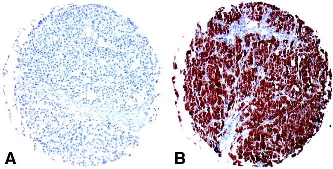
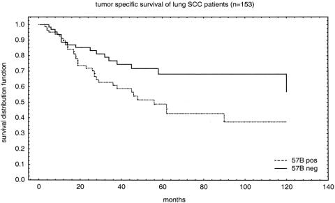
Methods: A TMA containing 3,520 samples from 197 different tissues and a non-small-cell lung cancer TMA including 301 specimens were stained using the MAGE TAA-specific monoclonal antibody 57B. For patients with squamous cell carcinoma of the lung, the dichotomous result (positive vs. negative) of MAGE TAA staining was used as a predictor variable along with other covariates in proportional hazard regression analysis of tumor-specific survival.
Results: MAGE TAAs are expressed with frequencies ranging between 22.7% (larynx) and 50% of cases (lung) in squamous cell carcinomas from different anatomic areas and in large cell carcinomas of the lung (37.9%). The authors provide here the first description of MAGE TAA expression in basalioma (48.1%). To investigate the clinical significance of MAGE expression in a frequently positive tumor type, a non-small-cell lung cancer, TMA was then studied. In this TMA 43.2% of tumors were 57B positive. In patients with squamous cell carcinoma, MAGE TAA positivity was significantly correlated with a shorter tumor-specific survival in the proportional hazard regression analysis model.
Conclusions: These data suggest novel potential therapeutic indications in different types of cancers. In lung squamous cell carcinoma, the significant association of MAGE TAA expression with poor prognosis suggests that patients with 57B-positive tumors may benefit from early, specific immunotherapy procedures.
Figures
References
-
- van der Bruggen P, Traversari C, Chomez P, et al. A gene encoding an antigen recognized by cytolytic T lymphocytes on a human melanoma. Science 1991; 254: 1643–1647. - PubMed
-
- Jungbluth AA, Busam KJ, Kolb D, et al. Expression of MAGE-antigens in normal tissues and cancer. Int J Cancer 2000; 85: 460–465. - PubMed
-
- Chomez P, De Backer O, Bertrand M, et al. An overview of the MAGE gene family with the identification of all human members of the family. Cancer Res 2001; 61: 5544–5551. - PubMed
-
- Duffour MT, Chaux P, Lurquin C, et al. A MAGE-A4 peptide presented by HLA-A2 is recognized by cytolytic T lymphocytes. Eur J Immunol 1999; 29: 3329–3337. - PubMed
Publication types
MeSH terms
Substances
LinkOut - more resources
Full Text Sources
Other Literature Sources
Medical
