

Dermatofibrosarcoma protuberans treated by micrographic surgery
- PMID: 12454766
- PMCID: PMC2376302
- DOI: 10.1038/sj.bjc.6600643
Dermatofibrosarcoma protuberans treated by micrographic surgery
Abstract
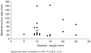
Dermatofibrosarcoma protuberans is an uncommon cutaneous tumour which rarely metastasises. However, local recurrence following apparently adequate surgical excision is well recognised, presumably as a result of sub-clinical contiguous growth, for which micrographically controlled excision would be a logical treatment. A retrospective study of all patients treated by micrographic surgery, from April 1995-March 2000, at a tertiary skin oncology centre. Twenty-one patients (11 males), age 14 to 71 years with dermatofibrosarcoma protuberans on the trunk (10 patients), groin (four), head and neck (four), and limbs (three) were treated. In 15 patients one micrographic layer cleared the tumour, and four were cleared with two layers. For one patient the second stage was completed by conventional excision guided by positive margins. Another patient with a multiply recurrent perineal dermatofibrosarcoma protuberans, not cleared in one area after two layers, died from a pulmonary embolus before total clearance could be achieved. There was no correlation between tumour size and lateral excision margin. No recurrence was observed during the follow-up, from 21 to 80 months, median 47 months. The study provides further support for micrographic surgery as the treatment of choice for dermatofibrosarcoma protuberans.
Copyright 2002 Cancer Research UK
Figures
Similar articles
-
Mohs micrographic surgery for the treatment of dermatofibrosarcoma protuberans. Results of a multiinstitutional series with an analysis of the extent of microscopic spread.J Am Acad Dermatol. 1997 Oct;37(4):600-13. doi: 10.1016/s0190-9622(97)70179-8. J Am Acad Dermatol. 1997. PMID: 9344201
-
A comparison between Mohs micrographic surgery and wide surgical excision for the treatment of dermatofibrosarcoma protuberans.J Am Acad Dermatol. 1996 Jul;35(1):82-7. J Am Acad Dermatol. 1996. PMID: 8682970 Review.
-
Dermatofibrosarcoma protuberans treated with Mohs micrographic surgery: cure rates and surgical margins.Dermatol Surg. 1996 Jun;22(6):530-4. Dermatol Surg. 1996. PMID: 8646467
-
Dermatofibrosarcoma Protuberans: Wide Local Excision Versus Mohs Micrographic Surgery.Surg Oncol Clin N Am. 2016 Oct;25(4):827-39. doi: 10.1016/j.soc.2016.05.011. Epub 2016 Aug 3. Surg Oncol Clin N Am. 2016. PMID: 27591501 Review.
-
Surgical margins for excision of dermatofibrosarcoma protuberans.J Am Acad Dermatol. 1995 Feb;32(2 Pt 1):233-6. doi: 10.1016/0190-9622(95)90132-9. J Am Acad Dermatol. 1995. PMID: 7829708
Cited by
-
Dermatofibrosarcoma protuberans in a pre-adolescent child.Int J Surg Case Rep. 2023 Sep;110:108761. doi: 10.1016/j.ijscr.2023.108761. Epub 2023 Aug 30. Int J Surg Case Rep. 2023. PMID: 37666158 Free PMC article.
-
Bifocal metachronous dermato fibrosarcoma protuberans in children: A case report.Clin Case Rep. 2025 Jan 31;13(2):e9497. doi: 10.1002/ccr3.9497. eCollection 2025 Feb. Clin Case Rep. 2025. PMID: 39895847 Free PMC article.
-
Dermatofibrosarcoma protuberans challenges: a case series and review of the literature.J Med Case Rep. 2023 Jan 19;17(1):18. doi: 10.1186/s13256-022-03728-6. J Med Case Rep. 2023. PMID: 36653860 Free PMC article. Review.
-
Management of Recurrent Dermatofibro Sarcoma Protuberance of Scalp-a Reconstructive Challenge.Indian J Surg Oncol. 2013 Mar;4(1):15-8. doi: 10.1007/s13193-012-0178-7. Epub 2012 Jul 31. Indian J Surg Oncol. 2013. PMID: 24426693 Free PMC article. No abstract available.
-
[Dermatofibrosarcoma protuberans, particular skin tumor: report of 32 cases and review of the literature].Pan Afr Med J. 2014 Oct 24;19:196. doi: 10.11604/pamj.2014.19.196.4470. eCollection 2014. Pan Afr Med J. 2014. PMID: 25821539 Free PMC article. Review. French.
References
-
- BarnesLColemanJA1984Dermatofibrosarcoma protuberans of the head and neck Arch Otolaryngol 110398404 - PubMed
-
- Bendix-HansenKMyhre-JensenOKaaeS1983Dermatofibrosarcoma protuberans: a clinic-pathological study of nineteen cases and review of world literature Scand J Plast Reconstr Surg 17247252 - PubMed
-
- ClaytonBDLeshinBHitchcockMGMarksMWhiteWL2000Utility of rush paraffin-embedded tangential sections in the management of cutaneous neoplasms Dermatol Surg 26671678 - PubMed
-
- DawesKWHankeCW1996Dermatofibrosarcoma protuberans treated with Mohs micrographic surgery: cure rates and surgical margins Dermatol Surg 22530534 - PubMed
-
- GlosterHMHarrisKRRoenigkRK1996A comparison between Mohs micrographic surgery and wide surgical excision for the treatment of dermatofibrosarcoma protuberans J Am Acad Dermatol 358287 - PubMed
Publication types
MeSH terms
LinkOut - more resources
Full Text Sources
Medical
Miscellaneous
