

Uptake and localisation of mTHPC (Foscan) and its 14C-labelled form in normal and tumour tissues of the hamster squamous cell carcinoma model: a comparative study
- PMID: 12454779
- PMCID: PMC2376296
- DOI: 10.1038/sj.bjc.6600651
Uptake and localisation of mTHPC (Foscan) and its 14C-labelled form in normal and tumour tissues of the hamster squamous cell carcinoma model: a comparative study
Abstract
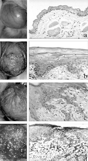
The aim of this study was to evaluate the pharmacokinetics of meta(tetrahydroxyphenyl)chlorin (mTHPC) on different tissues of interest in a hamster tumour model and to confirm our earlier animal studies on semi-quantitative fluorescence microscopy. The results obtained by three different evaluation methods were compared: in vivo spectrofluorometry, ex vivo fluorescence microscopy and chemical extraction of (14)C-labelled mTHPC. Following intracardiac injection of 0.5 mg kg(-1) mTHPC, groups of five tumour-bearing animals were used for in situ light-induced fluorescence spectroscopy. Afterwards, the biopsies were taken and snap frozen for fluorescence microscopy. The presence of radioactivity in serum and tissues was determined after chemical digestion in scintillation fluid using a scintillation counter. For each analysed tissue, a good correlation was observed between the three evaluation methods. The highest fluorescence intensity and quantities of mTHPC were observed between 12 and 24 h in liver, kidney, serum, vascular endothelium and advanced neoplasia. The majority of mTHPC was found at around 48 h in smooth muscle and at 96 h in healthy cheek pouch mucosa and early malignant lesions. The lowest level of mTHPC was noted in striated muscle at all times. No selectivity in dye localisation was observed between early squamous cell carcinoma and healthy mucosa. Soon after the injection, a significant selectivity was noted for advanced squamous cell carcinoma as compared to healthy cheek pouch mucosa or striated muscle. A significant difference in mTHPC localisation and quantity was also observed between striated and smooth muscle during the first 48 h following the injection. Finally, this study demonstrated the usefulness of non-invasive in situ spectroscopic measurements to be performed systematically prior to photodynamic therapy as a real-time monitoring for each treated patient in order to individualise and adapt the light dosimetry and avoid over or under treatments.
Copyright 2002 Cancer Research UK
Figures


Similar articles
-
Pharmacokinetics and pharmacodynamics of tetra(m-hydroxyphenyl)chlorin in the hamster cheek pouch tumor model: comparison with clinical measurements.J Photochem Photobiol B. 2000 Aug;57(1):22-32. doi: 10.1016/s1011-1344(00)00069-5. J Photochem Photobiol B. 2000. PMID: 11100834
-
Photodynamic therapy of early squamous cell carcinoma with tetra(m-hydroxyphenyl)chlorin: optimal drug-light interval.Br J Cancer. 1997;76(8):1021-8. doi: 10.1038/bjc.1997.502. Br J Cancer. 1997. PMID: 9376261 Free PMC article.
-
Localization of liposomal mTHPC formulations within normal epithelium, dysplastic tissue, and carcinoma of oral epithelium in the 4NQO-carcinogenesis rat model.Lasers Surg Med. 2013 Dec;45(10):668-78. doi: 10.1002/lsm.22197. Epub 2013 Oct 30. Lasers Surg Med. 2013. PMID: 24174342
-
Localization of tetra(m-hydroxyphenyl)chlorin (Foscan) in human healthy tissues and squamous cell carcinomas of the upper aero-digestive tract, the esophagus and the bronchi: a fluorescence microscopy study.J Photochem Photobiol B. 2001 Aug 15;61(1-2):1-9. doi: 10.1016/s1011-1344(01)00148-8. J Photochem Photobiol B. 2001. PMID: 11485842
-
mTHPC mediated, systemic photodynamic therapy (PDT) for nonmelanoma skin cancers: Case and literature review.Lasers Surg Med. 2015 Dec;47(10):779-87. doi: 10.1002/lsm.22429. Epub 2015 Oct 14. Lasers Surg Med. 2015. PMID: 26462858 Review.
Cited by
-
Photodynamic therapy effect of m-THPC (Foscan) in vivo: correlation with pharmacokinetics.Br J Cancer. 2003 Jul 21;89(2):398-404. doi: 10.1038/sj.bjc.6601101. Br J Cancer. 2003. PMID: 12865935 Free PMC article.
-
Photodynamic therapy with meta-tetrahydroxyphenylchlorin (Foscan) in the management of squamous cell carcinoma of the head and neck: experience with 35 patients.Eur Arch Otorhinolaryngol. 2009 Dec;266(12):1937-44. doi: 10.1007/s00405-009-0947-2. Epub 2009 Mar 17. Eur Arch Otorhinolaryngol. 2009. PMID: 19290535
-
Möglichkeiten und Grenzen der Fluoreszenzdiagnostik und photodynamischen Therapie : Teil 2: Photodynamische Therapie.HNO. 2004 Feb;52(2):175-192. doi: 10.1007/s00106-003-1029-1. HNO. 2004. PMID: 28246683 German.
References
-
- Andrejevic BlantSBalliniJvan den BerghHFontollietCWagnièresGMonnierP2000Time-dependent biodistribution of tetra(m-hydroxyphenyl)chlorin and benzoporphyrin derivative monoacid ring A in the hamster model: Comparative fluorescence microscopy study Photochem Photobiol 71333340 - PubMed
-
- Andrejevic BlantSWoodtliAWagnieresGFontollietCVan den BerghHMonnierP1998Interstitial photodynamic therapy with tetra(m-hydroxyphenyl)chlorin: tumour versus striated muscle damage Intl J Radiat Oncol Biol Phys 42403412 - PubMed
-
- AndrejevicSSavaryJFMonnierPFontollietCBraichotteDWagnieresGvan den BerghH1996Measurements by fluorescence microscopy of the time-dependent distribution of meso-tetra-hydroxyphenylchlorin in healthy tissues and chemically induced ‘early’ squamous cell carcinoma of the Syrian hamster cheek pouch J Photochem Photobiol B Biol 36143151 - PubMed
-
- BielMA1995Photodynamic therapy of head and neck cancers Sem Surg Oncol 11355359 - PubMed
-
- BraichotteDRSavaryJFMonnierPvan den BerghHE1996Optimizing light dosimetry in photodynamic therapy of early stage carcinomas of the esophagus using fluorescence spectroscopy Lasers Surg Med 19340346 - PubMed
Publication types
MeSH terms
Substances
LinkOut - more resources
Full Text Sources
Medical
