. 2002 Dec 23;454(4):373-82.
doi: 10.1002/cne.10423.

Localization of neuropeptide Y1 receptor immunoreactivity in the rat retina and the synaptic connectivity of Y1 immunoreactive cells

Affiliations

Localization of neuropeptide Y1 receptor immunoreactivity in the rat retina and the synaptic connectivity of Y1 immunoreactive cells

Iona D'Angelo et al. J Comp Neurol. .

Abstract

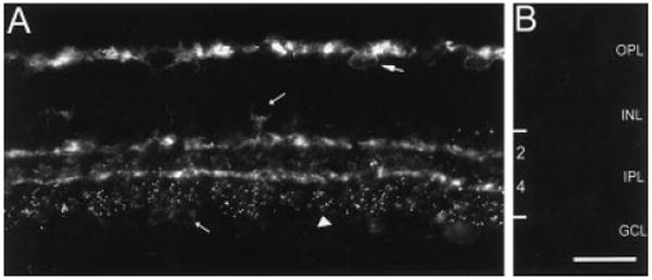
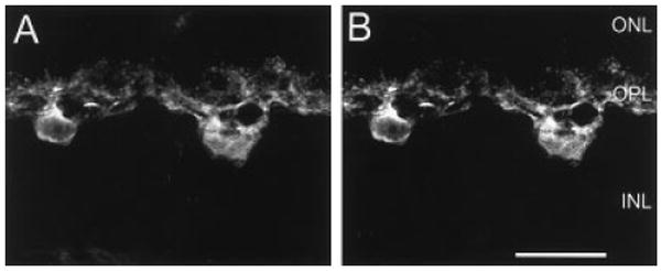
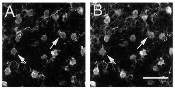
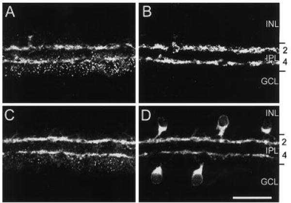
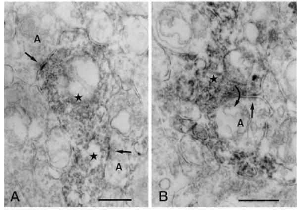
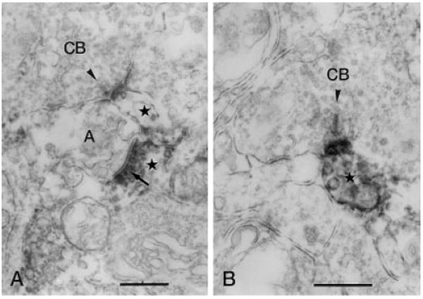
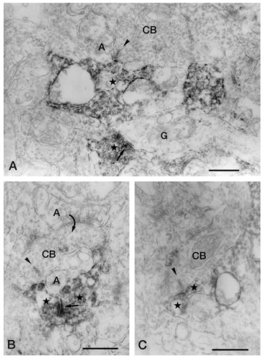
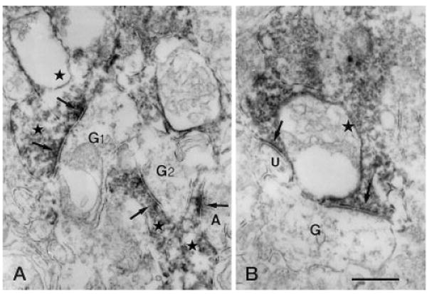
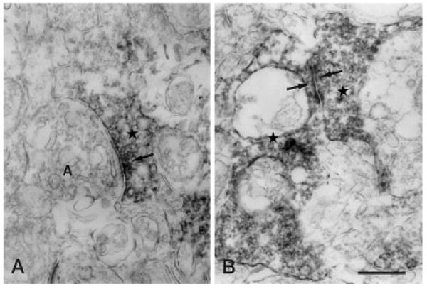
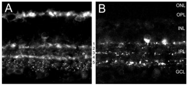
Neuropeptide Y (NPY), an inhibitory neuropeptide expressed by a moderately dense population of wide-field amacrine cells in the rat retina, acts through multiple (Y1-y6) G-protein-coupled receptors. This study determined the cellular localization of Y1 receptors and the synaptic connectivity of Y1 processes in the inner plexiform layer (IPL) of the rat retina. Specific Y1 immunoreactivity was localized to horizontal cell bodies in the distal inner nuclear layer and their processes in the outer plexiform layer. Immunoreactivity was also prominent in cell processes located in strata 2 and 4, and puncta in strata 4 and 5 of the IPL. Double-label immunohistochemical experiments with calbindin, a horizontal cell marker, confirmed Y1 immunostaining in all horizontal cells. Double-label immunohistochemical experiments, using antibodies to choline acetyltransferase and vesicular acetylcholine transporter to label cholinergic amacrine cell processes, demonstrated that Y1 immunoreactivity in strata 2 and 4 of the IPL was localized to cholinergic amacrine cell processes. Electron microscopic studies of the inner retina showed that Y1-immunostained amacrine cell processes and puncta received synaptic inputs from unlabeled amacrine cell processes (65.2%) and bipolar cell axon terminals (34.8%). Y1-immunoreactive amacrine cell processes most frequently formed synaptic outputs onto unlabeled amacrine cell processes (34.0%) and ganglion cell dendrites (54.1%). NPY immunoreactivity in the rat retina is distributed primarily to strata 1 and 5 of the IPL, and the present findings, thus, suggest that NPY acts in a paracrine manner on Y1 receptors to influence both horizontal and amacrine cells.


Figures

Fig. 1
Fig. 1
Y1 immunoreactivity in the rat retina. Fluorescence photomicrograph of a retinal section cut perpendicular to the vitreal surface at 12 μm. A: Y1 immunoreactivity in the outer retina is localized to horizontal cell bodies in the distal INL (large arrow) and to processes in the OPL. Immunoreactivity in the inner retina is localized to faintly immunoreactive cell bodies in the proximal INL and GCL (short arrows), strata 2 and 4 of the IPL, and to puncta spanning lamina 5 of the IPL (arrowhead). B: Y1 immunoreactivity is absent from a retinal section incubated overnight with Y1 antibody that was preincubated with 10-6 M synthetic Y1370-382 peptide. Cryostat section. OPL, outer plexiform layer; INL, inner nuclear layer; IPL, inner plexiform layer; GCL, ganglion cell layer. Scale bar = 30 μm in B (applies to A,B).
Fig. 2
Fig. 2
Confocal images of Y1 (A) and calbindin (CaBP; B) immunoreactivities. A,B: Y1 and CaBP immunoreactivities in the same retinal section. A: Y1 immunoreactivity is in horizontal cell bodies and processes in the OPL. B: CaBP immunoreactivity is in horizontal cell bodies and processes in the OPL. A,B are a stack of eight, 1-μm scans. ONL, outer nuclear layer; OPL, outer plexiform layer; INL, inner nuclear layer. Scale bar = 20 μm in B (applies to A,B).
Fig. 3
Fig. 3
Confocal images of Y1 (A) and CaBP (B) immunoreactivities in a horizontal section through the outer retina. Y1 (A) and CaBP (B) immunoreactivities are colocalized to horizontal cells (arrows indicate colocalization in two horizontal cells). Immunolabeled processes in the image plane are also illustrated in the top portion of A and B. A,B are a stack of five, 1-μm scans through the distal inner nuclear layer. Scale bar = 40 μm in B (applies to A,B).
Fig. 4
Fig. 4
Confocal images of Y1 (A,C), vesicular acetylcholine transporter (VAChT; B), and choline acetyltransferase (ChAT; C) immunoreactivities in the inner retina. A,B: Y1 and VAChT immunoreactivities in the same section. A: Y1 immunoreactivity is localized to processes in strata 2 and 4 of the IPL and puncta primarily distributed to strata 4 and 5. B: VAChT immunoreactivity, which marks cholinergic amacrine cell terminals and processes, is mainly localized to strata 2 and 4 of the IPL. C,D: Y1 and ChAT immunoreactivities in the same retinal section. C: Y1 immunoreactivity localized to processes in the inner retina. D: ChAT immunoreactivity localized to cell bodies in the INL and GCL and processes in strata 2 and 4 of the IPL. A–D are stacks of ten 1-μm scans. INL, inner nuclear layer; IPL, inner plexiform layer; GCL, ganglion cell layer. Scale bar = 30 μm.
Fig. 5
Fig. 5
Vertical sections through the IPL of a rat retina processed for Y1 immunoreactivity. A: Two immunoreactive amacrine cell processes (stars) receive synaptic input (arrows) from two unlabeled amacrine cell processes (A) in stratum 4 of the IPL, respectively. B: An immunoreactive amacrine cell process (star) receiving synaptic input (straight arrow) from an unlabeled amacrine cell process (A) in stratum 4 of the IPL. The immunoreactive process makes synaptic output (curved arrow) back onto the unlabeled amacrine cell process. IPL, inner plexiform layer. Scale bars = 0.5 μm in A,B.
Fig. 6
Fig. 6
Vertical sections through the IPL stained for Y1 immunoreactivity. A: Y1-immunoreactive process (stars) forms a postsynaptic dyad with an unlabeled amacrine cell process (A), postsynaptic (arrow) to an immunoreactive amacrine cell process, at the ribbon synapse (arrowhead) of a cone bipolar axon terminal in stratum 4 of the IPL. B: Immunoreactive amacrine cell process is postsynaptic to the ribbon synapse (arrowhead) of a CB in stratum 2 of the IPL. IPL, inner plexiform layer; CB, cone bipolar axon terminal. Scale bars = 0.5 μm in A,B.
Fig. 7
Fig. 7
Vertical sections through the IPL stained for Y1 immunoreactivity. A: Y1-immunoreactive amacrine cell process (star) forms a postsynaptic dyad with an unlabeled amacrine cell process (A) at the ribbon synapse (arrowhead) of a CB in stratum 4 of the IPL. In the lower part of the figure, a labeled amacrine cell processes (star) that is making synaptic output (arrow) onto a ganglion cell dendrite (G) can be seen. B: A CB, postsynaptic (curved arrow) to an unlabeled amacrine cell process (upper A), makes a ribbon synapse (arrowhead) onto a postsynaptic dyad consisting of a labeled amacrine cell process (left star) and an unlabeled amacrine cell process (lower A) in stratum 4 of the IPL. The labeled process receives synaptic input (arrow) from another labeled amacrine cell process (right star). C: Two labeled amacrine cell processes (stars) form a postsynaptic dyad at the ribbon synapse of an axon terminal of a CB) in stratum 4 of the IPL. IPL, inner plexiform layer; CB, cone bipolar axon terminal. Scale bar = 0.5 μm.
Fig. 8
Fig. 8
Vertical sections through the IPL of a rat retina processed for Y1 immunoreactivity. A: G1 is postsynaptic (left, upper arrows) to two immunoreactive amacrine cell processes (left, upper stars) in stratum 2 of the IPL. Another ganglion cell dendrite (G2) receiving synaptic input from an immunoreactive amacrine cell process (center star) is seen. In the right lower corner of the figure, an immunolabeled amacrine cell process (right, lower star) receives synaptic input (right, lower arrows) from an unlabeled amacrine cell process (A). B: Immunoreactive amacrine cell process (star) makes output synapses (arrow) onto an unlabeled G and an unidentified process (U) in stratum 4 of the IPL. G,G1,G2, ganglion cell dendrite; IPL, inner plexiform layer. Scale bar = 0.5 μm in B (applies to A,B).
Fig. 9
Fig. 9
Vertical sections through the IPL of a rat retina processed for Y1 immunoreactivity. A: Immunoreactive amacrine cell process (star) makes an output synapse (arrow) onto an amacrine cell process (A) in stratum 2 of the IPL. B: Synaptic contact (arrows) between two immunoreactive amacrine cell processes (stars) is seen in stratum 4 of the IPL. IPL, inner plexiform layer. Scale bar = 0.5 μm in B (applies to A,B).
Fig. 10
Fig. 10
Diagram comparing the distribution of NPY and Y1 immunoreactivity in the rat retina. NPY immunoreactivity is localized to amacrine cells and displaced amacrine cells and their processes that ramify primarily in strata 1 and 5 of the IPL. Some processes are also present in strata 3 of the IPL (D'Angelo and Brecha, 1999). Y1 immunoreactivity is localized to horizontal cells in the distal INL and their processes in the OPL, to cholinergic or “starburst” amacrine cell processes that ramify in strata 2 and 4 of the IPL, and to discrete puncta that are primarily localized to strata 4 and 5 of the IPL. NPY, neuropeptide Y; IPL, inner plexiform layer; OPL, outer plexiform layer.

Similar articles

Cited by

References

    1. Ammar DA, Hughes BA, Thompson DA. Neuropeptide Y and the retinal pigment epithelium: receptor subtypes, signaling, and bioelectrical responses. Invest Ophthalmol Vis Sci. 1998;39:1870–1878. - PubMed
    1. Arvidsson U, Riedl M, Elde R, Meister B. Vesicular acetylcholine transporter (VAChT) protein: a novel and unique marker for cholinergic neurons in the central and peripheral nervous systems. J Comp Neurol. 1997;378:454–467. - PubMed
    1. Balasubramaniam AA. Neuropeptide Y family of hormones: receptor subtypes and antagonists. Peptides. 1997;18:445–457. - PubMed
    1. Bao L, Kopp J, Zhang X, Xu ZQ, Zhang LF, Wong H, Walsh J, Hökfelt T. Localization of neuropeptide Y Y1 receptors in cerebral blood vessels. Proc Natl Acad Sci U S A. 1997;94:12661–12666. - PMC - PubMed
    1. Blomqvist AG, Herzog H. Y-receptor subtypes: how many more? Trends Biol Sci. 1997;20:294–298. - PubMed

Publication types

Substances