. 2002 Oct;1(5):725-35.
doi: 10.1128/EC.1.5.725-735.2002.

Aspergillus nidulans catalase-peroxidase gene (cpeA) is transcriptionally induced during sexual development through the transcription factor StuA

Affiliations

Aspergillus nidulans catalase-peroxidase gene (cpeA) is transcriptionally induced during sexual development through the transcription factor StuA

Mario Scherer et al. Eukaryot Cell. 2002 Oct.

Abstract

Catalases, peroxidases, and catalase-peroxidases are important enzymes to cope with reactive oxygen species in pro- and eukaryotic cells. In the filamentous fungus Aspergillus nidulans three monofunctional catalases have been described, and a fourth catalase activity was observed in native polyacrylamide gels. The latter activity is probably due to the bifunctional enzyme catalase-peroxidase, which we characterized here. The gene, named cpeA, encodes an 81-kDa polypeptide with a conserved motif for heme coordination. The enzyme comprises of two similar domains, suggesting gene duplication and fusion during evolution. The first 439 amino acids share 22% identical residues with the C terminus. Homologous proteins are found in several prokaryotes, such as Escherichia coli and Mycobacterium tuberculosis (both with 61% identity). In fungi the enzyme has been noted in Penicillium simplicissimum, Septoria tritici, and Neurospora crassa (69% identical amino acids) but is absent from Saccharomyces cerevisiae. Expression analysis in A. nidulans revealed that the gene is transcriptionally induced upon carbon starvation and during sexual development, but starvation is not sufficient to reach high levels of the transcript during development. Besides transcriptional activation, we present evidence for posttranscriptional regulation. A green fluorescent protein fusion protein localized to the cytoplasm of Hülle cells. The Hülle cell-specific expression was dependent on the developmental regulator StuA, suggesting an activating function of this helix-loop-helix transcription factor.


Figures

FIG.1.
FIG.1.
Sequence of the catalase-peroxidase (cpeA) and the pyrroline-5-carboxylate reductase (pcrA) locus. (A) A nucleotide sequence of the depicted area was obtained from both strands by using a 3-kb PstI, a 2.5-kb BamHI, and a 5.5-kb EcoRV restriction fragment as a template. The derived amino acid sequences, obtained after removal of one intron in the pcrA gene, are denoted below the DNA sequence in the single-letter code. Since the two genes are encoded on opposite strands of the DNA, we indicated the direction of translation at the start codons with arrows. The intron border sequences of pcrA are indicated. The amino acids of CpeA involved in heme coordination are boxed. The peptide sequences obtained previously from purified CpeA protein are shaded. The sequence used for primer extension is underlined (dotted line). The putative binding sites for the regulators StuA (StRE) and CreA are indicated in the cpeA promoter region by dark and light gray boxes, respectively. The striped boxes indicate the location of four putative AreA binding sites. The determined transcription start site is labeled with an asterisk above the sequence. The two primers (prom2 and prom1) used for the amplification of the promoter with or without the StuA binding site are indicated by a line above the DNA sequence. The displayed sequence of the cpeA and the pcrA genes are available under the accession numbers AJ305225 and AJ313094.
FIG. 2.
FIG. 2.
Disruption of the cpeA gene. (A) Scheme of the construct used for homologous integration. A 5′ XhoI-EcoRV fragment and a 3′ EcoRV-EcoRV fragment were cloned into pBluescript, and the argB marker was inserted as an EcoRI restriction fragment in between them. (B) Southern blot analysis to confirm the integration event. Genomic DNA of a wild type (WT) and a disruptant strain (SMH1) was digested with EcoRI and hybridized to the 5.3-kb XhoI-EcoRV fragment depicted in panel A. (C) Activity stain of peroxidase and catalase in a native 10% polyacrylamide gel. The wild type and the disruptant strain (SMH1) were induced for sexual development, and mycelia were harvested after 72 h of growth. The total protein (50 μg) was separated in a native polyacrylamide gel, and activities were determined by staining as described earlier (34). The asterisk indicates the catalase activity not present in the diruptant strain. The other catalase activities visible above this band are found in both strains and indicate equal loading of protein.
FIG. 3.
FIG. 3.
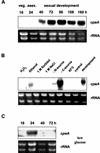
Transcriptional regulation of cpeA. (A) Northern blot analysis of RNA isolated at different time points during the life cycle of A. nidulans. A total of 105 conidiospores were inoculated onto membranes and exposed onto a minimal medium agar plate. The plates were incubated at 37°C, and mycelia were harvested after 16 h (veg.), 24 h when asexual development was completed (asex.), and during sexual development at the time points indicated. RNA was isolated from the mycelia and hybridized to a cpeA-specific probe (upper panel). Then, 2 μg of the same RNA preparation was loaded onto a gel and stained with ethidium bromide as loading controls for the gel. (B) Northern blot analysis of RNA isolated from mycelia after different treatments. Conidia of FGSCA4 were inoculated in complete liquid medium and incubated at 37°C for 20 h. The mycelium was harvested and divided into nine equal portions. One fraction was resuspended in minimal medium and incubated for 3 h before RNA isolation (lane “control”). One fraction was induced for development (lane “development”) and then used for RNA isolation. The other seven samples were resuspended in minimal medium containing 0.5 mM H2O2-4% ethanol instead of 2% glucose-100 mM glucose plus 1 M sorbitol or 1 M NaCl into medium lacking any nitrogen or carbon source. Heat shock treatment was performed at 42°C. The treatments are indicated above the lanes. RNA was processed as described in panel A. (C) Northern blot analysis of RNA from mycelia induced for development and grown at a low glucose concentration (0.8%) in which no sexual development occurred. The time points of harvesting are indicated above the lanes.
FIG. 4.
FIG. 4.
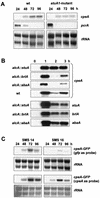
Posttranscriptional regulation of CpeA. To test for CpeA-GFP expression upon carbon starvation and during development, mycelia of SMS14 (CpeA-GFP) was grown for 20 h at 37°C and then shifted for 5 h to medium containing glucose (+) or without glucose (−) or induced for development. (A) RNA accumulation upon starvation was analyzed with a gfp-specific probe. (B) To detect the fusion protein in a Western blot, 40 μg of protein was analyzed with the anti-GFP antibody. Under starvation conditions, no protein band is visible (left two lanes). During development, the protein appears after 72 h, and the small form is detectable after 96 h.
FIG. 5.
FIG. 5.
Localization of CpeA-GFP during the course of sexual development. Strain SMS11 (CpeA-GFP) was induced for development and mature cleistothecia (A) with attached Hülle cells, and individual Hülle cells were analyzed under fluorescence conditions (B) or by phase-contrast microscopy (C). The scale bar represents 40 μm in panel A and 10 μm in panels B and C.
FIG. 6.
FIG. 6.
Transcriptional activation of cpeA is regulated through StuA during sexual development. (A) RNA was isolated from FGSCA4 and a stuA1 mutant strain (GO256) at the time points indicated above the lanes. RNA was hybridized to cpeA (upper panel)- and pcrA (middle panel)-specific probes. rRNA was stained on the membrane with methylene blue as a loading control (lower panel). (B) Forced expression of stuA, brlA, and abaA was achieved by growth of the strains for 20 h on glucose medium (repressing conditions) and then shifted to ethanol-containing medium for the times indicated above the panels. RNA isolated from wild-type strain FGSCA4 and the three transgenic strains UI132 (alcA::stuA), TPM1 (alcA::abaA), TTA292 (alcA::brlA) was processed for a Northern blot. The membrane was hybridized to a cpeA specific probe (upper 4 panels) and to a stuA, brlA and an abaA specific probe (lower 3 panels). (C) The transgenic strain SMS14 (cpeA::gfp) and SMS16 [cpeA(ΔStuA binding sites)::gfp] were analyzed for cpeA-gfp expression after the induction of development. As a probe for the Northern blot, we used gfp (upper panels) or cpeA (lower panels). rRNA staining was used as loading control. (D) Strains SMS14 and SMS16 were also analyzed for CpeA-GFP expression upon carbon starvation (lower panels). gfp or cpeA were used as probes as indicated.
FIG. 6.
FIG. 6.
Transcriptional activation of cpeA is regulated through StuA during sexual development. (A) RNA was isolated from FGSCA4 and a stuA1 mutant strain (GO256) at the time points indicated above the lanes. RNA was hybridized to cpeA (upper panel)- and pcrA (middle panel)-specific probes. rRNA was stained on the membrane with methylene blue as a loading control (lower panel). (B) Forced expression of stuA, brlA, and abaA was achieved by growth of the strains for 20 h on glucose medium (repressing conditions) and then shifted to ethanol-containing medium for the times indicated above the panels. RNA isolated from wild-type strain FGSCA4 and the three transgenic strains UI132 (alcA::stuA), TPM1 (alcA::abaA), TTA292 (alcA::brlA) was processed for a Northern blot. The membrane was hybridized to a cpeA specific probe (upper 4 panels) and to a stuA, brlA and an abaA specific probe (lower 3 panels). (C) The transgenic strain SMS14 (cpeA::gfp) and SMS16 [cpeA(ΔStuA binding sites)::gfp] were analyzed for cpeA-gfp expression after the induction of development. As a probe for the Northern blot, we used gfp (upper panels) or cpeA (lower panels). rRNA staining was used as loading control. (D) Strains SMS14 and SMS16 were also analyzed for CpeA-GFP expression upon carbon starvation (lower panels). gfp or cpeA were used as probes as indicated.
FIG. 7.
FIG. 7.
Visualization of CpeA-GFP expression in SMS14 (cpeA::gfp) (A, B, and C) and SMS16 [cpeA(ΔStuA binding sites)::gfp] (D, E, and F). (A) View of an agar plate grown with SMS14 with induced sexual development. Hülle cells (Hu) are brightly stained, conidiophores (Co) are not stained. The boxed area shows a cleistothecium. Exposure time, 0.8 s. (B and C) A cleistothecium was broken and observed under a coverslip under fluorescence (2-s exposure) or phase-contrast illumination. Hu, Hülle cells; dM, dikaryotic mycelium. (D) View onto an agar plate grown with SMS16 with induced sexual development. Almost no fluorescence is detectable (0.8-s exposure). (E and F) Observation of Hülle cells and dikaryotic mycelium under fluorescence or phase-contrast illumination. Hülle cells were not stained, but the dikaryotic mycelium shows fluorescence with a similar intensity to that seen in panel B. Exposure time, 4 s. The scale bars represent 100 μm in panels A and D and 10 μm in panels B, C, E, and F.

Similar articles

Cited by

References

    1. Adams, T. H., J. K. Wieser, and J.-H. Yu. 1998. Asexual sporulation in Aspergillus nidulans. Microbiol. Mol. Biol. Rev. 62:35-54. - PMC - PubMed
    1. Amati, B., and H. Land. 1994. Myc-Max-Mad: a transcription factor network controlling cell cycle progression, differentiation, and death. Curr. Opin. Genet. Dev. 4:102-108. - PubMed
    1. Andrianopoulos, A., and W. E. Timberlake. 1994. The Aspergillus nidulans abaA gene encodes a transcriptional activator that acts as a genetic switch to control development. Mol. Cell. Biol. 14:2503-2515. - PMC - PubMed
    1. Aramayo, R., Y. Peleg, R. Addison, and R. Metzenberg. 1996. Asm-1+, a Neurospora crassa gene related to transcriptional regulators of fungal development. Genetics 144:991-1003. - PMC - PubMed
    1. Berteaux-Lecellier, V., M. Picard, C. Thompson-Coffe, D. Zickler, A. Panvier-Adoutte, and J.-M. Simonet. 1995. A nonmammalian homolog of the PAF1 gene (Zellweger Syndrome) discovered as a gene involved in caryogamy in the fungus Podospora anserina. Cell 81:1043-1051. - PubMed

Publication types

MeSH terms

Associated data

LinkOut - more resources