. 2002 Oct;1(5):774-86.
doi: 10.1128/EC.1.5.774-786.2002.

Eng1p, an endo-1,3-beta-glucanase localized at the daughter side of the septum, is involved in cell separation in Saccharomyces cerevisiae

Affiliations

Eng1p, an endo-1,3-beta-glucanase localized at the daughter side of the septum, is involved in cell separation in Saccharomyces cerevisiae

Victoriano Baladrón et al. Eukaryot Cell. 2002 Oct.

Abstract

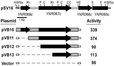
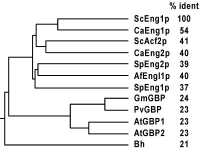
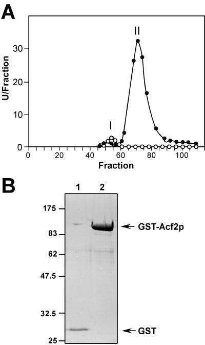
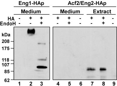
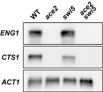
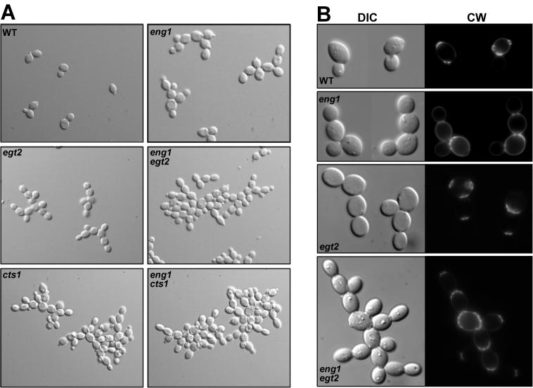
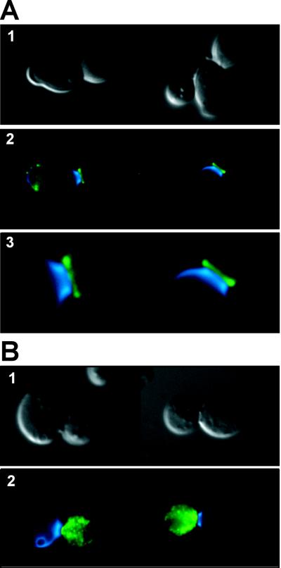
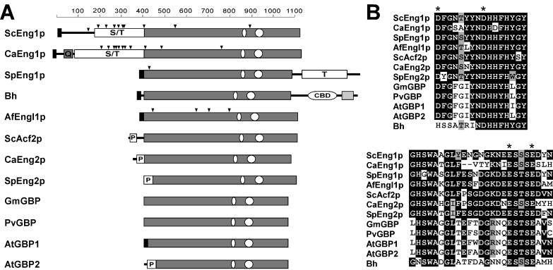
ENG1 (YNR067c), a gene encoding a new endo-1,3-beta-glucanase, was cloned by screening a genomic library with a DNA probe obtained by PCR with synthetic oligonucleotides designed according to conserved regions found between yeast exo-1,3-beta-glucanases (Exglp, Exg2p, and Ssglp). Eng1p shows strong sequence similarity to the product of the Saccharomyces cerevisiae ACF2 gene, involved in actin assembly "in vitro," and to proteins present in other yeast and fungal species. It is also related to plant glucan-binding elicitor proteins, which trigger the onset of a defense response upon fungal infection. Eng1p and Acf2p/Eng2p are glucan-hydrolyzing proteins that specifically act on 1,3-beta linkages, with an endolytic mode of action. Eng1p is an extracellular, heavily glycosylated protein, while Acf2p/Eng2p is an intracellular protein with no carbohydrate linked by N-glycosidic bonds. ENG1 transcription fluctuates periodically during the cell cycle; maximal accumulation occurs during the M/G1 transition and is dependent on the transcription factor Ace2p. Interestingly, eng1 deletion mutants show defects in cell separation, and Eng1p localizes asymmetrically to the daughter side of the septum, suggesting that this protein is involved, together with chitinase, in the dissolution of the mother-daughter septum.


Figures

FIG. 1.
FIG. 1.
Physical and functional maps of ENG1 (YNR067c). At the top is a map of plasmid pSV16, with arrows indicating the 5′ to 3′ orientation of the different coding sequences. Deletion constructs used to test β-glucanase activity are indicated below, with solid boxes designating the sequences present in each plasmid and dashed lines representing the fragments absent from each construct. To the right are the levels of 1,3-β-glucanase activity (in milliunits per milligram of protein) against laminarin detected in whole-cell lysates in the exg1 exg2 strain YPA84. Restriction sites: B, BamHI; C, ClaI; E, EcoRI; H, HindIII; P, PstI; Sa, Sau3A; Xh, XhoI.
FIG. 2.
FIG. 2.
Eng1p belongs to a family of conserved proteins. The dendrogram was generated by the Clustal program from the alignment of the protein sequences of yeast and fungal glucanases, including the S. cerevisiae Eng1p (ScEng1p, amino acids 400 to 1117) and Acf2p (ScAcf2p, amino acids 75 to 779), C. albicans Eng1p (CaEng1p, from 428 to 1145) and Eng2p (CaEng2p, from 51 to 734), S. pombe Eng1p (SpEng1p, from 1 to 738) and SpEng2p proteins, and the A. fumigatus protein Engl1p (AfEngl1p). The plant β-glucan elicitor binding proteins were from G. max (GmGBP), P. vulgaris (PvGBP), and A. thaliana (AtGBP1 and AtGBP2). The sequence from the B. halodurans protein (Bh) is also included. The percent identity between Eng1p and each protein (determined by pairwise alignments) is shown in the right column.
FIG. 3.
FIG. 3.
Purification of 1,3-β-glucanases encoded by ENG1 and ACF2/ENG2. (A) β-Glucanase activity against laminarin assayed in the fractions eluted from gel filtration chromatography on Biogel A 1.5. The plot represents the elution profile of the β-glucanase activity produced in strain YPA84 (exg1 exg2) carrying ENG1 on high-copy-number plasmid pVB10 (solid circles) or vector alone (open circles), expressed as units per fraction. (B) Purification of the GST-Acf2p fusion protein. A GST-Acf2p fusion protein (lane 2) or GST (lane 1) was expressed in S. cerevisiae under the control of a galactose-inducible promoter and purified on glutathione-Sepharose columns. Proteins used for enzymatic activity determination were separated by SDS-polyacrylamide gel electrophoresis (PAGE) and stained with Coomassie blue. Sizes are shown in kilodaltons.
FIG. 4.
FIG. 4.
Cellular localization and carbohydrate content of the Eng1p and Acf2p/Eng2p proteins. Strain YPA84 (exg1 exg2) was transformed with plasmids containing epitope-tagged versions of ENG1 and ACF2/ENG2 in which three copies of the HA epitope were inserted at the C terminus immediately before the stop codon. Culture supernatants (Medium, lanes 1 to 6) or whole-cell extracts (Extract, lanes 7 to 9) prepared from cells bearing ENG1-HA (lanes 2 and 3) or ACF2/ENG2-HA (lanes 4 to 5 and 7 to 8) were treated with endoglycosidase H (EndoH, lanes 3, 5, and 8) and separated by 3 to 15% gradient SDS-PAGE. Proteins were transferred to nitrocellulose membranes and probed with monoclonal anti-HA antibodies. Lane 1 contains supernatants from mutant cells carrying the wild-type ENG1 gene, and lanes 6 and 9 contain supernatants or whole-cell extracts, respectively, from transformants bearing wild-type ACF2/ENG2.
FIG. 5.
FIG. 5.
ENG1 expression is dependent on Ace2p. Polyadenylated RNA (3 μg) obtained from vegetatively growing wild-type (WT, strain YPA24) cells and the isogenic ace2 (LS61), swi5 (LS64), and ace2 swi5 (LS67) derivatives was denatured, separated by agarose gel electrophoresis, transferred to a nylon membrane, and hybridized with specific probes for ENG1, CTS1, and ACT1.
FIG. 6.
FIG. 6.
ENG1 is repressed but ACF2/ENG2 is induced during the sporulation process. (A) Polyadenylated RNA prepared from strain AP1 (a/α) growing vegetatively (VEG) or at 10 h after transfer to sporulation medium (SPO) was denatured, fractionated by electrophoresis (3 μg per lane), and transferred to a nylon membrane. Immobilized RNA was hybridized with radioactively labeled probes specific for ENG1 and SSG1. The ACT1 gene was used as a loading control. (B) Total RNA from strain YPA24 (12 μg per lane) growing vegetatively (VEG) or at 10 h after transfer to sporulation medium (SPO) was hybridized with specific probes for ACF2/ENG2, SSG1, and ACT1. (C) Meiotic time course of ENG2 expression. Total RNA was purified from wild-type cells (strain YPA24) growing vegetatively (time zero) and at the indicated times (in hours) after transfer to sporulation medium. The RNA (12 μg per lane) was hybridized sequentially with the following radioactively labeled gene-specific probes: ACF2/ENG2, HOP1, SPO12, SSG1, and SPS100. The ACT1 gene was used to test for equal loading of RNA in all lanes.
FIG. 7.
FIG. 7.
eng1 cells have a defect in cell separation. (A) Diploid wild-type (WT, YPA24), eng1 (VB28), egt2 (LS93), eng1 egt2 (LS94), cts1 (LS79), and cts1 eng1 (LE30) cells from exponentially growing cultures were mounted directly on glass slides and photographed with differential interference contrast optics. (B) The same strains grown to mid-log phase were fixed with formaldehyde and stained with Calcofluor White to visualize the chitin. Photographs of differential interference contrast microscopy (DIC, left panels) and fluorescence microscopy (CW, right panels) are shown.
FIG. 8.
FIG. 8.
Eng1p localizes to the daughter side of the septum. Diploid wild-type cells (CEN.PK2 strain) transformed with plasmid pVB35 carrying the ENG1-HA epitope were grown to mid-log phase, fixed, and stained with anti-HA antibodies (HA.11; Babco) to visualize Eng1p by indirect immunofluorescence. Cells were briefly treated with zymolyase 20T and glusulase to partially digest the cell wall (B) or not treated (A) before the primary and secondary antibodies were added. For both panels, images are as follows: 1. differential interference contrast microscopy; 2, overlay of Eng1p-HA fluorescence (green) and Calcofluor White staining of chitin (blue); 3. enlarged view of the mother-daughter neck region (only shown in A).
FIG. 9.
FIG. 9.
Schematic representation of endo-1,3-β-glucanases and plant β-glucan elicitor binding proteins. (A) The structure of each protein is shown at the same scale (indicated at the top as number of amino acids), with a gray rectangle indicating the region conserved among all the proteins. The abbreviations used to name the proteins are the same as those indicated in Fig. 2. A black box in the N-terminal region indicates the predicted secretory signal sequence, while triangles mark the positions of putative N-glycosylation sites. White boxes represent Ser/Thr-rich regions (indicated by S/T), the Thr-rich domain (marked with a T), Pro-rich regions (P), or a cellulose-binding domain (CBD). A gray box in the sequence of C. albicans Eng1p indicates the presence of a poly-Gln stretch (Q) and a region common to other bacterial xylanases in the sequence of Bh. The oval regions mark the relative positions of the most conserved segments between the proteins whose alignments are shown in panel B, with identities indicated as black boxes and conservative substitutions in gray. The asterisks indicate the conserved Asp and Glu residues that could be part of the catalytic center.

References

    1. Byers, B. 1981. Cytology of the yeast life cycle, p. 59-96. In J. N. Strathern, E. W. Jones, and J. R. Broach (ed.), The molecular and cellular biology of the yeast Saccharomyces cerevisiae. Cold Spring Harbor Laboratory, Cold Spring Harbor, N.Y.
    1. Cabib, E., D. H. Roh, M. Schmidt, L. B. Crotti, and A. Varma. 2001. The yeast cell wall and septum as paradigms of cell growth morphogenesis. J. Biol. Chem. 276:19679-19682. - PubMed
    1. Cabib, E., S. J. Silverman, and J. A. Shaw. 1992. Chitinase and chitin synthase 1: counterbalancing activities in cell separation of Saccharomyces cerevisiae. J. Gen. Microbiol. 138:97-102. - PubMed
    1. Cappellaro, C., V. Mrsa, and W. Tanner. 1998. New potential cell wall glucanases of Saccharomyces cerevisiae and their involvement in mating. J. Bacteriol. 180:5030-5037. - PMC - PubMed
    1. Carlson, M., and D. Botstein. 1982. Two differentially regulated mRNAs with different 5′ ends encode secreted and intracellular forms of yeast invertase. Cell 28:145-154. - PubMed

Publication types

MeSH terms

Substances

Associated data