. 2002 Jun;1(3):378-90.
doi: 10.1128/EC.1.3.378-390.2002.

A G-protein beta subunit required for sexual and vegetative development and maintenance of normal G alpha protein levels in Neurospora crassa

Affiliations

A G-protein beta subunit required for sexual and vegetative development and maintenance of normal G alpha protein levels in Neurospora crassa

Qi Yang et al. Eukaryot Cell. 2002 Jun.

Abstract

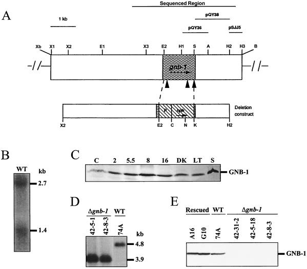
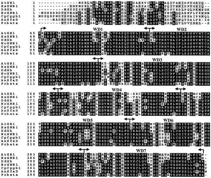
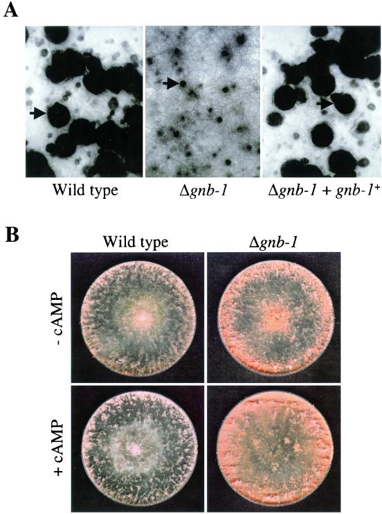
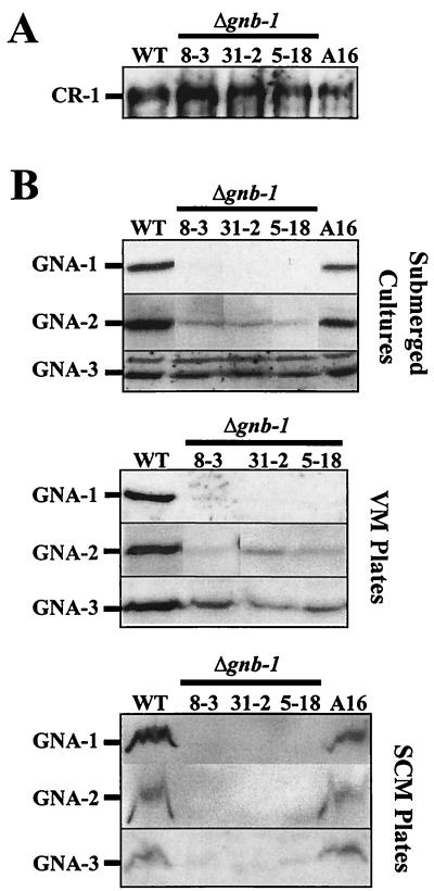
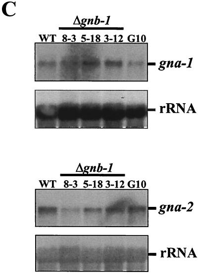
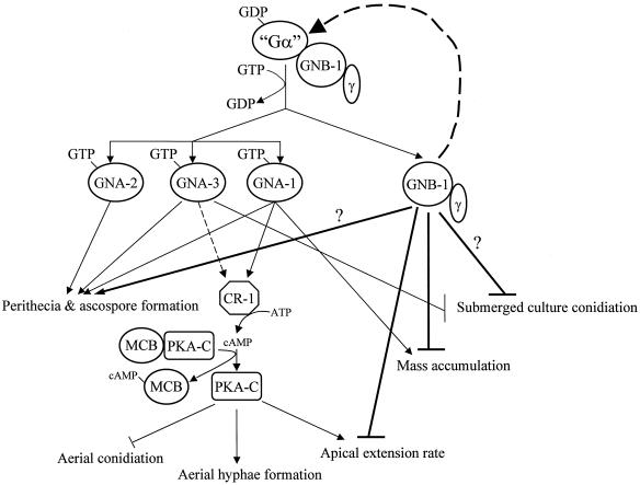
The genome of the filamentous fungus Neurospora crassa contains a single gene encoding a heterotrimeric G-protein beta subunit, gnb-1. The predicted GNB-1 protein sequence is most identical to G beta proteins from the filamentous fungi Cryphonectria parasitica and Aspergillus nidulans. N. crassa GNB-1 is also 65% identical to the human GNB-1 protein but only 38 and 45% identical to G beta proteins from budding and fission yeasts. Previous studies in animal and fungal systems have elucidated phenotypes of G beta null mutants, but little is known about the effects of G beta loss on G alpha levels. In this study, we analyzed a gnb-1 deletion mutant for cellular phenotypes and levels of the three G alpha proteins. Delta gnb-1 strains are female-sterile, with production of aberrant fertilized reproductive structures. Delta gnb-1 strains conidiate more profusely and have altered mass on solid medium. Loss of gnb-1 leads to inappropriate conidiation and expression of a conidiation-specific gene during growth in submerged culture. Intracellular cyclic AMP levels are reduced by 60% in vegetative plate cultures of delta gnb-1 mutants. Loss of gnb-1 leads to lower levels of the three G alpha proteins under a variety of conditions. Analysis of transcript levels for the gna-1 and gna-2 G alpha genes in submerged cultures indicates that regulation of G alpha protein levels by gnb-1 is posttranscriptional. The results suggest that GNB-1 directly regulates apical extension rate and mass accumulation. In contrast, many other delta gnb-1 phenotypes, including female sterility and defective conidiation, can be explained by altered levels of the three N. crassa G alpha proteins.


Figures

FIG. 1.
FIG. 1.
Structure of the N. crassa gnb-1 genomic region and construction of Δgnb-1 and rescued strains. (A) gnb-1 genomic clone. The unique XbaI and BamHI sites are artifacts of cloning. The region that was sequenced, as well as the fragments used as probes for Southern and Northern analysis, is shown at the top of the figure. The shaded area depicts the ORF, and the closed triangles correspond to introns which are (left to right) 106, 80, and 67 bp in length. The gene replacement construct using the hph marker is shown below the genomic clone. P, A. nidulans trpC promoter. Enzyme abbreviations: A, AatII; B, BamHI, C, ClaI; E, EcoRI; H, HindIII; K, KpnI; N, NcoI; S, StyI; X, XhoI; Xb, XbaI. In the case of multiple sites for a given enzyme, the number following the abbreviation indicates order (from left to right) in the figure. (B) gnb-1 mRNA species. Samples containing 20 μg of total RNA isolated from 5.5-h-submerged cultures inoculated at 107 conidia of wild type (WT) strain 74A per ml were subjected to Northern analysis using a 1.0-kb HindIII-AatII fragment from pQY36 as a probe (see panel A). (C) Expression of GNB-1 during the N. crassa life cycle. Samples from wild-type strain 74A (30 μg of the plasma membrane protein fraction) were conidia (C); 2-, 5.5-, 8-, and 16-h-submerged cultures (inoculated at 107 conidia/ml); cultures grown at 30°C on solid VM in the dark (DK); cultures grown at room temperature on solid VM under light (LT); and cultures grown at room temperature on SCM under light (S). (D) Southern analysis. Genomic DNA was digested with NcoI and HindIII. The 600-bp HindIII fragment from plasmid pSJJ5 was used as a probe (see panel A). 42-5-1 and 42-8-3 are purified homokaryotic Δgnb-1 mutants, while 74A is the wild-type (WT) strain. (E) GNB-1 protein levels. Samples containing 30 μg of protein from plasma membrane fractions of 16-h-submerged cultures inoculated at 3 × 10⁶ conidia/ml were subjected to Western analysis using the GNB-1 antibody. Rescued, Δgnb-1, gnb-1+ complemented strains; WT, wild type.
FIG. 2.
FIG. 2.
Alignment of GNB-1 with its five closest Gβ relatives. The amino acid sequence of GNB-1 (NcGNB1) was aligned with Gβ proteins from A. tigrinum (AtGB1; accession no. AF277161), Homo sapiens (HsGNB1; XM 010575.1), D. discoideum (DdGb; DDGPBS), C. parasitica (CpCpgb1; CPU95139), A. nidulans (AnSfaD; AF056182), and P. carinii (Pcbeta; AF306565) using the Genetics Computer Group PILEUP program, followed by shading using Boxshade. Black shading indicates identical sequences. The positions of the seven WD repeats are labeled and indicated by arrows.
FIG. 3.
FIG. 3.
Fertility and conidiation on solid medium. (A) Strains 42-8-3 (Δgnb-1), 74A (wild type), and A16 (Δgnb-1, gnb-1+) were cultured on SCM plates for 6 days in light prior to fertilization with wild-type strain 73a conidia. Plates were photographed 6 days after fertilization. Arrows indicate normal perithecia in wild-type and Δgnb-1, gnb-1+ strains and an aberrant perithecium in the Δgnb-1 strain. (B) Strains 42-8-3 and 74A were cultured on solid VM plates in the absence or presence of 1 mM cAMP for 3 days at 30°C in the dark. The more abundant conidium production in the Δgnb-1 strain is visible as the darker-orange, dense area at the edge of the plate.
FIG. 4.
FIG. 4.
Growth in submerged culture. (A) Microscopic observation. Wild-type (74A), Δgnb-1 (42-8-3 and 42-5-18), and Δgnb-1, gnb-1+ (A16) strains were cultured for 16 h in liquid VM in the absence or presence of 2% peptone. Representative conidiophores in the Δgnb-1 (no peptone) culture are indicated by arrows. (B) Levels of con-10 message. Samples containing 20 μg of total RNA obtained from the cultures in panel A were subjected to Northern analysis using the 250-bp BamHI-EcoRI insert from pW100 (con-10 gene) as a probe. Blots were reprobed with an rRNA gene to check for equal loading and transfer of RNA.
FIG. 5.
FIG. 5.
Analysis of Gα and adenylyl cyclase levels. (A) CR-1 protein levels in submerged cultures. Whole-cell extracts were prepared from 16-h-submerged cultures of the indicated strains, and 30 μg of protein was examined by Western analysis with CR-1 antiserum. (B) GNA-1, GNA-2 and GNA-3 protein levels under various growth conditions. Plasma membrane fractions were obtained from 16-h-submerged (inoculated at 3 × 10⁶ conidia/ml), VM plate, or SCM plate cultures. Samples containing 30 μg (submerged or VM plate cultures) or 15 μg (SCM plate cultures) of protein were subjected to Western analysis with specific antisera. The curved nature of the cross-reacting protein bands in the SCM preparations presumably results from residual cell wall components that are not removed during the plasma membrane isolation procedure. (C) Levels of the gna-1 and gna-2 messages. Total RNA was extracted from 16-h-submerged cultures, and 20 μg was subjected to Northern analysis using the EcoRI-ClaI fragment of pPNO5 or the BamHI insert of p13 M2A5-2 as a probe to detect gna-1 and gna-2 expression, respectively. The N. crassa rRNA gene probe was used as an internal standard to check for relative amounts of RNA blotted onto the membrane. WT, wild-type strain 74A.
FIG. 5.
FIG. 5.
Analysis of Gα and adenylyl cyclase levels. (A) CR-1 protein levels in submerged cultures. Whole-cell extracts were prepared from 16-h-submerged cultures of the indicated strains, and 30 μg of protein was examined by Western analysis with CR-1 antiserum. (B) GNA-1, GNA-2 and GNA-3 protein levels under various growth conditions. Plasma membrane fractions were obtained from 16-h-submerged (inoculated at 3 × 10⁶ conidia/ml), VM plate, or SCM plate cultures. Samples containing 30 μg (submerged or VM plate cultures) or 15 μg (SCM plate cultures) of protein were subjected to Western analysis with specific antisera. The curved nature of the cross-reacting protein bands in the SCM preparations presumably results from residual cell wall components that are not removed during the plasma membrane isolation procedure. (C) Levels of the gna-1 and gna-2 messages. Total RNA was extracted from 16-h-submerged cultures, and 20 μg was subjected to Northern analysis using the EcoRI-ClaI fragment of pPNO5 or the BamHI insert of p13 M2A5-2 as a probe to detect gna-1 and gna-2 expression, respectively. The N. crassa rRNA gene probe was used as an internal standard to check for relative amounts of RNA blotted onto the membrane. WT, wild-type strain 74A.
FIG. 6.
FIG. 6.
Contributions of GNB-1 and the three Gα subunits to N. crassa growth and development. It is presumed that each of the three GDP-bound Gα subunits (GNA-1, -2, and -3) can form a complex with GNB-1 and a yet-uncharacterized Gγ subunit in N. crassa; however, physical association has not been directly demonstrated. Ligand binding to a receptor(s) triggers GDP/GTP exchange on a Gα protein(s) and dissociation from the GNB-1/Gγ heterodimer. GNB-1 plays a global, positive role in maintaining normal levels of all three Gα proteins. During sexual development, the three Gα proteins (and perhaps GNB-1) regulate perithecial and ascospore formation via a cAMP-independent mechanism. GNA-1 and GNA-3 positively regulate adenylyl cyclase (CR-1) activity and protein levels, respectively. cAMP binds the regulatory subunit of protein kinase A (MCB) (4), leading to dissociation of MCB from the catalytic subunit (PKA-C). PKA-C activity promotes aerial hypha formation and apical extension while negatively regulating aerial conidiation. Apical extension rate and mass accumulation are each negatively regulated by GNB-1. Both GNA-3 and GNB-1 block conidiation in submerged culture. Bold lines indicate pathways regulated by GNB-1. Dashed lines depict regulation of Gα or CR-1 protein levels. “Gα” is GNA-1, -2, and -3. A question mark represents an uncertain contribution.

Similar articles

Cited by

References

    1. Altschul, S. F., T. L. Madden, A. A. Schäffer, J. Zhang, Z. Zhang, W. Miller, and D. J. Lipman. 1997. Gapped BLAST and PSI-BLAST: a new generation of protein database search programs. Nucleic Acids Res. 25:3389-3492. - PMC - PubMed
    1. Baasiri, R. A., X. Lu, P. S. Rowley, G. E. Turner, and K. A. Borkovich. 1997. Overlapping functions for two G protein α subunits in Neurospora crassa. Genetics 147:137-145. - PMC - PubMed
    1. Baxevanis, A. D., and D. Landsman. 1995. The HMG-1 box protein family: classification and functional relationships. Nucleic Acids Res. 23:1604-1613. - PMC - PubMed
    1. Bruno, K. S., R. Aramayo, P. F. Minke, R. L. Metzenberg, and M. Plamann. 1996. Loss of growth polarity and mislocalization of septa in a Neurospora mutant altered in the regulatory subunit of cAMP-dependent protein kinase. EMBO J. 15:5772-5782. - PMC - PubMed
    1. Case, M. E., M. Schweizer, S. R. Kushner, and N. H. Giles. 1979. Efficient transformation of Neurospora crassa by utilizing hybrid plasmid DNA. Proc. Natl. Acad. Sci. USA 76:5259-5263. - PMC - PubMed

Publication types

MeSH terms

Associated data

LinkOut - more resources