

Review
doi: 10.1128/EC.1.4.495-502.2002.
Kinetoplast DNA network: evolution of an improbable structure
Affiliations
- PMID: 12455998
- PMCID: PMC117999
- DOI: 10.1128/EC.1.4.495-502.2002
Item in Clipboard
Review
Kinetoplast DNA network: evolution of an improbable structure
Eukaryot Cell.
2002 Aug.
No abstract available
Figures
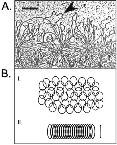
kDNA network structure. (A) Electron micrograph of the periphery of an isolated kDNA network from T. avium. Loops represent interlocked minicircles (the arrowhead indicates a clear example). Bar, 500 nm. (B) Diagrams showing the organization of minicircles. (I) Segment of an isolated network showing interlocked minicircles in a planar array. (II) Section through a condensed network disk in vivo showing stretched-out minicircles. The double-headed arrow indicates the thickness of the disk, which is about half the circumference of a minicircle.
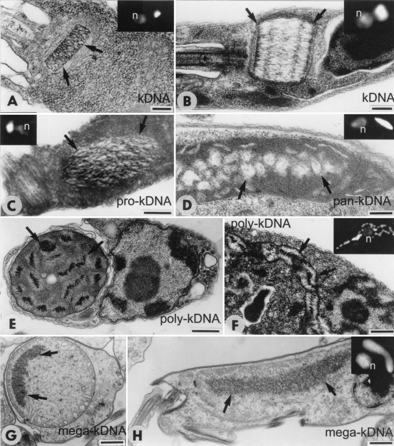
Images of trypanosomatid and bodonid cells showing kDNA. For light microscopy, cells were fixed in 4% paraformaldehyde for 3 min at room temperature, incubated in phosphate-buffered saline containing 0.1 μg of DAPI/ml for 3 min at room temperature, and examined with a Zeiss Axioplan 100 microscope. For electron microscopy, cells were fixed in 2% glutaraldehyde in 0.2 M cacodylate buffer at 4°C overnight, postfixed in 2% osmium tetroxide for 1 h at room temperature, and embedded in Epon-Araldite. Thin sections stained with uranyl acetate and lead citrate were examined in a JEOL 1010 microscope. Arrows in electron micrographs indicate kDNA. Insets show DAPI-stained cells (n, nucleus); kDNA is stained brightly. (A) Longitudinal section through the classical disk-shaped kDNA of C. fasciculata. The disk thickness is about half the minicircle circumference (2.5 kb). (B) Longitudinal section through the kDNA disk of T. avium. The disk appears cylindrical due to the large minicircle size (10 kb), but the organization is similar to that of C. fasciculata. (C) Pro-kDNA bundle of B. saltans in a dilated region of the mitochondrion, close to the basal bodies of the flagella. (D) Pan-kDNA of C. helicis, composed of multiple electron-lucent loci in the mitochondrial lumen. (E and F) Transverse (E) and longitudinal (F) sections of the mitochondrion of D. trypaniformis showing multiple poly-kDNA nucleoids with the DNA fibrils radiating from a dense core. (G and H) Transverse (G) and longitudinal (H) sections of T. borreli in which a dense body of mega-kDNA is spread throughout the mitochondrial lumen. Bars, 200 nm in panels A to F and 1 μm in panels G and H. Cells in insets are all at the same scale.
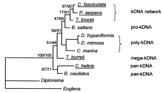
Kinetoplastid phylogenetic tree. Majority-consensus maximum-likelihood tree constructed by using a small-subunit rRNA alignment (alignment 10 at http://www.rna.ucla.edu/trypanosome/alignments.html ) narrowed to species for which kDNA structural information is available (see the text). Bootstrap analysis was performed with 1,000 replicates. Bootstrap values for maximum likelihood and maximum parsimony are shown (to left and right of slashes, respectively). Parasitic species are underlined. Pro-kDNA contains monomeric relaxed minicircles condensed in a single region of the mitochondrial matrix. Poly-kDNA contains monomeric relaxed minicircles condensed in multiple foci. Pan-kDNA contains monomeric supercoiled minicircles distributed throughout a large region of the mitochondrial matrix. Mega-kDNA contains molecules with tandemly linked minicircle-like sequences. The B. caudatus strain used by Hajduk et al. (20) appears to have pan-kDNA, although electron microscopy data on thin sections is lacking. See the text for further discussion.
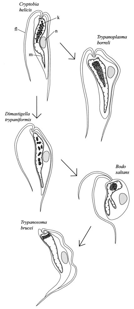
Proposed evolution of kinetoplastids, emphasizing differences in kDNA organization and compaction. kDNA (k) is the structure within the mitochondrial matrix. fl, flagellum; m, mitochondrion; n, nucleus. kDNA in C. helicis is pan-kDNA, that in T. borreli is mega-kDNA, that in D. trypaniformis is poly-kDNA, that in B. saltans is pro-kDNA, and that in T. brucei is a kDNA network.
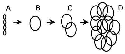
Proposed stages in the evolution of a kDNA network. (A) Supercoiled noncatenated minicircles present in early-branching bodonids. (B) Relaxed noncatenated minicircles also found in early-branching bodonids. (C) Small catenanes of relaxed minicircles, relatively abundant in some late-branching bodonids. (D) kDNA network, present only in late-emerging trypanosomatids.
References
-
- Borst, P. 1991. Why kinetoplast DNA networks? Trends Genet. 7:139-141. - PubMed
-
- Breunig, A., G. Brugerolle, K. Vickerman, H. Hertel, and H. König. 1993. Isolation and ultrastructural features of a new strain of Dimastigella trypaniformis Sandon 1928 (Bodonina, Kinetoplastida) and comparison with a previously isolated strain. Eur. J. Protistol. 29:416-424. - PubMed
Publication types
MeSH terms
Substances
Grants and funding
LinkOut - more resources
Full Text Sources
