. 2002 Aug;1(4):568-82.
doi: 10.1128/EC.1.4.568-582.2002.

Sgt1p contributes to cyclic AMP pathway activity and physically interacts with the adenylyl cyclase Cyr1p/Cdc35p in budding yeast

Affiliations

Sgt1p contributes to cyclic AMP pathway activity and physically interacts with the adenylyl cyclase Cyr1p/Cdc35p in budding yeast

Caroline Dubacq et al. Eukaryot Cell. 2002 Aug.

Abstract

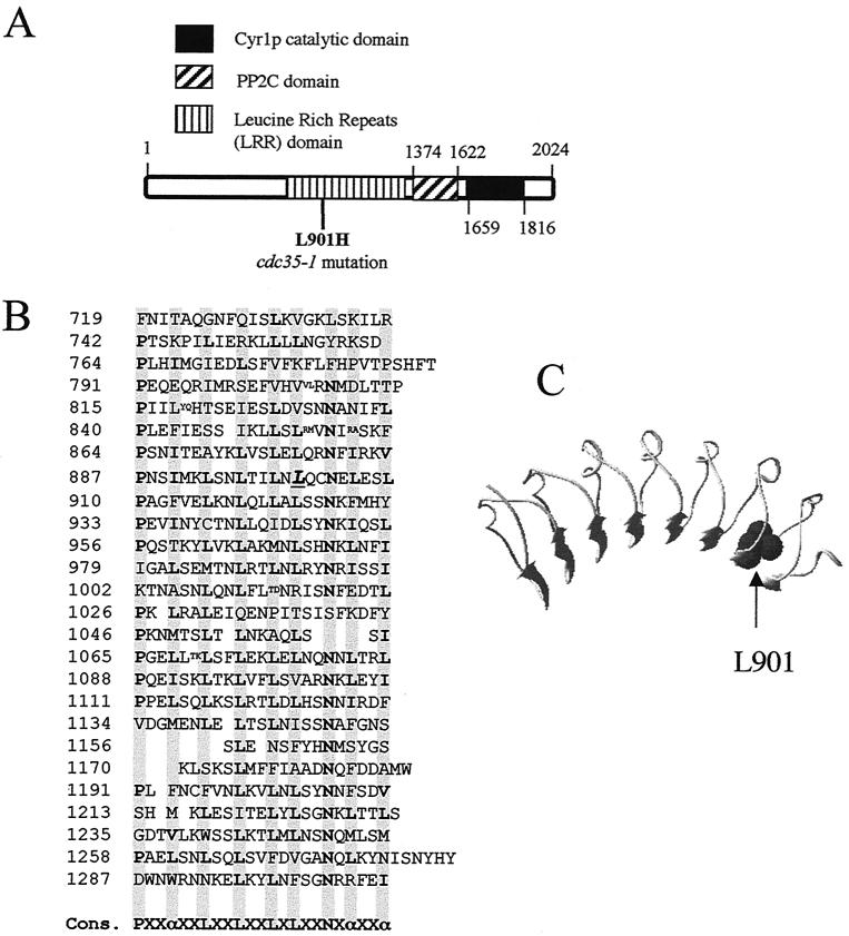
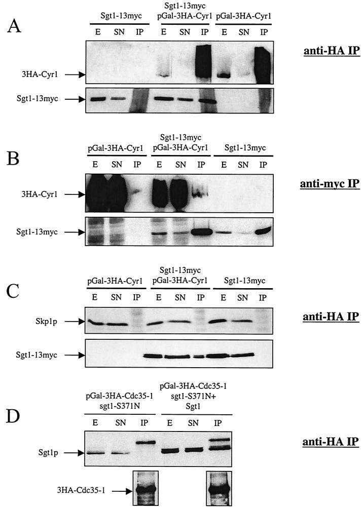
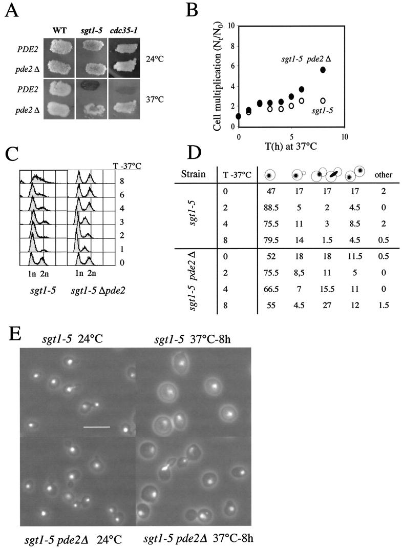
Sgt1p is a highly conserved eucaryotic protein that is required for both SCF (Skp1p/Cdc53p-Cullin-F-box)-mediated ubiquitination and kinetochore function in yeast. We show here that Sgtlp is also involved in the cyclic AMP (cAMP) pathway in Saccharomyces cerevisiae. SGT1 is an allele-specific suppressor of cdc35-1, a thermosensitive mutation in the leucine-rich repeat domain of the adenylyl cyclase Cyrlp/Cdc35p. We demonstrate that Sgt1p and Cyrlp/Cdc35p physically interact and that the activity of the cAMP pathway is affected in an sgt1 conditional mutant. Sequence analysis suggests that Sgtlp has features of a cochaperone. Thus, Sgt1p is a novel activator of adenylyl cyclase in S. cerevisiae and may function in the assembly or the conformational activation of specific multiprotein complexes.


Figures

FIG. 1.
FIG. 1.
Isolation of SGT1 and structure of the encoded protein. (A) SGT1 is an allele-specific suppressor of cdc35-1. cdc35-1 (CMY282), cdc35-10 (CMY391), and cyr1-2 (CMY389) mutants were transformed with the indicated plasmids at 23°C, with selection for growth on synthetic medium without uracil. Patches of cells were then replica plated and incubated at 23 and 36°C to test for the suppression of thermosensitive growth. Ycp50-CYR1 and Ycp50 are positive and negative controls, respectively, for complementation. (B) Schematic diagram of domains found within the Sgt1p sequence. The TPR and Sgt1 CHORD (CS domain) motifs were previously described (38, 63), and the highly conserved C-terminal sequence may be a domain that interacts with adenylyl cyclase (AC domain) in yeast cells. (C) Sequence and secondary structure prediction of the highly conserved C-terminal domain of Sgt1p from a series of eucaryotic organisms (S.c., S. cerevisiae; S.p., S. pombe; N.c., N. crassa; C.e., Caenorhabditis elegans; O.s., O. sativa, H.s., H. sapiens, D.m., Drosophila melanogaster) showing the positions of the sgt1-S371N (A364a) mutation and one of the two sgt1-5 mutations. The secondary structure prediction (Pred) (H, alpha helix; E, β strand; C, coil) was obtained with the Psi-Pred2 algorithm (34) (http://bioinf.cs.ucl.ac.uk/psipred/). The height of the bars is an estimate of the confidence (Conf) of the prediction. The region of Sgt1p between amino acids (AA) 338 and 365 is predicted with a high level of confidence to adopt a helix-turn-helix structure. Amino acid residues that are identical for at least five of the seven Sgt1p homologs are shown in bold.
FIG. 2.
FIG. 2.
The cdc35-1 mutation affects a conserved leucine in the LRR domain of adenylyl cyclase. (A) Schematic diagram of the Cyr1p/Cdc35p sequence showing the position of the L901H substitution within the LRR domain of the cdc35-1 mutant and the positions of the protein phosphatase 2C (PP2C) and adenylyl cyclase catalytic domains, as proposed by the Pfam database (3). (B) Sequence of the Cyr1p LRR domain showing the position of Leu-901 (underlined and in italic type). Note that this sequence corresponds to that of the wild-type CYR1/CDC35 gene in the A364a background, which differs at a number of positions from the published sequence of the gene in the S288C background (see Materials and Methods). Cons., consensus; α, preferred hydrophobic residue. Shaded columns show the positions of preferred amino acids in the leucine-rich repeats. Small letters show sites of amino acid insertions within the repeats. (C) Structural model of the Cyr1p LRR domain in the environment of Leu-901.
FIG. 3.
FIG. 3.
Sgt1p, but not Skp1p, coimmunoprecipitates with Cyr1p. (A) 3HA-Cyr1p overexpressed from a galactose-inducible promoter in strains CDY33 and CDY35 was immunoprecipitated with anti-HA antibody 12CA5, and the immunoprecipitates were analyzed by Western blotting to determine the efficiency of 3HA-Cyr1p IP and the fraction of coprecipitated Sgt1-13myc. Ten micrograms of the crude extract (E) and 10 μg of the supernatant (SN) are shown along with the total amount of the immunoprecipitated material (IP) from 1 mg of protein extract. (B) Sgt1-13myc expressed from its endogenous promoter in strains CDY33 and CDY34 was immunoprecipitated with anti-myc antibody 9E10, and the immu-noprecipitates were analyzed by Western blotting to determine the efficiency of Sgt1-13myc IP and the fraction of coprecipitated overexpressed 3HA-Cyr1p. Ten micrograms of the crude extract (E) and 10 μg of the supernatant (SN) are shown along with the total amount of the immunoprecipitated material (IP) from 4 mg of protein extract. (C) 3HA-Cyr1p overexpressed from a galactose-inducible promoter was immunoprecipitated with anti-HA antibody 12CA5, and the immunoprecipitates were analyzed by Western blotting to determine the fraction of coprecipitating Sgt1-13myc and Skp1p. Forty micrograms of the crude extract (E) and 40 μg of the supernatant (SN) are shown along with the total amount of the immunoprecipitated material (IP) from 4 mg of protein extract. Rabbit anti-Skp1p antibodies were kindly provided by Wade Harper. (D) 3HA-Cdc35-1p overexpressed from a galactose-inducible promoter in strain CDY116 with or without pSGT1 was immunoprecipitated from 1 mg of protein extract with protein A-Sepharose beads containing covalently coupled anti-HA antibody 12CA5. The immunoprecipitated material was then analyzed by immunoblotting with rabbit anti-Sgt1p polyclonal antibodies (upper panel) to test for coprecipitation of Sgt1-S371N or wild-type Sgt1p with Cdc35-1p or anti-HA antibodies to determine the level of 3HA-Cdc35-1p immunoprecipitated from each cellular extract (lower panels). In the upper panel, 20 μg each of crude extract (E) and supernatant (SN) was loaded next to the total amount of the immunoprecipitated material (IP).
FIG. 4.
FIG. 4.
Indirect immunofluorescence analysis of strain CDY34 done to visualize the intracellular localization of Sgt1-13myc with purified mouse anti-myc monoclonal antibody 9E10. DNA was visualized with DAPI staining. Three consecutive optical sections separated by 150 nm are shown from a z-section stack after deconvolution of the stack in order to remove out-of-plane fluorescence from each section. Sgt1-13myc fluorescence is shown in red, DNA is shown in blue, andcolocalization is shown in magenta. Under identical conditions, no immunofluorescence signal was observed for a congenic strain that did not express a myc-tagged protein. N, nucleus; V, vacuole. The yeast cells shown are about 5 μm in diameter.
FIG.5.
FIG.5.
Elevation of intracellular cAMP levels affects sgt1-5 phenotypes. (A) Wild-type (WT; YPH499), sgt1-5 (YKK57), cdc35-1 (CMY282), pde2Δ (CDY1), sgt1-5 pde2Δ (CDY3), and cdc35-1 pde2Δ (CDY5) strains were stained with iodine to visualize the accumulation of glycogen in patches of yeast colonies replicated on YPD plates at 24 and 37°C. The darkly staining sgt1-5 and cdc35-1 patches of cells incubated at 37°C indicate a high level of glycogen in these cells. (B) Exponentially growing sgt1-5 cells (open circles) and sgt1-5 pde2Δ cells (filled circles) on YPD plates at 24°C were transferred to 37°C, and the cell multiplication factor (number of cells at various times [Nt] divided by the number of cells at time zero [N0]) was determined at the indicated times (T). (C) Fluorescence-activated cell sorting analysis of DNA content. Times (T) are given in hours. 1n, G1-phase DNA content; 2n, G2-phase DNA content. (D) Morphological analysis of sgt1-5 and sgt1-5 pde2Δ cells at 24°C (time zero) and after transfer to 37°C for the indicated times (T, in hours). (E) Fluorescent phase-contrast images of sgt1-5 and sgt1-5 pde2Δ cells stained with propidium iodide to visualize nuclear DNA. Bar, 10 μm.
FIG. 6.
FIG. 6.
Induced proteolysis of N-degron-Sgt1p leads to arrest of cell growth and activation of the expression of a STRE-LacZ reporter construction. (A) Schematic diagram of the N-degron system (49) used to create a conditional null mutant of Sgt1p by induced proteolysis. (B to E) The addition of 0.5 mM CuSO4 to strain CDY26 (N-degron-Sgt1p) growing on synthetic complete medium at 30°C but not parental strain ZMY60 blocks cell growth (B); induces the proteolysis of N-degron-4HA-Sgt1p after 2, 4, and 6 h of incubation with CuSO4, as determined by immunoblotting with anti-HA antibodies (arrow, position of R-LacI-4HA-Sgt1p) (C); activates the expression of STRE-LacZ (β-gal., β-galactosidase) (D); and triggers glycogen accumulation specifically in strain CDY26 after 8 h of incubation, as shown by iodine staining of patches of cells replicated on agar medium plates (E).
FIG. 7.
FIG. 7.
Sequence threading suggesting that the C5 domain of Sgt1p adopts a fold similar to that of the Hsp90 cochaperone p23. (A) Sequences of the CS domains of S. cerevisiae Sgt1 (Sgt1_sc), H. sapiens Sgt1 (Sgt1_hs), and SIP that align with that of the Hsp90 cochaperone p23 (PDB no. 1EJF), validated by using fold recognition methods. The positions of the hydrophobic residues buried in the structure of p23 (73) are highlighted in gray (solvent-accessible surface, below 20%). The arrows indicate the positions of the β strands in the structure of p23, and their colors correspond to those of the β strands in panel B. The red asterisk indicates conserved N213. (B) The structural model derived from the fold recognition methods is shown as a ribbon representation. The colors of the strands indicate the order of the secondary structure elements from the N terminus (blue) to the C terminus (red). (C) The accessible surface of a set of residues close to N213 (implicated in binding Skp1p) is shown and colored with respect to the sequence diversity (blue for conserved, green for similar, and yellow for nonconserved positions). Their corresponding positions in the sequence alignment are indicated. (D) The accessible surface of the set of residues close to N213 is shown with a color code related to the amino acid type (gray for hydrophobic, yellow for polar, and blue for positively charged amino acids).

Similar articles

Cited by

References

    1. Bai, C., P. Sen, K. Hofmann, L. Ma, M. Goebl, J. W. Harper, and S. J. Elledge. 1996. SKP1 connects cell cycle regulators to the ubiquitin proteolysis machinery through a novel motif, the F-box. Cell 86:263-274. - PubMed
    1. Baroni, M. D., P. Monti, and L. Alberghina. 1994. Repression of growth-regulated G1 cyclin expression by cyclic AMP in budding yeast. Nature 371:339-342. - PubMed
    1. Bateman, A., E. Birney, R. Durbin, S. R. Eddy, K. L. Howe, and E. L. Sonnhammer. 2000. The Pfam protein families database. Nucleic Acids Res. 28:263-266. - PMC - PubMed
    1. Beckner, S. K., S. Hattori, and T. Y. Shih. 1985. The ras oncogene product p21 is not a regulatory component of adenylate cyclase. Nature 317:71-72. - PubMed
    1. Beebe, S. J. 1994. The cAMP-dependent protein kinases and cAMP signal transduction. Semin. Cancer Biol. 5:285-294. - PubMed

Publication types

MeSH terms