. 2002 Aug;1(4):583-93.
doi: 10.1128/EC.1.4.583-593.2002.

New class of cargo protein in Tetrahymena thermophila dense core secretory granules

Affiliations

New class of cargo protein in Tetrahymena thermophila dense core secretory granules

Alex Haddad et al. Eukaryot Cell. 2002 Aug.

Abstract

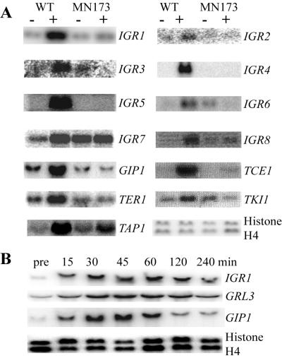
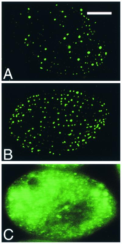
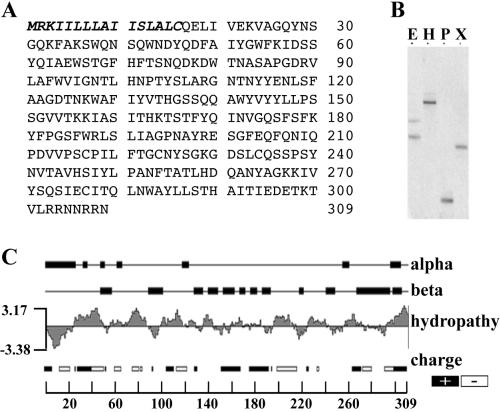
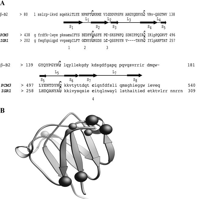
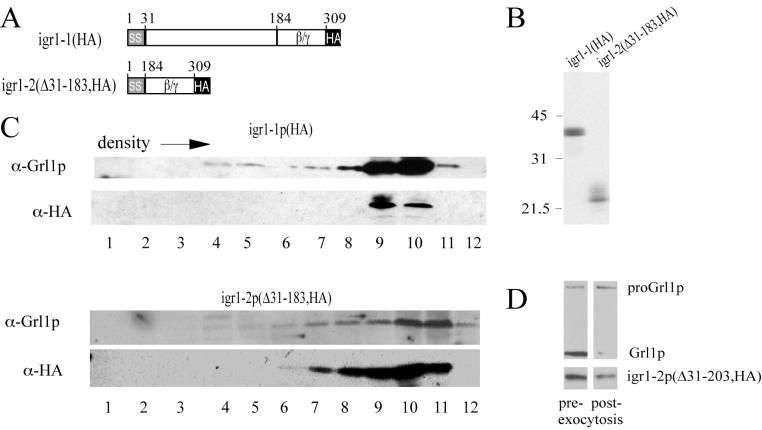
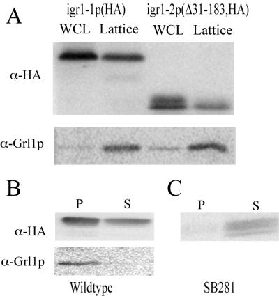
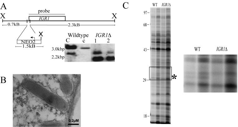
Regulated exocytosis of dense core secretory granules releases biologically active proteins in a stimulus-dependent fashion. The packaging of the cargo within newly forming granules involves a transition: soluble polypeptides condense to form water-insoluble aggregates that constitute the granule cores. Following exocytosis, the cores generally disassemble to diffuse in the cell environment. The ciliates Tetrahymena thermophila and Paramecium tetraurelia have been advanced as genetically manipulatable systems for studying exocytosis via dense core granules. However, all of the known granule proteins in these organisms condense to form the architectural units of lattices that are insoluble both before and after exocytosis. Using an approach designed to detect new granule proteins, we have now identified Igr1p (induced during granule regeneration). By structural criteria, it is unrelated to the previously characterized lattice-forming proteins. It is distinct in that it is capable of dissociating from the insoluble lattice following secretion and therefore represents the first diffusible protein identified in ciliate granules.


Figures

FIG. 1.
FIG. 1.
Isolation of genes induced during DCG biogenesis in T. thermophila. (A) Each unique amplicon identified by differential-display PCR was used as a probe to assay the abundance of the corresponding mRNA in total RNA prepared from cells before (−) and after (+) exocytosis. A subset of the positive clones is shown, with the histone H4 transcripts serving as a loading control. Approximate transcript sizes are as follows: IGR1, 1.0 kb; IGR2, 1.2 kb; IGR3, 0.8 kb; IGR4, 0.8 kb; IGR5, 1.6 kb; IGR6, 1.0 kb; IGR7, ∼6 to 8 kb; IGR8, 1.0 kb; GIP1, 1.1 kb; TCE1, 2.0 kb; TER1, 0.9 kb; TKI1, 0.8 kb; TAP1, 1.9 kb. WT, wild type. (B) Northern blots of T. thermophila total RNA prepared prior to and at various times during granule replacement. The mRNAs for both GRL3 and IGR1 show identical patterns of accumulation, rapidly accumulating within 60 min of secretagogue exposure. The GIP1 transcript accumulates rapidly following stimulation but decreases more rapidly than GRL3 or IGR1. The level of histone H4 transcripts remains unchanged throughout the time course.
FIG. 2.
FIG. 2.
Expression of GFP fusion proteins reveals that the IGR1 product localizes to DCGs in vivo. Confocal micrographs of T. thermophila strains harboring GFP-tagged constructs. Tangential optical sections are shown. Bar, 10 μM. (A) Cells expressing igr1-4p(GFP) show punctate fluorescence along the cell surface. The periodic pattern is due to the fact that DCG docking sites are regularly spaced along a well-ordered cortical cytoskeletal network (1 and 2° meridians) in these cells. (B) A similar pattern of fluorescence is seen in cells expressing a fusion to Grl1p, a DCG lattice protein. (C) Cells expressing GFP linked to the signal sequence of Grl1p. GFP fluorescence appears in the form of heterogeneous puncta and is not detected within DCGs.
FIG. 3.
FIG. 3.
Sequence and predicted features of Igr1p. (A) The 930-bp IGR1 ORF encodes a 309-amino-acid protein, whose sequence is shown. The predicted amino-terminal signal sequence is in italics. (B) Southern blot of T. thermophila genomic DNA digested with EcoRI (E), HindIII (H), PacI (P), and XbaI (X). A single strong band is present in all digestions, with an additional weaker reactive band present in lane E. (C) Predicted features of Igr1p. The sole extended stretch of hydrophobic residues corresponds to the amino-terminal signal sequence. In contrast to the acidic Grlps, Igr1p is a slightly basic protein and is not predicted to fold as α-helical coiled coils.
FIG. 4.
FIG. 4.
Tentative alignment of ciliate proteins Igr1p and Pcm3p with bovine β B2-crystallin, domain 2. (A) Residues indicated by double-height letters are absolutely conserved among β/γ-crystallins (G36, S60, and G80) and were used as guides for the alignment. A linear representation of the β/γ-crystallin 2° structure, immediately below the sequence, shows the relative positions of β-strands (S) and loops (L). Four cysteine residues present in Igr1p and Pcm3p but not in β/γ-crystallins are in boldface and were assigned arbitrary numbers. A four-residue deletion in the Igr1p sequence, relative to the other two sequences shown, falls within an extended loop (L3). (B) Modeling the carboxy termini of the ciliate proteins on the tertiary structure of a β/γ-crystallin domain (bovine β B2-crystallin). Small balls: the three residues conserved between Igr1p, the Pcmps, and the vertebrate β/γ-crystallins, which are positioned at tight turns where the range of acceptable side chains is constrained; large balls: four cysteines found in the ciliate, but not the vertebrate, proteins. When the ciliate sequences are mapped on the β/γ-crystallin domain structure, the cysteines appear well situated to form two disulfide bonds.
FIG. 5.
FIG. 5.
Expression and characterization of HA-tagged full-length and truncated Igr1p. (A) The HA nonapeptide was fused to the extreme carboxy termini of Igr1p and a truncated derivative. The former is igr1-1p(HA). In the latter, igr1-2p(Δ31-183,HA), the N-terminal 153 amino acid residues (not including the signal sequence, residues 1 to 30) are deleted, leaving the carboxy-terminal region linked to the signal sequence. (B) Western blots of whole-cell lysates expressing these constructs reveal anti-HA-reactive bands near the predicted molecular weights. (C) Fractionation of cell homogenates by equilibrium density ultracentifugation on continuous Nycodenz gradients. Harvested fractions were subjected to SDS-PAGE and analyzed by antibody blotting using antibodies against HA or Grl1p. (Top) Both igr1-1p(HA) and Grl1p are found in the same high-density fractions. However, Grl1p is most concentrated in fraction 10, while igr1-1p(HA) is most concentrated in fraction 9. (Bottom) Equivalent fractionation of cells expressing igr1-2p(Δ31-183,HA). On this particular gradient, the Grl1p-containing DCG peak is shifted one fraction relative to that above. As for the holoprotein, the distribution of igr1-2p(Δ31-183,HA) is subtly different from that of Grl1p. (D) Depletion of igr1-2p(Δ31-183,HA) and Grl1p from cells following exocytic release. Cells expressing igr1-2p(Δ31-183,HA) were stimulated with dibucaine for 15 s and separated from the released granule contents. Cells were depleted of ∼60% of igr1-2p(Δ31-183,HA) and ∼90% of Grl1p. ProGrl1p serves as an internal control, since the proprotein is not released upon stimulation (52).
FIG. 6.
FIG. 6.
Igr1p is not stably associated with the Grl-based DCG lattice. (A) The insoluble contents of DCGs were enriched from stimulated cell supernatants as described in Materials and Methods. Grl1p and igr1-1p(HA) were detected by Western blotting of whole-cell lysates prepared from prestimulated cells (WCL) or a fraction corresponding to the insoluble fraction of enriched granule lattices, isolated from stimulated-cell supernatants (lattice). The igr1-1p(HA) was detected in both whole-cell lysates and in the granule contents but was depleted approximately twofold in the latter. In contrast, Grl1p is enriched ∼20-fold in the granule content fraction. Similar results were obtained for cells expressing igr1-2p(Δ31-183,HA). SDS-PAGE samples were prepared for equal protein loading. Antibodies were detected with 125I-protein A. (B) Wild-type cells expressing igr1-1p(HA) were solubilized with detergent, and the insoluble fraction was pelleted as described in Materials and Methods. The relative levels of HA-tagged proteins and Grl1p in pellets (P) and supernatants (S) were determined by Western blotting. While more than 95% of Grl1p is found in the pellet fraction, igr1-1p(HA) is present in both the pellet (∼70%) and supernatant (∼30%). (C) igr1-1p(HA) was expressed in T. thermophila exo strain SB281, which is defective in processing DCG proproteins and in assembling insoluble granule cores. Detergent lysates were prepared and fractionated as described for panel A. The igr1-1p(HA) was present almost exclusively in the soluble fraction.
FIG. 7.
FIG. 7.
Disruption of the macronuclear IGR1 gene. (A) The construct used for interruption of macronuclear IGR1. A 256-bp fragment including the translational start site of IGR1 was replaced with the NEO2 cassette and used to transform cells. Correct targeting and replacement of the endogenous macronuclear copies of IGR1 were confirmed by Southern blotting of untransformed strains (lanes C and c) and two different IGR1Δ strains (lanes 1 and 2) using an IGR1 probe. The 3.0-kb XbaI fragment in untransformed cells is replaced by a 2.3-kb fragment, as expected for complete replacement of the endogenous allele. Lane c contains a reduced amount of DNA; the signal at this dilution is due to the two transcriptionally silent micronuclear copies of IGR1, which are not replaced in such transformants. A weaker secondary band is common to all three strains. (B) DCGs in IGR1Δ strains are indistinguishable from those in wild-type cells. Electron microscope thin section of an IGR1Δ cell showing a DCG docked at the plasma membrane. (C) Comparison of the secreted proteins in wild-type versus IGR1Δ cells by SDS-PAGE and Coomassie blue staining demonstrated the absence of a minor polypeptide (molecular mass, ∼34 kDa) in the IGR1Δ sample. ∗, position of the polypeptide. The right panel is a magnified view of the boxed region of the gel shown on the left.

Similar articles

Cited by

References

    1. Allen, S. L., M. I. Altschuler, P. J. Bruns, J. Cohen, F. P. Doerder, J. Gaertig, M. Gorovsky, E. Orias, and A. Turkewitz. 1998. Proposed genetic nomenclature rules for Tetrahymena thermophila, Paramecium primaurelia and Paramecium tetraurelia. Genetics 149:459-462. - PMC - PubMed
    1. Arvan, P., and D. Castle. 1998. Sorting and storage during secretory granule biogenesis: looking backward and looking forward. Biochem. J. 332:593-610. - PMC - PubMed
    1. Blazquez, M., C. Thiele, W. B. Huttner, K. Docherty, and K. I. Shennan. 2000. Involvement of the membrane lipid bilayer in sorting prohormone convertase 2 into the regulated secretory pathway. Biochem. J. 349:843-852. - PMC - PubMed
    1. Cassidy-Hanley, D., J. Bowen, J. H. Lee, E. Cole, L. A. VerPlank, J. Gaertig, M. A. Gorovsky, and P. J. Bruns. 1997. Germline and somatic transformation of mating Tetrahymena thermophila by particle bombardment. Genetics 146:135-147. - PMC - PubMed
    1. Chan, C. W., Y. Saimi, and C. Kung. 1999. A new multigene family encoding calcium-dependent calmodulin-binding membrane proteins of Paramecium tetraurelia. Gene 231:21-32. - PubMed

Publication types

MeSH terms

LinkOut - more resources