

Molecular mechanisms of irradiation-induced apoptosis
- PMID: 12456331
- PMCID: PMC2585024
- DOI: 10.2741/927
Molecular mechanisms of irradiation-induced apoptosis
Abstract
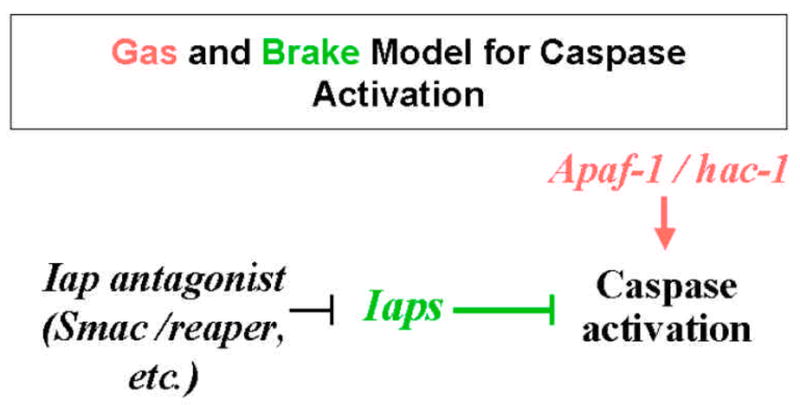
The following review focuses on our current knowledge as to how the cell death regulatory machinery is activated to mediate irradiation-induced cell death. In particular, we will address recent developments related to the following questions: 1.) Which cell death regulatory genes mediate irradiation-induced cell death? 2.) What is the mechanism of irradiation-induced activation or suppression of cell death regulatory genes (proteins)? 3.) How does the condition of the cell death regulatory machinery affect the cell's sensitivity or resistance to irradiation? Now more than ever, it seems clear that irradiation -induced apoptosis is a complex process involving all three major cell death regulatory pathways: the mitochondria pathway (Bcl-2/Apaf-1), the Iap pathway, and the death receptor pathway. Depending on the cellular context, one or multiple pathways may be activated to mediate irradiation-induced cell death. Therefore, a comprehensive understanding of these processes demands systematic strategies in contrast to traditional approaches that focused on one gene/protein. For this reason, we will also examine recent studies applying genomic (proteomic) methods in this area.
Figures

References
-
- Zhou BB, Elledge SJ. The DNA damage response: putting checkpoints in perspective. Nature. 2000;408:433–9. - PubMed
-
- Rich T, Allen RL, Wyllie AH. Defying death after DNA damage. Nature. 2000;407:777–83. - PubMed
-
- Vaux DL, Korsmeyer SJ. Cell Death in Development. Cell. 1999;96:245–254. - PubMed
-
- Horvitz HR. Genetic control of programmed cell death in the nematode Caenorhabditis elegans. Cancer Res. 1999;59:1701s–1706s. - PubMed
-
- Bergmann A, Agapite J, Steller H. Mechanisms and control of programmed cell death in invertebrates. Oncogene. 1998;17:3215–3223. - PubMed
Publication types
MeSH terms
Grants and funding
LinkOut - more resources
Full Text Sources
Research Materials