. 2002 Dec 2;21(23):6377-86.
doi: 10.1093/emboj/cdf640.

Structure of eukaryotic prefoldin and of its complexes with unfolded actin and the cytosolic chaperonin CCT

Affiliations

Structure of eukaryotic prefoldin and of its complexes with unfolded actin and the cytosolic chaperonin CCT

Jaime Martín-Benito et al. EMBO J. .

Abstract

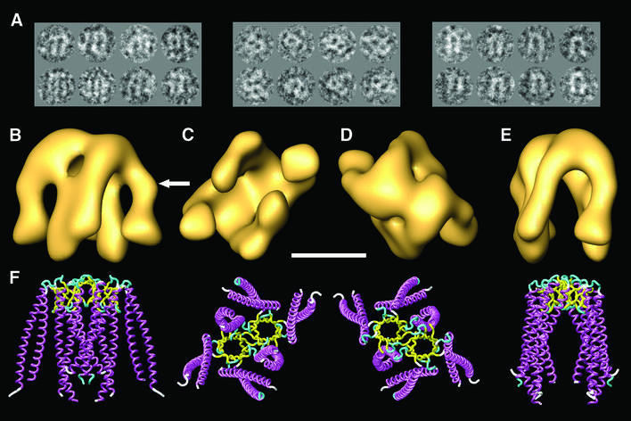
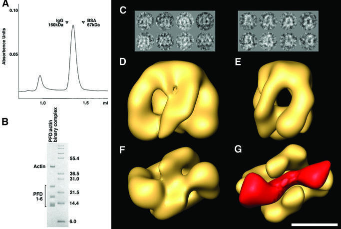
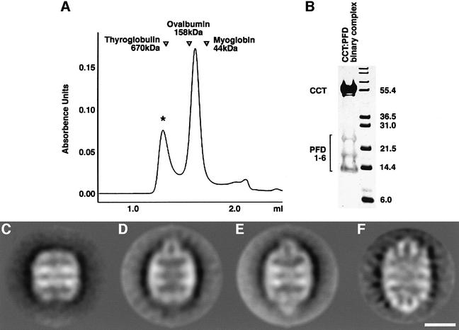
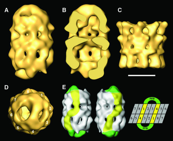
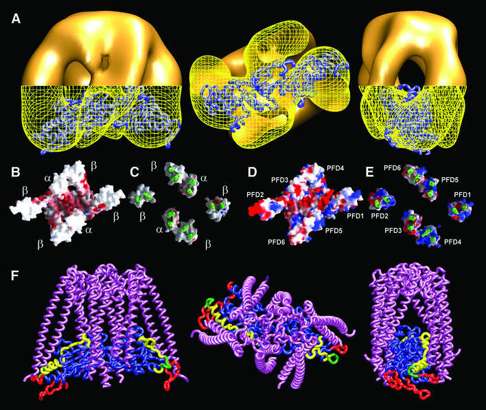
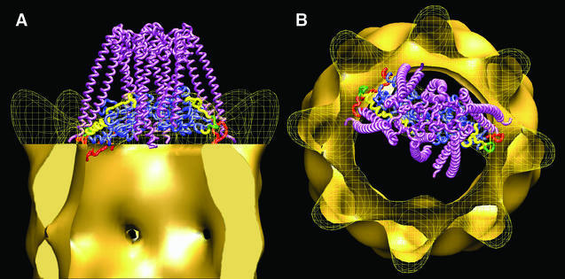
The biogenesis of the cytoskeletal proteins actin and tubulin involves interaction of nascent chains of each of the two proteins with the oligomeric protein prefoldin (PFD) and their subsequent transfer to the cytosolic chaperonin CCT (chaperonin containing TCP-1). Here we show by electron microscopy that eukaryotic PFD, which has a similar structure to its archaeal counterpart, interacts with unfolded actin along the tips of its projecting arms. In its PFD-bound state, actin seems to acquire a conformation similar to that adopted when it is bound to CCT. Three-dimensional reconstruction of the CCT:PFD complex based on cryoelectron microscopy reveals that PFD binds to each of the CCT rings in a unique conformation through two specific CCT subunits that are placed in a 1,4 arrangement. This defines the phasing of the CCT rings and suggests a handoff mechanism for PFD.


Figures

None
Fig. 1. Three-dimensional reconstruction of human PFD. (A) A gallery of negatively stained images similar to those used for the three-dimensional reconstruction of PFD. The three groups of images shown are archetypes of the three main views, which are orthogonal. (BE) Several views of the three-dimensional reconstruction of human PFD. (F) Similar views of the atomic structure of the archaeal homolog of PFD (MtGimC from M.thermoautotrophicum; Siegert et al., 2000); PDB accession code 1FXK. The arrow points to a kink in the eukaryotic structure of PFD that is found reproducibly in all the three-dimensional reconstructions generated, and which is not present in the atomic structure of the archaeal PFD. Scale bar = 50 Å.
None
Fig. 2. Generation of the PFD:actin complex and its three-dimensional reconstruction. (A) Isolation of PFD:actin complexes by gel filtration on Superdex 200. The locations of molecular mass markers run on the same column are shown. (B) Analysis by SDS–PAGE of material in the peak shown in (A) migrating with an apparent Mr of ∼100 kDa. Molecular mass markers (in kDa) are shown on the right. (C) A gallery of negatively stained images similar to those used for the three-dimensional reconstruction of the PFD:actin complex. The two main views are depicted. (DF) Front, side and bottom views, respectively, of the three-dimensional reconstruction of the PFD:actin complex. (G) The volume of substrate-free PFD (solid yellow) placed in the same bottom view as the PFD:actin complex depicted in (F). The mass shown in red corresponds to the difference between the PFD:actin complex and substrate-free PFD, and is very suggestive of actin existing in an open conformation, traversing the PFD cavity and interacting with the tips of the tentacles. Scale bar = 50 Å.
None
Fig. 3. Two-dimensional averages of CCT:PFD complexes. (A) Gel filtration on Superose 6 of the products resulting from co-incubation of CCT and PFD. The location of molecular size markers run on the same column under identical conditions is shown. (B) Analysis by SDS–PAGE of the products contained in the peak [marked with an asterisk in (A)] emerging from the column with an apparent Mr of ∼700 kDa. Note the presence of polypeptides characteristic of both CCT (in the size range around 55 kDa) and PFD (in the size range 14–25 kDa). (C) Average image obtained from negatively stained CCT particles in the absence of PFD (average image obtained from 350 particles; 27 Å resolution). (D) Average image obtained from negatively stained particles of CCT:PFD asymmetric complexes and (E) symmetric complexes (average images of 561 and 1128 particles; 27 and 24 Å resolution, respectively) (F) Average image of the CCT:PFD symmetric complex obtained from frozen-hydrated specimens (715 particles; 25 Å resolution). Scale bar = 100 Å.
None
Fig. 4. Three-dimensional reconstruction of the CCT:PFD symmetric complex. (A) Side view of the CCT:PFD symmetric complex. (B) A section along the longitudinal axis of the same volume. (C) Side view of the three-dimensional reconstruction of apo-CCT (Llorca et al, 2000). (D) Top view of the three-dimensional reconstruction showing the 1,4 interaction between PFD and the CCT subunits. (E) A scheme showing the interaction of the two PFD oligomers with the CCT subunits of the two rings. The two volumes on the left represent two opposing views of the CCT:PFD complex. The PFD oligomers are highlighted in green and the CCT subunits interacting with PFD are shown in yellow. The topology of the CCT subunits depicted on the right is for illustrative purposes only, and the numbers have the sole purpose of discriminating among the subunits. Scale bar = 100 Å in (A–D).
None
Fig. 5. Model of the interaction between denatured actin and PFD. (A) Several views of the three-dimensional reconstruction of the PFD:actin complex in which the atomic model of unfolded actin generated upon its docking into the three-dimensional reconstruction of actin bound to nucleotide-free CCT (Llorca et al., 1999, 2000) has been fitted into the actin part of the PFD:actin complex. The envelope of the tips of the PFD:actin complex has been drawn as a yellow grid to allow visualization of the atomic model of unfolded actin. (B and C) Bottom and top views, respectively, of the tips of the tentacles of the atomic structure of the archaeal PFD from M.thermoautotrophicum (Siegert et al., 2000). (D and E) The same views of the structural model generated with human PFD subunits. In these images, the α and β archaeal subunits were used as templates, and the human α- and β-like subunits were placed in the center and outer positions, respectively. The β-like subunits are placed randomly. Human PFD was modeled using the homology modeling programs Swiss-PdbViewer and Swiss-Model (Guex and Peitsch, 1997; Schwede et al., 2000). In (C) and (E), the base of the structure and part of the tentacles have been removed to allow visualization of the outer surface of the tentacle tips. Surfaces (negatively charged residues in red, positively charged residues in blue) were generated using GRASP (Nicholls et al., 1991). (F) Several views of the modeled interaction between unfolded actin and PFD. The atomic structure of the archaeal PFD and the atomic model of unfolded actin shown in (A) were used as structures for the modeling of this interaction. Actin residues putatively involved in the interaction of PFD with CCT are highlighted as follows: residues involved in the interaction with CCT (Llorca et al., 2000, 2001b) (red); residues involved in PFD binding (Rommelaere et al., 2001) (yellow); residues involved in both functions (green).
None
Fig. 6. Model of the interaction between unfolded actin, PFD and CCT. (A and B) Side view and top views, respectively, of the proposed ternary interaction between actin, PFD and CCT. The PFD:actin complex shown in Figure 5 interacts with specific CCT subunits through the tips of some of the PFD subunits and presents the actin molecule to the CCT subunits that interact with this molecule (Llorca et al., 1999). The actin molecule has been fitted into the actin part of the three-dimensional reconstruction of the CCT:actin complex used to generate the model (Llorca et al., 2000). The color code of the actin molecule is as in Figure 5.

Similar articles

Cited by

References

    1. Buckle A.M., Zahn,R. and Fersht,A.R. (1997) A structural model for GroEL-polypeptide recognition. Proc. Natl Acad. Sci. USA, 94, 3571–3575. - PMC - PubMed
    1. Bukau B. and Horwich,A.L. (1998) The Hsp70 and Hsp60 chaperone machines. Cell, 92, 351–380. - PubMed
    1. Bukau B., Deuerling,E., Pfund,C. and Craig,E.A. (2000) Getting newly synthesized proteins into shape. Cell, 101, 119–122. - PubMed
    1. Chen X., Sullivan,D.S. and Huffaker,T.C. (1994) Two yeast genes with similarity to TCP-1 are required for microtubule and actin function in vivo. Proc. Natl Acad. Sci. USA, 91, 9111–9115. - PMC - PubMed
    1. Cowan N.J.and.Lewis.,S.A. (2002) Type II chaperonins, prefoldin and the tubulin-specific chaperones. Adv. Protein Chem., 59, 73–104. - PubMed

Publication types