. 2002 Dec 2;21(23):6429-39.
doi: 10.1093/emboj/cdf644.

XPak3 promotes cell cycle withdrawal during primary neurogenesis in Xenopus laevis

Affiliations

XPak3 promotes cell cycle withdrawal during primary neurogenesis in Xenopus laevis

Jacob Souopgui et al. EMBO J. .

Abstract

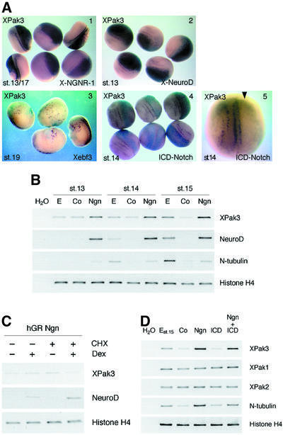
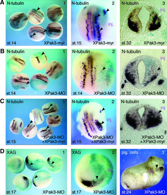
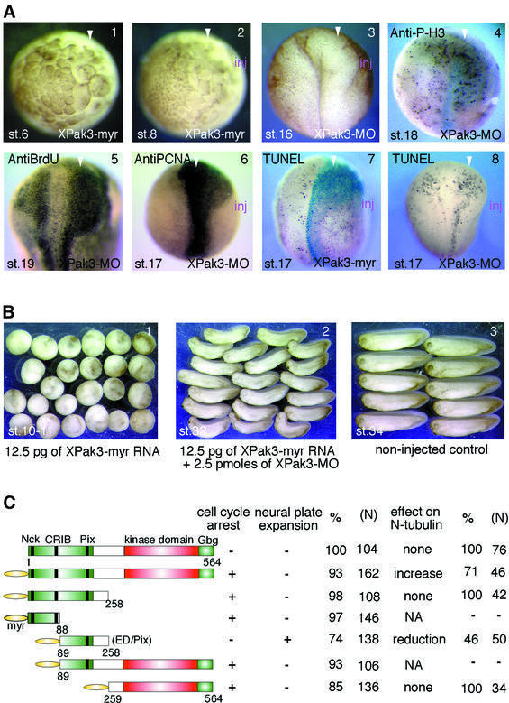
We have isolated the Xenopus p21-activated kinase 3 (XPak3) by virtue of its expression in the territory of primary neurogenesis in the developing embryo. XPak3, but not the other Pak variants, responds positively to X-Ngnr-1 and negatively to X-Notch-1. A constitutively active form of XPak3, generated by fusing a myristylation signal to the N-terminus (XPak3-myr), induces early cell cycle arrest at high concentrations, while ectopic expression of low amounts induces premature neuronal differentiation. Conversely, XPak3 loss of function achieved by use of an antisense morpholino oligonucleotide increases cell proliferation and inhibits neuronal differentiation; this phenotype is rescued by co-injection of XPak3-myr. We conclude that XPak3 is a novel member of the proneural pathway, functioning downstream of neurogenin to withdraw neuronally programmed cells from the mitotic cell cycle, thus allowing for their differentiation.


Figures

None
Fig. 1. XPak3 sequence analysis. The XPak3 protein sequence, as predicted from the cDNA, was aligned with the murine (m) and human (h) orthologues, as well as with the two other Xenopus variants, XPak1 and XPak2 (Bagrodia et al., 1995; Faure et al., 1997; Allen et al., 1998; Cau et al., 2000). Dashes represent identical amino acids while dots indicate the absence of amino acids. The kinase domain is shaded in grey. The Cdc42/Rac1-interacting binding (CRIB) domain is indicated and partially overlaps with the autoinhibitory domain (AID) indicated in bold. The proline-rich motifs (PXXP and PRM), specific for Nck and Pix interactions, are boxed. The G-β/γ-binding domain (Gbg) is conserved at the C-terminal edge. The percentages of sequence identity and similarity are indicated.
None
Fig. 2. Differential expression patterns of XPak genes during Xenopus development. (A) Spatial and temporal expression of XPak3 was analysed at different stages (st.) of neurogenesis using whole-mount in situ hybridization. (1–5) XPak3 transcripts are found in three bilateral stripes, lateral (l), intermediate (i) and medial (m), in the posterior neural plate. Embryos are shown in a dorsal (d) view with anterior up. (6) In a transverse (T) section of a stage 17 embryo at the level shown in panel 5 (white dotted line), XPak3 transcripts are detectable in the deep layer of the neuroectoderm in regions corresponding to the medial, intermediate and lateral stripes. Panels 7–10 show the expression in the anterior neural plate; (8) XPak3-expressing cells first appear in the trigeminal placode (tp), then (9) in a centrally located, semicircular array corresponding to a ventral area within the prospective fore-/midbrain where neurons first develop (vfmb) and (10) in the cement gland (cg). (11–14) At later stages of neurogenesis, XPak3 is expressed in the forebrain (fb), midbrain (mb), hindbrain (hb), retina (r), optic nerve (on), olfactory placode (op), otic vesicle (ov) and cement gland (cg): (11 and 12) lateral view (l), (13) horizontal (H) section of the head region at the level indicated in panel 12, (14) anterior view (a). (B) Differential expression of neuronal marker genes in the neural tube of stage 32 embryos. (1) X-Ngnr-1 is expressed in the ventricular zone (vz), (3) N-tubulin in the marginal zone (mz) and (2) XPak3 in the marginal and subventricular zone (svz). The level of the transverse (T) sections is shown in (A) panel 12. (C) Expression patterns of XPak1 and XPak2. (1) XPak1 transcripts are present in the dorsal midline (dml) and (2 and 5–8) later in the otic vesicle, mandibular arch (ma), branchial arch (ba), midbrain–hindbrain boundary (mhb), stomodeal–hypophyseal anlage (sha), lateral placode (lp), notochord (no) and dorsal foregut (dfg). (3) Early on, XPak2 is expressed ubiquitously in the neural plate and (4) later the expression becomes stronger in the brain, eye and tailbud. Embryos are shown in (1 and 3) dorsal view with anterior up and (2, 4 and 5) lateral view. (6–8) Levels of the transverse (T) sections (T1–3) are indicated in panel 5.
None
Fig. 3. Regulation of XPak3 expression in both embryos and animal caps. (A) XPak3 expression is positively regulated by proneural genes and negatively regulated by X-Notch-1. Xenopus albino embryos were injected into one cell at the two-cell stage with synthetic RNA encoding X-Ngnr-1, X-NeuroD, Xebf3 and ICD-Notch, along with LacZ RNA as tracer. Embryos were fixed at the neurula stage, then stained with X-gal (light blue) and analysed by whole-mount in situ hybridization for XPak3 expression (purple). (1) X-Ngnr-1 and (2) X-NeuroD injection results in strong ectopic expression of XPak3 (100 and 96%, n = 76 and 64, respectively). (3) Xebf3 is sufficient to turn on ectopically the expression of XPak3 but in a weak and scattered manner (62%, n = 48). (4 and 5) The activated form of X-Notch-1, ICD-Notch, blocks XPak3 expression (100%, n = 56). (B) X-Ngnr-1 is sufficient to activate XPak3 expression in animal caps. Pigmented embryos were injected with 50 pg of X-Ngnr-1 mRNA. Animal caps were isolated and cultivated to the indicated stages. Total RNAs were prepared from the caps and analysed by RT–PCR for XPak3 expression, using NeuroD, N-tubulin and histone H4 as controls. For each stage, E (embryos), Co (control caps) and Ngn (neurogenin-injected caps) are shown. (C) XPak3 is not induced directly by X-Ngnr-1. Embryos were injected into two blastomeres of the two-cell stage with hGR Ngn. Cycloheximide (CHX) treatment was from stage 11.5 followed by 2.5 h treatment with or without dexamethasone (Dex). Total RNAs were prepared from these caps and analysed by RT–PCR for XPak3, NeuroD and histone H4. (D) XPak3, but not XPak1 and XPak2, is activated by X-Ngnr-1 in animal cap explants. Pigmented embryos were injected with X-Ngnr-1 RNA and/or ICD-Notch RNA and allowed to grow up to stage 9. Animal caps were isolated and cultivated to stage 15. Total RNAs were prepared from the caps and analysed by RT–PCR for the genes indicated. From left to right, the columns represent: water control (H2O), uninjected control embryos stage 15 (E), uninjected control caps (Co), X-Ngnr-1-injected caps (Ngn), ICD-Notch-injected caps (ICD) and X-Ngnr-1/ICD-Notch-co-injected caps (Ngn + ICD).
None
Fig. 4. XPak3 activation induces premature neuronal differentiation. (AC) Modulation of XPak3 activity influences neuronal differentiation. Xenopus albino embryos were injected into one blastomere at the two-cell stage with (A) 10 pg of XPak3-myr RNA, (B) 2.5 pmol of XPak3-MO and (C) 2.5 pmol of XPak3-MO/12.5 pg XPak3-myr RNA, all with LacZ RNA as tracer. Embryos were fixed at neurula and tailbud stages, then stained with X-gal (light blue) and analysed for neuronal differentiation, as marked by N-tubulin expression. (A3, B3 and C3) Transverse sections of stage 32 embryos were prepared at the level of the hindbrain. (A1 and 2) XPak3-myr injection results in increased N-tubulin expression within the trigeminal placode and within the territories of primary neurons, as indicated by arrowheads (71%, n = 46). (A3) In the neural tube, XPak3-myr injections expand/shift the expression domain of N-tubulin from the marginal zone towards the ventricular zone (white arrowhead). (B1–3) XPak3-MO injections strongly inhibit N-tubulin expression within both the trigeminal placode and the territories of primary neurons (100%, n = 92), and within the neural tube. (C1 and 2) Co-injection of XPak3-MO and XPak3-myr RNA results in rescue of N-tubulin expression in a pattern that is not identical to, but reminiscent of the normal one (84%, n = 73). (C3) In the neural tube, co-injection increases/expands the expression domain of N-tubulin similarly to XPak3-myr injection (see A, panel 3). (D) XPak3 is necessary for cement gland formation. Embryos were injected into one blastomere of two-cell stage embryos with 2.5 pmol of XPak3-MO and LacZ RNA as tracer. One batch of these embryos was fixed at stage 17, stained for β-galactosidase activity (light blue) and bleached. The other batch was fixed at stage 24. (1 and 2) By whole-mount in situ hybridization, XAG is found to be suppressed, as marked by arrowheads (100%, n = 71). (3) In stage 24 embryos, pigmented cement gland cells are missing on the injected side.
None
Fig. 5. XPak3 is involved in cell cycle regulation. (A) Pigmented embryos were injected into one blastomere of two- or four-cell stage embryos with either XPak3-myr RNA or XPak3-MO; these embryos were allowed to develop to the stages indicated. The arrowhead shows the injected side: (1) 50 pg of Xpak3-myr induce cell cycle inhibition before MBT (100%, n = 75); (2) 25 pg give a similar phenotype after MBT (100%, n = 83). (3) Microinjection of 2.5–5 pmol of XPak3-MO induces neural plate expansion (67%, n = 116); (4–6) immunostained embryos showing increased signal on the injected side (arrowhead) for phosphorylated histone H3 (54%, n = 54), BrdU (58%, n = 48) and PCNA (79%, n = 62). (7 and 8) TUNEL staining of embryos injected with 10 pg of XPak3-myr RNA or 2.5 pmol of XPak3-MO shows no significant effect on apoptosis; rather, apoptotic cell death appears to correlate with increased proliferation. (B) Embryos were injected into one blastomere of two-cell stage embryos with either XPak3-myr RNA alone or XPak3-myr RNA and XPak3-MO together. By stage 10–11, XPak3-myr-injected embryos stopped growing (100%, n = 107), while XPak3-myr/XPak3-MO-co-injected ones continued to develop (79%, n = 95). Embryos were fixed at the stages indicated. (C) Deletion mutants of XPak3 were generated and tested for their influence on cell cycle regulation and neuronal differentiation. The corresponding phenotypes are indicated.

Similar articles

Cited by

References

    1. Allen C.M., Gleeson,J.G., Bagrodia,S., Partington,M.W., MacMillan,J.C., Cerione,R.A., Mulley,J.C. and Walsh,C.A. (1998) Pak3 mutation in nonsyndromic X-linked mental retardation. Nat. Genet., 20, 25–30. - PubMed
    1. Bagrodia S. and Cerione,R.A. (1999) Pak to the future. Trends Cell Biol., 9, 350–355. - PubMed
    1. Bagrodia S., Taylor,S.J., Creasy,C.L., Chernoff,J. and Cerione,R.A. (1995) Identification of a mouse p21Cdc42/Rac activated kinase. J. Biol. Chem., 270, 22731–22737. - PubMed
    1. Bellefroid E.J., Bourguignon,C., Holleman,T., Ma,Q., Anderson,D.J., Kintner,C. and Pieler,T. (1996) X-MyT1, a Xenopus C2HC-type zinc finger protein with a regulatory function in neuronal differentiation. Cell, 87, 1191–1202. - PubMed
    1. Bienvenu T. et al. (2000) Missense mutation in Pak3, R67C, causes X-linked nonspecific mental retardation. Am. J. Med. Genet., 93, 294–298. - PubMed

Publication types

MeSH terms