. 2002 Dec 2;21(23):6461-72.
doi: 10.1093/emboj/cdf658.

BLNK: molecular scaffolding through 'cis'-mediated organization of signaling proteins

Affiliations

BLNK: molecular scaffolding through 'cis'-mediated organization of signaling proteins

Christopher W Chiu et al. EMBO J. .

Abstract

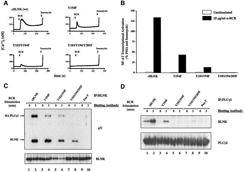
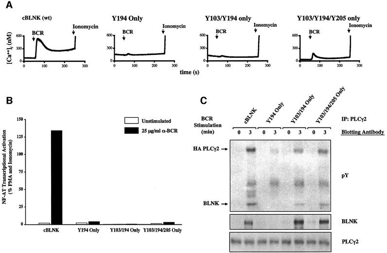
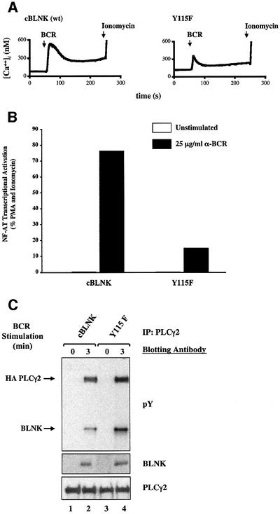
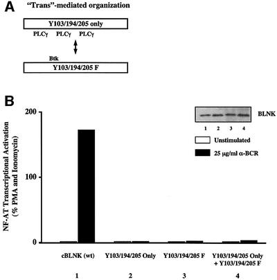
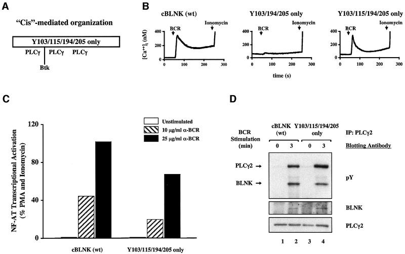
Assembly of intracellular macromolecular complexes is thought to provide an important mechanism to coordinate the generation of second messengers upon receptor activation. We have previously identified a B cell linker protein, termed BLNK, which serves such a scaffolding function in B cells. We demonstrate here that phosphorylation of five tyrosine residues within human BLNK nucleates distinct signaling effectors following B cell antigen receptor activation. The phosphorylation of multiple tyrosine residues not only amplifies PLCgamma-mediated signaling but also supports 'cis'-mediated interaction between distinct signaling effectors within a large molecular complex. These data demonstrate the importance of coordinate phosphorylation of molecular scaffolds, and provide insights into how assembly of macromolecular complexes is required for normal receptor function.


Figures

None
Fig. 1. Identification of phosphorylated tyrosine sites in BLNK. (A) Phosphopeptide maps of in vivo labeled BLNK. Trypsin digested 32P-labeled hBLNK was isolated from resting (left) or BCR-activated (right) Daudi B cells and analyzed by 2-dimensional electrophoresis and TLC, as described in Materials and methods. The first dimension of electrophoretic separation is represented on the x-axis and ascending chromatography is represented on the y-axis. The bottom panel represents a schematic diagram of the resultant peptides. Each peptide was eluted from the TLC plate and further analyzed for phosphoamino acid content (data not shown). This data is summarized in the bottom panel with black spots representing peptides that contain pY, pS and pT, while the open spots represent peptides that contain only phosphoserine and phosphothreonine. These maps are representative of a minimum of five independent labeling experiments for each sample. (B) Phosphopeptide maps of in vitro labeled BLNK. Purified wild-type hBLNK (left) and hBLNK(Y3F) in which Ys 72, 84 and 96 are mutated to F (right) were phosphorylated by Syk in vitro in the presence of [γ32P]-ATP, digested with trypsin and analyzed by 2-dimensional TLC, as described in Materials and methods. These maps are representative of a minimum of six independent experiments for each sample. (C) Schematic diagram of predicted peptides. The three predicted tryptic peptides containing the seven N-terminal tyrosine residues of hBLNK are depicted. The first four tyrosine residues (Ys 52, 72, 84 and 96) are encoded within a 56 aa tryptic fragment. Y118 is encoded within a 7 aa peptide. Ys 178 and 189 are encoded within a 27 aa peptide. The cycle number of 32P released for the tryptic peptides by Edman degradation is depicted above each Y. The cycle number of 32P released for peptides additionally digested with endo-gluC is shown below each Y.
None
Fig. 2. Generation of cells expressing wild-type and mutant BLNK molecules. (A) Analysis of the phosphorylation of hBLNK utilizing phosphospecific antibodies. hBLNK was immunoprecipitated from resting or BCR-activated cells and analyzed by immunoblotting with antiserum raised against each of the phosphorylated tyrosine residues in hBLNK (lanes 1–10), an anti-pY mAb (4G10, lanes 11–12), or an anti-BLNK antiserum (1761, lanes 13–14). The phosphospecific antibodies, as described in the Materials and methods, were used at 0.5 µg/ml in 0.5% BSA/TBST (0.05% Tween-20, 10 mM Tris pH 8.0 and 150 mM NaCl) and incubated for 1 h. The blots were washed three times for 15 min with TBST and incubated with HRP-conjugated anti-rabbit antisera (Pierce) diluted to 1:20 000 for 1 h. The blots were washed as before and developed by ECL according to manufacturer’s instructions (Pierce). (B) Comparison of hBLNK and cBLNK. Schematic diagrams of the N-terminal Ys are depicted. The Y residues that are within the conserved Syk phosphorylation sequence are underlined. (C) cBLNK(Pan F) is not phosphorylated on tyrosine residues following BCR crosslinking. Wild-type cBLNK or cBLNK(Pan F) were immunoprecipitated from resting or BCR-activated cells (M4, 4 µg/ml for 2 min at 37°C) and analyzed by immunoblotting with an anti-pY mAb (top) or an anti-BLNK antiserum (1761, bottom). (DIn vivo phosphorylation of cBLNK mutants expressing Y91, Y103, Y115, Y194 or Y205. Stable clones expressing wild-type or mutant cBLNK molecules were established as described in Materials and methods. CBLNK(wt) or the mutant BLNK molecules were immunoprecipitated from resting or BCR-activated cells (M4, 4 µg/ml for 2 or 5 min at 37°C) and analyzed by immunoblotting with an anti-pY mAb (top) or an anti-BLNK antiserum (1761, bottom).
None
Fig. 3. Requirement for tyrosine phosphorylation of BLNK in [Ca2+]i and MAP activation. (A) Association and phosphorylation of PLCγ2 requires BLNK tyrosine phosphorylation. Transiently transfected PLCγ2 was immunoprecipitated from the blnk–/–, wild-type cBLNK or cBLNK(Pan F)-expressing cells from resting and BCR-activated cells (M4, 4 µg/ml for 2 min at 37°C) and analyzed by immunoblotting with anti-pY mAb (top), an anti-BLNK antiserum (1761, middle), or anti-PLCγ2 antiserum (bottom). (B) Absence of BCR induced [Ca2+]i in cells expressing cBLNK(Pan F). blnk–/– DT40 cells (left) or blnk–/– cells reconstituted with wild-type cBLNK (middle) or cBLNK(Pan F) (right) were analyzed for their ability to increase [Ca2+]i following BCR crosslinking (BCR arrow) or ionomycin (ionomycin arrow), as described in Materials and methods. (C) Transcriptional activation of an NF-AT/AP-1 reporter gene is dependent upon tyrosine phosphorylation of BLNK. blnk–/– DT40 cells (left) and cells reconstituted with wild-type cBLNK (middle) or cBLNK(Pan F) (right) were analyzed for their ability to activate an NF-AT/AP-1 responsive element. Reporter activity was analyzed for cells incubated with media alone, in media containing anti-BCR M4 mAb (5 or 10 µg/ml, shaded or filled bars, respectively), or media containing PMA and ionomycin. These data are representative of five independent experiments and of at least two independent clones. (DF) Efficient activation of all three families of MAPKs is dependent on tyrosine phosphorylation of BLNK. blnk–/– DT40 cells (lanes 1–6) and cells reconstituted with wild-type cBLNK (lanes 7–12) or cBLNK(Pan F) (lanes 13–18) were analyzed for their ability to activate the Erk2 (panel D), JNK (panel E) and p38 (panel F) pathways. Cells were stimulated for the time periods denoted above each lane, lysed and immunoblotted with antibodies specific for the activated forms of each of the MAPKs. Equal loading of cell lysates was confirmed by blotting with antibodies for each of the MAPKs, as described in Materials and methods. Quantitation of bands was performed using UN-SCAN-IT software and the fold activation, as compared with resting cells (lanes 1, 7 and 13), is reported below each lane. This analysis is representative of a minimum of four independent experiments.
None
Fig. 4. Binding specificity of effector molecules to BLNK tyrosine phosphorylation sites. (A) Schematic diagram of preferential binding sites of PLCγ2 Btk, Nck and Vav on cBLNK. (B) Binding specificity of PLCγ2, Btk, Vav and Nck to cBLNK tyrosine residues. Daudi lysates were incubated with each of the tyrosine phosphorylated peptides corresponding to the phosphorylated tyrosine residues of chicken BLNK and analyzed by immunoblotting with anti-PLCγ2 (top), anti-Btk (top middle), anti-Vav (bottom middle) and anti-Nck (bottom). A phosphorylated peptide corresponding to a Y residue that would not normally be tyrosine phosphorylated was used as a control (Y138).
None
Fig. 5. Reduction of PLCγ-mediated signaling pathways with mutation of PLCγ binding sites. (A) Reduction in [Ca2+]i. blnk–/– DT40 cells expressing wild-type cBLNK, Y194F, Y103/194F or Y103/194/205F were analyzed for their ability to induce [Ca2+]i mobilization following BCR crosslinking or ionomycin. (B) Reduction in NF-AT transcriptional activation. blnk–/– DT40 cells expressing wild-type cBLNK, Y194F, Y103/194F or Y103/194/205F were analyzed for their ability to activate an NF-AT/AP-1 responsive element. Reporter activity was analyzed for cells incubated with media alone, in media containing anti-BCR mAb (M4, 25 µg/ml) or media containing PMA and ionomycin. (C) Reduced tyrosine phosphorylation and association of PLCγ2. BLNK was immunoprecipitated from PLCγ2 infected wild-type cBLNK-, Y194F-, Y103/194F- or Y103/194/205F-expressing cells from resting and BCR-activated cells (M4, 4 µg/ml for 3 min at 37°C) and analyzed by immunoblotting with anti-pY mAb (top) or an anti-BLNK antiserum (1761, bottom). Immunoprecipitation of PLCγ2 with an anti-HA mAb demonstrated similar graded reduction in PLCγ2 tyrosine phosphorylation (data not shown). (D) Reduced association of PLCγ2 with BLNK. Retrovirally infected PLCγ2 was immunoprecipitated from wild-type cBLNK-, Y194F-, Y103/194F- or Y103/194/205F-expressing cells from resting and BCR-activated cells (M4, 4 µg/ml for 3 min at 37°C) and analyzed by immunoblotting with an anti-BLNK antiserum (1761, top) or an anti-PLCγ2 antiserum (bottom).
None
Fig. 6. Restoration of PLCγ binding sites does not reconstitute normal activation of PLCγ-mediated signaling pathways. (A) Expression of PLCγ-binding cBLNK does not restore normal calcium mobilization. blnk–/– DT40 cells expressing wild-type cBLNK, Y194 only, Y103/194 only, or Y103/194/205 only were analyzed for their ability to induce [Ca2+]i following BCR crosslinking or ionomycin. (B) Failure to restore NF-AT transcriptional activation. blnk–/– DT40 cells expressing wild-type cBLNK, Y194 only, Y103/194 only or Y103/194/205 only were analyzed for their ability to activate an NF-AT/AP-1 responsive element. Reporter activity was analyzed for cells incubated with media alone, in media containing anti-BCR mAb (M4, 25 µg/ml) or media containing PMA and ionomycin. (C) Reconstitution of BLNK-PLCγ2 interaction. PLCγ2 was immunoprecipitated from PLCγ2 infected wild-type cBLNK, blnk–/–, Y194 only, Y103/194 only, or Y103/194/205- expressing cells from resting and BCR-activated cells (M4, 4 µg/ml for 3 min at 37°C) and analyzed by immunoblotting with anti-pY mAb (top), anti-BLNK antiserum (1761, middle) or PLCγ2 (bottom).
None
Fig. 7. Reduction of PLCγ-mediated signaling pathways with mutation of the Btk binding site. (A) Reduction in [Ca2+]i. blnk–/– DT40 cells expressing wild-type cBLNK or Y115F were analyzed for their ability to induce [Ca2+]i mobilization following BCR crosslinking or ionomycin. (B) Reduction in NF-AT transcriptional activation. blnk–/– DT40 cells expressing wild-type cBLNK or Y115F were analyzed for their ability to activate an NF-AT/AP-1 responsive element following incubation with media alone, in media containing anti-BCR mAb (M4, 25 µg/ml) or media containing PMA and ionomycin. (C) Normal tyrosine phosphorylation of PLCγ2. Retrovirally infected PLCγ2 was immunoprecipitated from wild-type cBLNK or Y115F-expressing cells from resting and BCR-activated cells (M4, 4 µg/ml for 3 min at 37°C) and analyzed by immunoblotting with anti-pY mAb (top), anti-BLNK antiserum (1761, middle) or an anti-PLCγ2 antiserum (bottom).
None
Fig. 8.Trans’-mediated organization of signaling proteins with cBLNK does not reconstitute BCR activation. (A) Model for ‘trans’-mediated organization of signaling proteins with cBLNK. In the trans model, the PLCγ2-binding mutant cBLNK (Y103/194/205 only) can cooperate with a non-PLCγ2-binding cBLNK mutant (Y103/194/205F) to regulate NF-AT transcriptional activation. (B) Lack of complementation by PLCγ and non-PLCγ binding cBLNK mutants in trans. blnk–/– DT40 cells were transiently transfected with 25 µg of wild-type cBLNK, Y103/Y194/Y205 only, Y103/Y194/Y205F, or both BLNK mutant cDNAs (12.5 µg each). The cells were analyzed for their ability to activate an NF-AT/AP-1 responsive element following incubation with media alone, in media containing anti-BCR mAb (M4, 25 µg/ml) or media containing PMA and ionomycin. Expression of BLNK was monitored by immunoblotting with an anti-BLNK antiserum (inset).
None
Fig. 9. ‘Cis’-mediated organization of signaling proteins with cBLNK. (A) Model for ‘cis’-mediated organization of signaling proteins with cBLNK. In the ‘cis’ model, tyrosine phosphorylation of a single BLNK molecule provides docking sites for both PLCγ2 and Btk to regulate NF-AT transcriptional activation. (B) Restoration of [Ca2+]i by PLCγ and Btk binding cBLNK in cis. blnk–/– DT40 cells expressing wild-type cBLNK or Y103/115/194/205 only, were analyzed for their ability to induce [Ca2+]i mobilization following BCR crosslinking or ionomycin. (C) Restoration of NF-AT transcriptional activation by PLCγ and Btk binding cBLNK in cis. blnk–/– DT40 cells expressing wild-type cBLNK or Y103/115/194/205 only, were analyzed for their ability to activate an NF-AT/AP-1 responsive element following incubation with media alone, in media containing anti-BCR mAb (M4, 10 or 25 µg/ml, hatched or filled bar, respectively) or media containing PMA and ionomycin. (D) Normal tyrosine phosphorylation of PLCγ2. Transiently transfected PLCγ2 was immunoprecipitated from wild-type cBLNK or Y103/115/194/205 only expressing cells from resting and BCR- activated cells (M4, 4 µg/ml for 3 min at 37°C) and analyzed by immunoblotting with anti-pY mAb (top) or an anti-PLCγ2 antiserum (bottom).

References

    1. Baba Y., Hashimoto,S., Matsushita,M., Watanabe,D., Kishimoto,T., Kurosaki,T. and Tsukada,S. (2001) BLNK mediates Syk-dependent Btk activation. Proc. Natl Acad. Sci. USA, 98, 2582–2586. - PMC - PubMed
    1. Benschop R. and Cambier,J. (1999) B cell development: signal transduction by antigen receptors and their surrogates. Curr. Opin. Immunol., 11, 143–151. - PubMed
    1. Chan A.C., Dalton,M., Johnson,R., Kong,G.-h., Wang,T., Thoma,R. and Kurosaki,T. (1995) Activation of ZAP-70 kinase activity by phosphorylation of tyrosine 493 is required for lymphocyte antigen receptor function. EMBO J., 14, 2499–2508. - PMC - PubMed
    1. Cheng A.M., Rowley,R.B., Pao,W., Hayday,A., Bolen,J.B. and Pawson,T. (1995) Syk tyrosine kinase required for mouse viability and B-cell development. Nature, 378, 303–306. - PubMed
    1. Costello P., Walters,A., Mee,P., Turner,M., Reynolds,L., Prisco,A., Sarner,N., Zamoyska,R. and Tybulewicz,V. (1999) The Rho-family GTP exchange factor Vav is a critical transducer of T cell receptor signals to the calcium, ERK, and NF-κ B pathways. Proc. Natl Acad. Sci. USA, 96, 3035–3040. - PMC - PubMed

MeSH terms