. 2002 Dec 2;21(23):6571-80.
doi: 10.1093/emboj/cdf635.

Discontinuous and non-discontinuous subgenomic RNA transcription in a nidovirus

Affiliations

Discontinuous and non-discontinuous subgenomic RNA transcription in a nidovirus

A L W van Vliet et al. EMBO J. .

Abstract

Arteri-, corona-, toro- and roniviruses are evolutionarily related positive-strand RNA viruses, united in the order Nidovirales. The best studied nidoviruses, the corona- and arteriviruses, employ a unique transcription mechanism, which involves discontinuous RNA synthesis, a process resembling similarity-assisted copy-choice RNA recombination. During infection, multiple subgenomic (sg) mRNAs are transcribed from a mirror set of sg negative-strand RNA templates. The sg mRNAs all possess a short 5' common leader sequence, derived from the 5' end of the genomic RNA. The joining of the non-contiguous 'leader' and 'body' sequences presumably occurs during minus-strand synthesis. To study whether toroviruses use a similar transcription mechanism, we characterized the 5' termini of the genome and the four sg mRNAs of Berne virus (BEV). We show that BEV mRNAs 3-5 lack a leader sequence. Surprisingly, however, RNA 2 does contain a leader, identical to the 5'-terminal 18 residues of the genome. Apparently, BEV combines discontinuous and non-discontinuous RNA synthesis to produce its sg mRNAs. Our findings have important implications for the understanding of the mechanism and evolution of nidovirus transcription.


Figures

None
Fig. 1. A schematic diagram of the BEV genome organization and expression. Indicated are the cap (black dot) and the poly(A) tail (An); boxes represent the genes for the replicase (ORFs 1a and 1b) and for the spike (S), membrane (M), hemagglutinin-esterase (H) and nucleocapsid proteins (N). Black arrows indicate transcription-regulating sequences. The nested set of the five BEV mRNAs (genome/mRNA 1 and sg mRNAs 2–5) is depicted below.
None
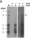
Fig. 2. (A) Immunoaffinity purification of BEV mRNAs with a cap-specific mAb. Total cytoplasmic RNA extracted from BEV-infected cells was incubated with protein G–Sepharose beads, coupled with either the m7G-cross-reactive mAb H20 (αCap) or, as a negative control, the isotype-matched mAb R78 (αTcR). RNA, bound to the mAb-coupled beads, was eluted, separated in denaturing 1% agarose gels and hybridized to a radiolabeled oligonucleotide probe (294; Table I) complimentary to the 3′ end of the genome. Untreated intracellular BEV RNAs served as a marker (M). Fluorographic exposures of 16 (left) and 192 h (right) are shown. (B) Xrn1p treatment of BEV RNAs. Total cytoplasmic RNA extracted from BEV-infected cells was incubated with purified Xrn1p at 37°C for 30 min, in either the presence (+) or absence (–) of EDTA. Samples were analyzed in parallel in non-denaturing ethidium bromide-stained 1% agarose gels (right hand panel), or in denaturing formaldehyde–1% agarose gels by hybridization to radiolabeled oligonucleotide 294 (left hand panel) or the equine 18S rRNA-specific oligonucleotide 1723 (middle panel). The locations of the BEV mRNAs and rRNAs are indicated.
None
Fig. 3. RLM-RACE analysis of the BEV genome and sg mRNAs. (A) RLM-RACE products obtained for the purified viral genome (1) and for sg mRNAs 2–5 were separated in 2% agarose gels. (B) Purification of BEV genomic RNA. Tissue culture supernatant of BEV-infected Ederm cells was harvested and cleared by low speed centrifugation. Subsequently, virions were pelleted through a 10% (w/v) sucrose cushion. Genomic RNA was extracted and analyzed by RNA hybridization with oligonucleotide 294 (V); intracellular BEV mRNAs (IC) served as a marker.
None
Fig. 4. Primer extension and RLM-RACE analysis of mRNA 1 and of genomic virion RNA. Left and middle panel: BEV genomic RNA, extracted from pelleted virions, and intracellular mRNA 1 were subjected to RLM-RACE. The resulting RT–PCR products were cloned and sequenced with oligonucleotide 1407 as a primer. Reaction mixtures were analyzed in 6% urea–polyacrylamide gels. Arrows indicate the 5′-most BEV nucleotide fused to the adaptor. The nucleotide sequence is presented, with adaptor-derived residues shaded in gray. Right panel: primer extension analysis of mRNA 1 was performed with oligonucleotide primer 1407. The 1407-primed dideoxy-sequencing samples of pDI-1000, a cloned full-length cDNA copy of the BEV DI-1000 RNA (Snijder et al., 1991), served as a molecular weight marker. The DI-1000 sequence, corresponding to the 5′ terminus of the BEV genome, is presented; the arrow indicates the run-off product.
None
Fig. 5. Primer extension and RLM-RACE analysis of BEV sg RNAs 2–5. RLM-RACE products obtained for mRNAs 2–5 (see Figure 3) were subjected to sequence analysis with oligonucleotide primers 1403, 1404, 1405 and 1406, respectively. In addition, primer extension analysis was performed with the respective oligonucleotides on mRNAs 2–4. Sequence reaction mixtures and primer extension products were run alongside in 6% urea–polyacrylamide gels. Arrows indicate the 5′-most residues of each mRNA as predicted by primer extension and/or RLM-RACE. The nucleotide sequence is presented, with adaptor- derived residues shaded in gray.
None
Fig. 6. Comparison of the 5′ termini of the BEV genome and mRNAs 3–5. Upper panel: alignment of the 5′ termini. Residues identical in three of the four sequences are shown in bold. Initiation codons of the M, HE and N genes are underlined. Lower panel: alignments of the 5′ termini from the BEV genome and from mRNAs 3–5 with the predicted 5′ end of DI-1000 (Snijder et al., 1991) and with the IGRs between the S and M genes (2/3 IGR), the M and HE genes (3/4 IGR) and the HE and N genes (4/5 IGR), respectively. The extended TRSs are shown in bold. The positions of the regions on the BEV genome (in nucleotides) are given on the right. Termination codons of the S and M genes are boxed; initiation codons of the genes for M, HE and N are underlined.
None
Fig. 7. Hybridization analysis of the 5′ terminus of mRNA 2. Upper panel: total cytoplasmic RNA, extracted from BEV-infected cells, was separated in denaturing formaldehyde–1% agarose gels and hybridized to the indicated radiolabeled oligonucleotide probes. Hybridization with probes 294 and 1746 was performed at 5°C below the Tm. Hybridization with probe 1553 was performed at the indicated temperatures. Lower panel: nucleotide sequence comparison of the mRNA 2 RLM-RACE product, the corresponding ORF1b region and the 5′ terminus of the BEV genome. Asterisks indicate identical residues. Also given are the sequences of the oligonucleotide probes. Mismatches with the BEV 5′ terminus or the ORF1b sequence are italicized. Nucleotide positions on the viral genome are indicated.
None
Fig. 8. (A) Nucleotide sequence comparison of the 5′ terminus of the BEV genome and the ORF1b fusion region (residues 21 118–21 161). The arrowhead indicates the mRNA 2 leader–body fusion site, comprising residues G21 133–U21 136 (italicized). As illustrated schematically, the residues upstream of the fusion site can adopt a hairpin structure. Asterisks indicate residues identical between ORF1b and the 5′ terminus of the BEV genome. Shown below is a comparison of the corresponding regions in PoTV. At present, only a partial sequence of the 5′ end of the PoTV genome is available. (B) Schematic hypothetical model for the discontinuous synthesis of BEV mRNA 2 and for the non-discontinuous transcription of mRNAs 3–5. For the latter RNAs, the TRSs on the (+)strand template (indicated in red) function as termination signals for (–)strand synthesis. The resulting sg (–)strands act in turn as templates for (+)strand sg RNA synthesis, with the anti-TRSs on the (–)strand template now functioning as transcription initiation signals. The viral RdRp is indicated as a gray circle. For the synthesis of mRNA 2, the stem–loop structure causes attenuation of (–)strand synthesis. Subsequently, a template switch occurs, during which the transcription complex is transferred to the 5′ end of the BEV genome, assisted by base pairing between ORF1b-derived sequences in the nascent (–) strand RNA and 5′-NTR sequences (indicated in blue). RNA synthesis resumes, resulting in the production of a transcription-competent sg RNA species, carrying a non-contiguous, genome-derived anti-TRS.

Similar articles

Cited by

References

    1. Baric R.S. and Yount,B. (2000) Subgenomic negative-strand RNA function during mouse hepatitis virus infection. J. Virol., 74, 4039–4046. - PMC - PubMed
    1. Baric R.S., Stohlman,S.A. and Lai,M.M. (1983) Characterization of replicative intermediate RNA of mouse hepatitis virus: presence of leader RNA sequences on nascent chains. J. Virol., 48, 633–640. - PMC - PubMed
    1. Baric R.S., Stohlman,S.A., Razavi,M.K. and Lai,M.M. (1985) Characterization of leader-related small RNAs in coronavirus-infected cells: further evidence for leader-primed mechanism of transcription. Virus Res., 3, 19–33. - PMC - PubMed
    1. Baric R.S., Shieh,C.K., Stohlman,S.A. and Lai,M.M. (1987) Analysis of intracellular small RNAs of mouse hepatitis virus: evidence for discontinuous transcription. Virology, 156, 342–354. - PMC - PubMed
    1. Bochnig P., Reuter,R., Bringmann,P. and Luhrmann,R. (1987) A monoclonal antibody against 2,2,7-trimethylguanosine that reacts with intact, class U, small nuclear ribonucleoproteins as well as with 7-methylguanosine-capped RNAs. Eur. J. Biochem., 168, 461–467. - PubMed