

Review
doi: 10.1136/mp.55.6.337.
Demystified... Molecular pathology in oncology
Affiliations
- PMID: 12456768
- PMCID: PMC1187267
- DOI: 10.1136/mp.55.6.337
Item in Clipboard
Review
Demystified... Molecular pathology in oncology
Mol Pathol.
2002 Dec.
Abstract
In the past 10 years, molecular biology has found major applications in pathology, particularly in oncology. This has been a field of enormous expansion, where pure science has found a place in clinical practice and is now of everyday use in any academic unit. This demystified review will discuss the techniques used in molecular pathology and then provide examples of how these can be used in oncology.
Figures

A schematic view of Southern blotting. The diagram shows how native DNA is cleaved, the restriction fragments are separated by electrophoresis, “blotted”, and then labelled with probe by hybridisation. Artwork courtesy of Mrs S McGrory, Department of Medical Illustration, Birmingham Heartlands Hospital, UK.

An example of western blot analysis of expression of the Epstein-Barr virus encoded latent membrane protein 1 (LMP-1) in an inducible system. The Hodgkin’s disease cell line, L428, was transfected with the LMP-1 gene and expression was induced by the addition of 6 mmol/litre cadmium chloride. Each lane represents increases in induction time, ranging from time zero (lane 1), through to 24 hours (lane 6). Figure courtesy of Dr K Baumforth, School of Health Sciences, University of Wolverhampton, UK.

A schematic illustration of fluorescent in situ hybridisation, showing how metaphase and interphase chromosomal structures are localised. The top part of the diagram shows centromeric labelling in both types of preparation and below that whole chromosomes are labelled. Next, short arm deletion is depicted, and at the bottom there is an illustration of the use of two gene probes in combination. Artwork courtesy of Mrs S McGrory, Department of Medical Illustration, Birmingham Heartlands Hospital, UK.
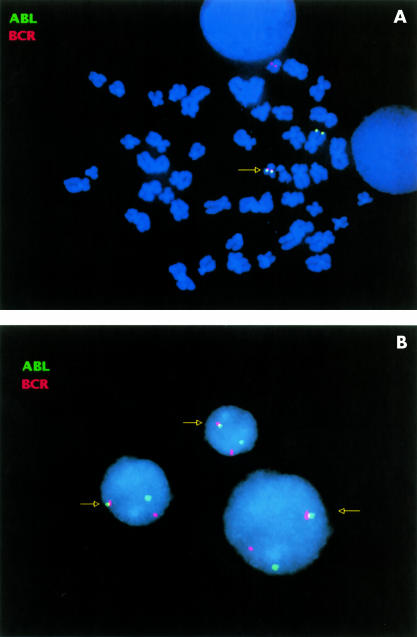
Fluorescent in situ hybridisation demonstrates BCR/ABL gene fusion in chronic myeloid leukaemia. (A) Metaphase: a BCR probe (red signal) hybridises to a complementary sequence on the normal chromosome 22 and on the der(22)t(9;22) translocation, known as the Philadelphia (Ph1) chromosome; arrowed). An ABL probe (green signal) hybridises to the normal chromosome 9 and the derived 22 (arrowed). (B) Interphase: for each informative interphase, a BCR probe (red signal) hybridises to a complementary sequence on normal chromosome 22 and on the der(22)t(9;22) Ph1 chromosome (arrowed). An ABL probe (green signal) hybridises to the normal chromosome 22 and to the derived 22 (arrowed). Note that with this probe set, a single fusion event is usually seen as a result of BCR/ABL fusion on the derivative Ph1 chromosome. This fusion event usually leads to a fluorescence colour change (red/green to yellow).

A depiction of the solution phase polymerase chain reaction, showing the amplification sequence induced by denaturation, annealing, and extension, followed by repeated cycles. Artwork by courtesy of Mrs S McGrory, Department of Medical Illustration, Birmingham Heartlands Hospital, UK.
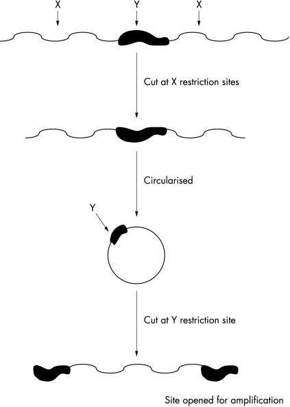
The inverse polymerase chain reaction, showing how unknown DNA sequences can be detected by selective cutting by enzymes, then circularised and “opened” again for analysis. Artwork courtesy of Mrs S McGrory, Department of Medical Illustration, Birmingham Heartlands Hospital, UK.

The sequence for in cell amplification. The cells or section undergo in situ amplification, eventually leading to indirect visualisation of the target nucleic acid. Artwork courtesy of Mrs S McGrory, Department of Medical Illustration, Birmingham Hospital, UK.

Gene sequence corresponding to phospholipase D2 (hPLD2). The different coloured peaks represent the different nucleotides. This enzyme is one of those responsible for the hydrolysis of phosphatidylcholine to give the signalling molecule phosphatidate. It has been shown to be upregulated in cancer cells. Plate courtesy of Dr D Power, Cancer Research Institute, Birmingham University, UK.

A typical polymerase chain reaction gel, showing strict DNA bands for the immunoglobulin heavy chain gene (IgH) rearrangement in a series of chronic lymphocytic leukaemias (CLL). The band on the far right is a molecular weight marker, the next two are negative controls, and the six to the left are from CLL cases with distinct bands, showing the IgH rearrangement. Gel courtesy of Mrs J Starczynski, Birmingham Heartlands Hospital, UK.

A polymerase chain reaction gel, showing, on the right, a molecular weight marker and, in the lane to the left of it, a band representing a cell line known to posses the t(14;18) translocation. Further to the left, there is a series of bands from several cases of follicular, follicle centre lymphoma, all with the 14;18 band. Gel courtesy of Mrs J Starczynski, Birmingham Hospital, UK.
References
-
- Bradley J, Johnson D, Rubenstein D, eds. Lecture notes on molecular medicine. Oxford: Blackwell Science, 1995.
-
- Crocker J, ed. Molecular biology in histopathology. Chichester: John Wiley and Sons, 1994.
-
- Latchman DS, ed. Basic molecular and cell biology. London: BMJ Publishing Group, 1997.
Publication types
MeSH terms
LinkOut - more resources
Full Text Sources
Miscellaneous