Review
. 2002 Dec;66(4):702-38, table of contents.
doi: 10.1128/MMBR.66.4.702-738.2002.

Biochemistry and comparative genomics of SxxK superfamily acyltransferases offer a clue to the mycobacterial paradox: presence of penicillin-susceptible target proteins versus lack of efficiency of penicillin as therapeutic agent

Affiliations
Review

Biochemistry and comparative genomics of SxxK superfamily acyltransferases offer a clue to the mycobacterial paradox: presence of penicillin-susceptible target proteins versus lack of efficiency of penicillin as therapeutic agent

Colette Goffin et al. Microbiol Mol Biol Rev. 2002 Dec.

Abstract

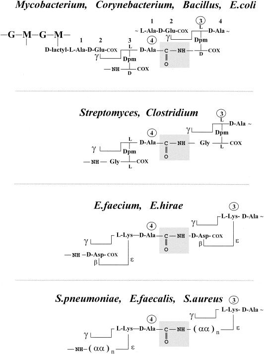
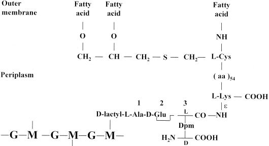
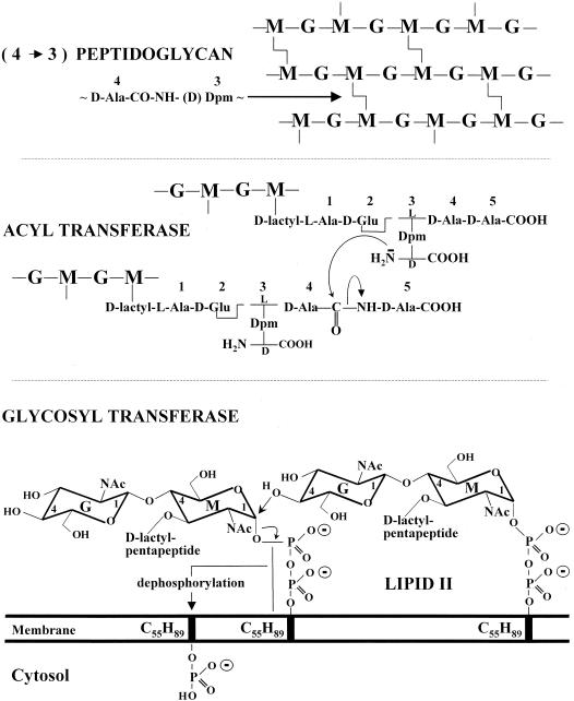
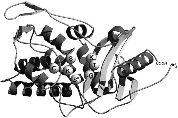
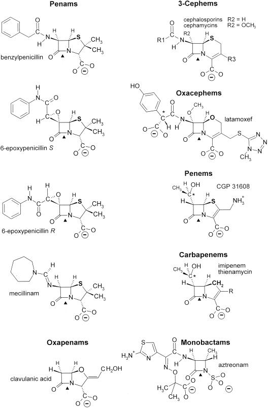
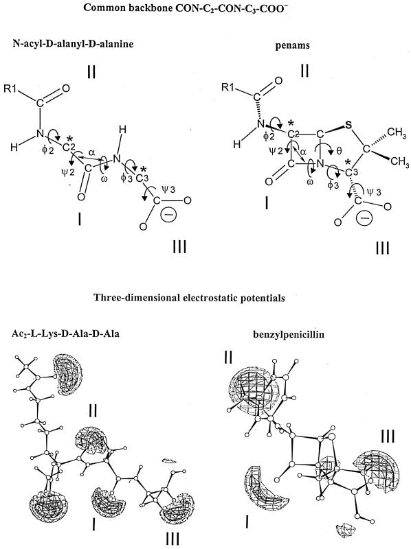
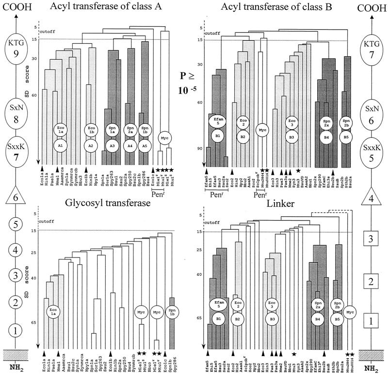
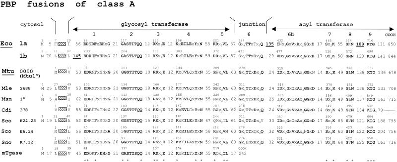
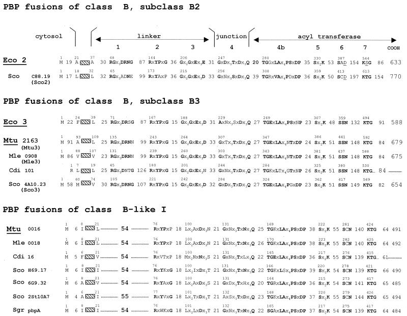
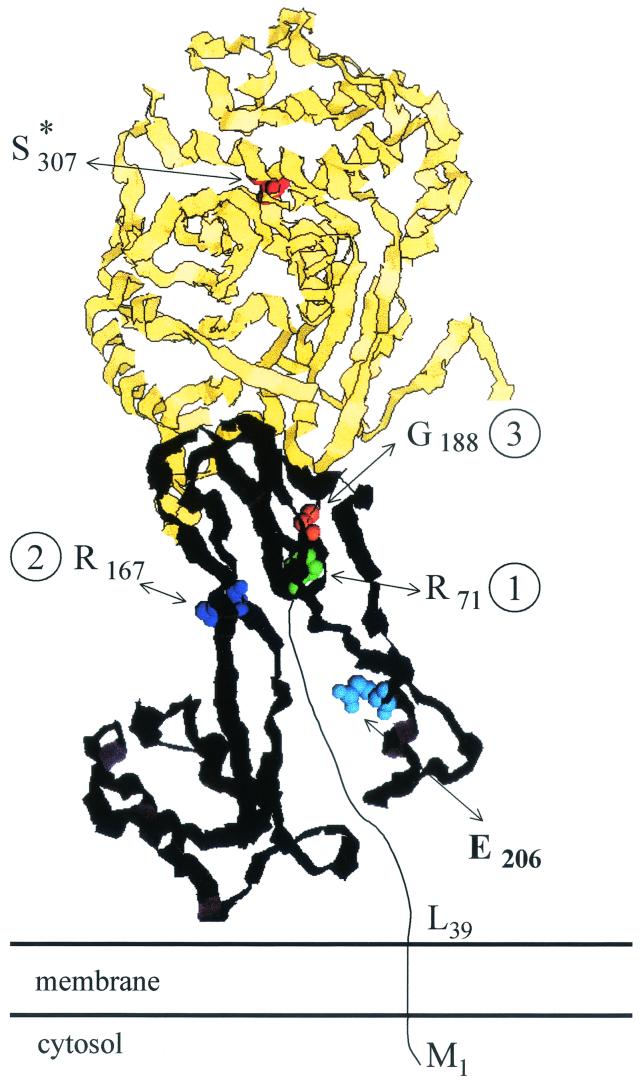
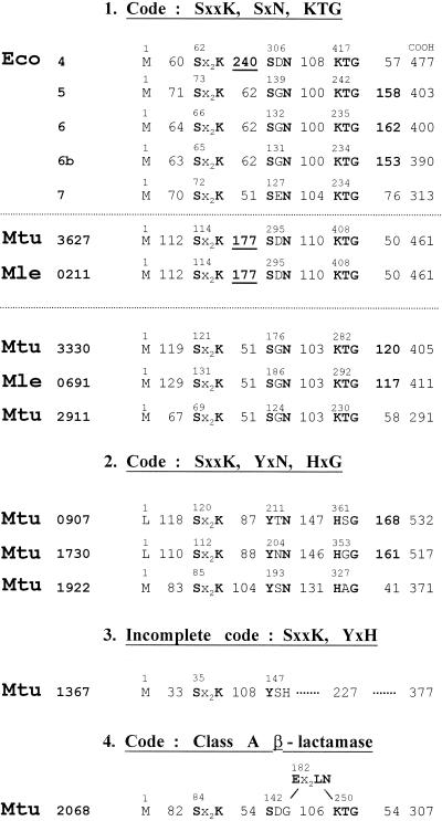
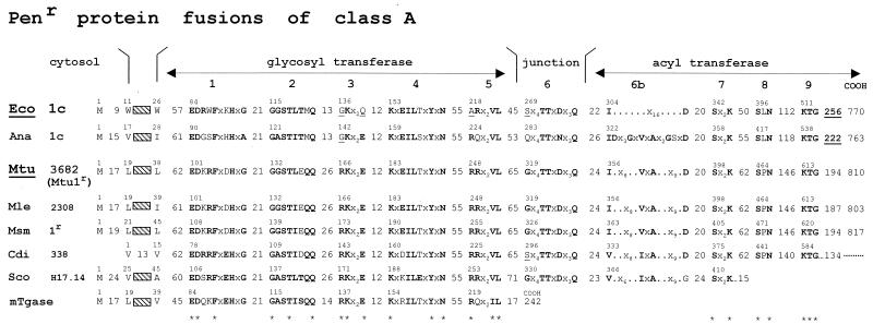
The bacterial acyltransferases of the SxxK superfamily vary enormously in sequence and function, with conservation of particular amino acid groups and all-alpha and alpha/beta folds. They occur as independent entities (free-standing polypeptides) and as modules linked to other polypeptides (protein fusions). They can be classified into three groups. The group I SxxK D,D-acyltransferases are ubiquitous in the bacterial world. They invariably bear the motifs SxxK, SxN(D), and KT(S)G. Anchored in the plasma membrane with the bulk of the polypeptide chain exposed on the outer face of it, they are implicated in the synthesis of wall peptidoglycans of the most frequently encountered (4-->3) type. They are inactivated by penicillin and other beta-lactam antibiotics acting as suicide carbonyl donors in the form of penicillin-binding proteins (PBPs). They are components of a morphogenetic apparatus which, as a whole, controls multiple parameters such as shape and size and allows the bacterial cells to enlarge and duplicate their particular pattern. Class A PBP fusions comprise a glycosyltransferase module fused to an SxxK acyltransferase of class A. Class B PBP fusions comprise a linker, i.e., protein recognition, module fused to an SxxK acyltransferase of class B. They ensure the remodeling of the (4-->3) peptidoglycans in a cell cycle-dependent manner. The free-standing PBPs hydrolyze D,D peptide bonds. The group II SxxK acyltransferases frequently have a partially modified bar code, but the SxxK motif is invariant. They react with penicillin in various ways and illustrate the great plasticity of the catalytic centers. The secreted free-standing PBPs, the serine beta-lactamases, and the penicillin sensors of several penicillin sensory transducers help the D,D-acyltransferases of group I escape penicillin action. The group III SxxK acyltransferases are indistinguishable from the PBP fusion proteins of group I in motifs and membrane topology, but they resist penicillin. They are referred to as Pen(r) protein fusions. Plausible hypotheses are put forward on the roles that the Pen(r) protein fusions, acting as L,D-acyltransferases, may play in the (3-->3) peptidoglycan-synthesizing molecular machines. Shifting the wall peptidoglycan from the (4-->3) type to the (3-->3) type could help Mycobacterium tuberculosis and Mycobacterium leprae survive by making them penicillin resistant.


Figures

FIG. 1.
FIG. 1.
(4→3) peptidoglycans. d-Alanyl-diaminoacyl interpeptide linkages and cross-bridges (boxed) between l-Ala-γ-d-Glu-(l)-diaminoacyl-d-alanine stem peptides. The diamino acid residues are meso-diaminopimelic acid (Dpm), l, l-diaminopimelic acid, or l-lysine. The stem peptides are unsubstituted at position 3 in Mycobacterium, Corynebacterium, and Bacillus spp. and E. coli. They are substituted at position 3 by one or several additional amino acid residues (αα) in the other organisms shown. G, N-acetylglucosamine; M, N-acetylmuramic acid (see Fig. 4); COX = COOH or CONH2. In S. pneumoniae, the cross-bridges are Nɛ-(l-alanyl-l-alanyl)- or (l-alanyl-l-seryl)-l-lysine. In S. aureus, the cross-bridges comprise five glycine residues or three glycine and two l-serine residues.
FIG. 2.
FIG. 2.
(3→3) peptidoglycans. Diaminoacyl-diaminoacyl interpeptide linkages and cross-bridges (boxed) between l-Ala-γ-d-Glu-(l)-diaminoacyl-d-alanine stem peptides. The stem peptides are unsubstituted at position 3 in Mycobacterium spp. and E. coli. They are substituted at position 3 by Gly in Streptomyces spp. and Clostridium perfringens and by β-d-Asp in E. faecium
FIG. 3.
FIG. 3.
Braun's lipoprotein-peptidoglycan complex of E. coli.
FIG. 4.
FIG. 4.
Lipid II precursor (bottom) and polymeric (4→3) peptidoglycan (top) of E. coli. Reactions catalyzed by the glycosyl- and acyltransferases.
FIG. 5.
FIG. 5.
Organization of the division cell wall (dcw) gene clusters of M. tuberculosis (Mtu), M. leprae (Mle), Streptomyces coelicolor (Sco), and E. coli (Eco). All the coding sequences are on the same DNA strands. Open arrows, mtu3, mle3, sco3, and eco3 (synonymous to ftsI), coding for cell septation SxxK PBP fusions. Shaded arrows, with fts standing for the filamentous phenotype of thermosensitive E. coli mutants, ftsW, ftsQ, ftsA, and ftsZ code for cell septation-related, non-penicillin-binding proteins. Genes murE, murF, murX (synonymous to mraY), murD, murG, and murC code for enzymes of the lipid II-synthesizing pathway. In S. coelicolor, murC (shaded rectangle) is not in the dcw cluster. Stippled grey arrows, genes of a dcw cluster having no homologues in the other clusters. In M. tuberculosis, the genes inserted between mtu3 and murE probably code for a glycine-rich protein of repetitive structure (Rv2162c), a protein of the lincomycin-synthesizing pathway (Rv2161c), and proteins of unknown function (Rv2160c and Rv2159c). A gene homologous to E. coli ddl (coding for the ligase which catalyzes the formation of d-alanyl-d-alanine) is located elsewhere in the chromosomes of M. tuberculosis, M. leprae, and S. coelicolor. E. coli ftsA codes for a cell division, actin-like ATPase. Solid, dashed, and dotted lines connecting the dcw genes indicate the extents, in decreasing order, of similarity between homologous genes. The E. coli cell septation genes mraZ, mraW, and ftsL (not shown) are upstream from eco3.
FIG. 6.
FIG. 6.
Acyl transfer reactions. Formation of (4→3) peptidoglycan interpeptide linkages by penicillin-susceptible d,d-N-acyl-d-alanyl-d-alanine transpeptidases (reaction I). Formation of (3→3) peptidoglycan interpeptide linkages by penicillin-resistant l,d-N-acyl-l-diaminoacyl-d-dipeptidyl transpeptidases (reaction II) and l,d-N-acyl-l-diaminoacyl-d-alanine transpeptidases (reaction III). Hydrolysis of stem pentapeptides into stem tetrapeptides by penicillin-resistant d,d-N-acyl-d-alanyl-d-alanine carboxypeptidases (reaction IIIa).
FIG. 7.
FIG. 7.
Basic polypeptide fold of the acyltransferases of the SxxK superfamily and spatial disposition of the three catalytic center-defining amino acid groupings. The structure shown is that of the 262-amino-acid dd-transpeptidase-PBP of Streptomyces sp. strain K15. The SxxK acyltransferases comprise an all-α domain (left side) and an α/β domain (right side). The invariant motif 1, S*xxK, where S* is the essential serine nucleophile, forms the amino-terminal turn of helix α2 of the all-α domain. Motif 2, most often SxN or SxD (here SxC), is on a loop connecting two helices of the all-α domain. Motif 3, most often KTG or KSG, is on strand β3 of the α/β domain. The structure was built with Molscript (130) and Raster3D (155). Illustration courtesy of Paulette Charlier, Eveline Fonzé, Michaël Delmarcelle, and André Piette, Center for Protein Engineering.
FIG. 8.
FIG. 8.
β-Lactam antibiotic family. Only 6-epoxypenicillin S is active. *, d-configured carbon atom. (“Mecillinam” is another name for amdinocillin.)
FIG. 9.
FIG. 9.
Common backbone of extended N-acyl-d-alanyl-d-alanine peptides and penams (top) and ab initio electrostatic potentials of bisacetyl-l-lysyl-d-alanyl-d-alanine and benzylpenicillin (bottom) optimized at level AM1 (the terminal carboxylates are protonated). The electrostatic negative wells I, II, and II are shown at levels of −40 kcal (solid contours) and −30 kcal (dotted contours). They are coplanar. The negative wells generated by the acetyl substituents of the α- and ɛ-amino groups of the l-lysine residue of bisacetyl-l-lysyl-d-alanyl-d-alanine are above and below the plane, respectively. The negative well of small amplitude seen between well I and well III of benzylpenicillin is due to the pair of free electrons of the nitrogen atom of the azetidinone ring. Illustration courtesy of Georges Dive, Center for Protein Engineering.
FIG. 10.
FIG. 10.
Hierarchical distribution of the SxxK PBP fusions and Penr protein fusions of classes A and B. The occurrence of class-specific motifs 1 to 9 (class A) and 1 to 7 (class B) along the polypeptide chains is shown. Adapted from reference . Scores (vertical axis of the dendrograms) are the standard deviation values above that expected from a run of 100 randomized pairs of sequences with the same amino acid composition as the two sequences under comparison. The protein identifiers (bottom of the dendrograms) are defined in Table 1. The clusters are labeled by two circles, one of which defines a particular subclass (A1 to A5 and B1 to B5) and the other the prototypic protein. Solid arrowheads help identify proteins that are discussed in the text. Solid stars help identify the mycobacterial proteins. The Penr acyltransferase modules are underlined.
FIG. 11.
FIG. 11.
Class A PBP fusions. Bar code, motifs 1 to 9 and inserts (underlined). For protein identifiers, see Table 1. mTgase, free-standing transglycosylase of E. coli.
FIG. 12.
FIG. 12.
Class B PBP fusions. Bar code, motifs 1 to 7. For protein identifiers, see Table 1.
FIG. 13.
FIG. 13.
Schematic structure of the membrane-bound SxxK subclass B3 PBP fusion protein Eco3 of E. coli. Spatial disposition along the polypeptide chain of the essential S*307 of the SxxK motif of the acyltransferase module (see Fig. 7) and of motifs 1, 2, and 3 (identified by the residue at the amino side of the sequences) and segment E206 to V217 of the linker module. The acyltransferase module is in yellow, with S*307 of the SxxK motif in red. The linker module is in black with motif 1 (R71 to G79) in green, motif 2 (R167 to G172) in dark blue, motif 3 (G188 to D197) in orange, and segment E206 to V217 in light blue. Motifs 1, 2, and 3 and other peptide segments form the core of the linker module in interaction with a noncatalytic groove of the acyltransferase module. The peptide segment M1 to R71 is of unknown structure. Adapted from reference . Illustration courtesy of Robert Brasseur, Faculté Universitaire des Sciences Agronomiques, Gembloux.
FIG. 14.
FIG. 14.
Occurrence of catalytic center-defining motifs along the sequences of free-standing SxxK polypeptides of E. coli, M. tuberculosis, and M. leprae. Polypeptides of group 1 bear the bar codes SxxK, SxN, and KTG. They are auxiliary cell cycle PBPs in E. coli. Inserts are underlined. The M. tuberculosis polypeptides of groups 2 and 3 have no equivalent in E. coli. Their bar codes are modified or incomplete. It is likely that they are not implicated in wall peptidoglycan metabolism. The M. tuberculosis β-lactamase of class A (group 4) has a class-specific Ex2LN motif. E. coli can produce two β-lactamases, one of which (plasmid coded) is of class A (Fig. 15).
FIG. 15.
FIG. 15.
Acyltransferases of the SxxK superfamily of diverse functions not related to wall peptidoglycan metabolism. The proteins are free-standing polypeptides except BlaR of B. licheniformis and DAP of O. anthropi, which are protein fusions. The SxxK acyltransferase module forms the carboxy-terminal domain of BlaR and the amino-terminal domain of DAP. The three-dimensional structures of the proteins marked with an asterisk are known. Motif 1, SxxK, is invariant. Motifs 2 and/or 3 are modified except in PBP R39. Amino acid changes and inserts are underlined. Motif 2 of BlaR is ambiguous.
FIG. 16.
FIG. 16.
Class B Penr protein fusions. Bar code, motifs 1 to 7 and inserts (framed). For protein identifiers, see Table 1.
FIG. 17.
FIG. 17.
Class A Penr protein fusions. Bar code, motifs 1 to 9 and extensions (underlined). For protein identifiers, see Table 1. mTgase, free-standing glycosyltransferase of E. coli.
FIG. 18.
FIG. 18.
β-Lactam antibiotics with side chains terminating in a d-configured NH2-C*HR-COOH grouping.

References

    1. Adam, M., C. Fraipont, N. Rhazi, M. Nguyen-Distèche, B. Lakaye, J. M. Frère, B. Devreese, J. Van Beeumen, Y. van Heijenoort, J. van Heijenoort, and J. M. Ghuysen. 1997. The bimodular G57-V577 polypeptide chain of the class B penicillin-binding protein 3 of Escherichia coli catalyzes peptide bond formation from thiolesters and does not catalyze glycan chain polymerization from lipid II intermediate. J. Bacteriol. 179:6005-6009. - PMC - PubMed
    1. Addinall, S. G., and J. Lutkenhaus. 1996. FtsZ-spirals and -arcs determine the shape of the invaginating septa in some mutants of Escherichia coli. Mol. Microbiol. 22:231-237. - PubMed
    1. Altschul, S. F., and W. Gish. 1996. Local alignment statistics. Methods Enzymol. 266:460-480. - PubMed
    1. Altschul, S. F., T. L. Madden, A. A. Schaffer, J. Zhang, Z. Zhang, W. Miller, and D. J. Lipman. 1997. Gapped BLAST and PSI-BLAST: a new generation of protein database search programs. Nucleic Acids Res. 25:3389-3402. - PMC - PubMed
    1. Ambler, R. P., A. F. W. Coulson, J. M. Frère, J. M. Ghuysen, B. Joris, M. Forsman, R. C. Levesque, G. Tiraby, and S. G. Waley. 1991. A standard numbering scheme for the class A β-lactamases. Biochem. J. 276:269-272. - PMC - PubMed

Publication types

MeSH terms