. 2002 Dec 1;545(2):355-73.
doi: 10.1113/jphysiol.2002.032151.

Gating properties of GIRK channels activated by Galpha(o)- and Galpha(i)-coupled muscarinic m2 receptors in Xenopus oocytes: the role of receptor precoupling in RGS modulation

Affiliations

Gating properties of GIRK channels activated by Galpha(o)- and Galpha(i)-coupled muscarinic m2 receptors in Xenopus oocytes: the role of receptor precoupling in RGS modulation

Qingli Zhang et al. J Physiol. .

Abstract

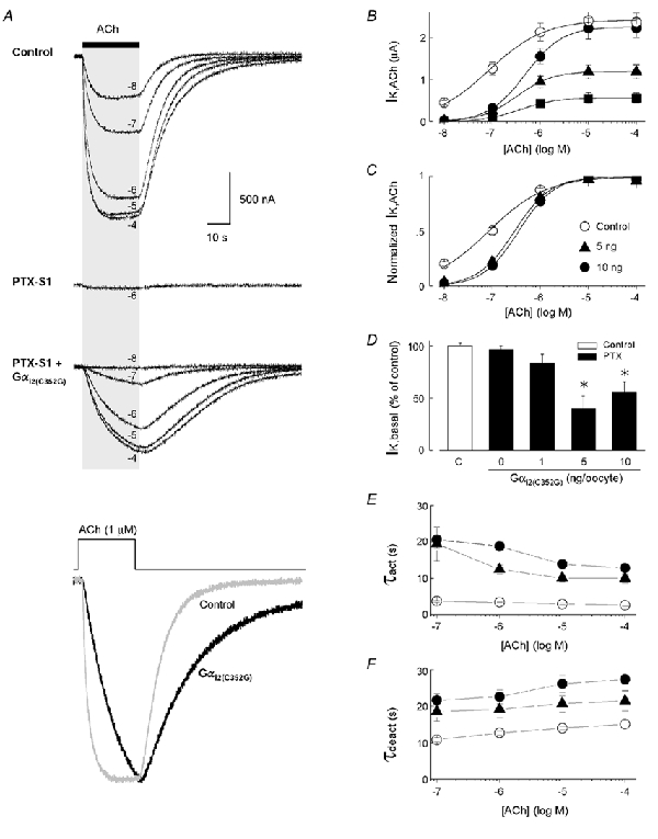
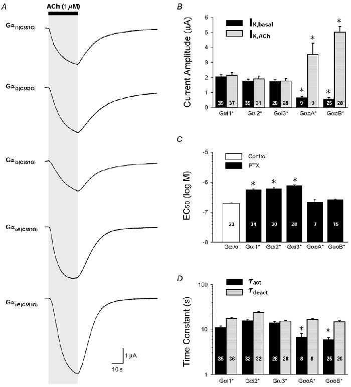
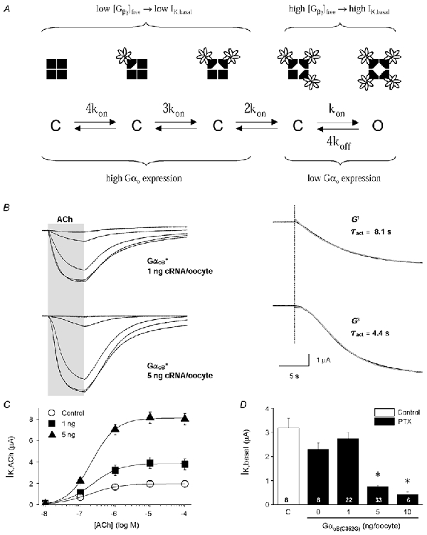
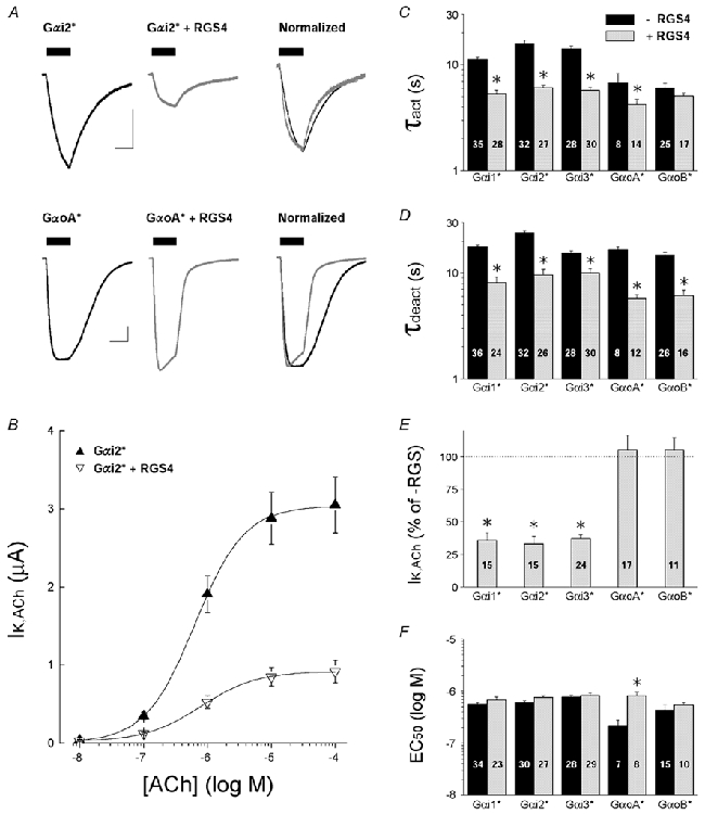
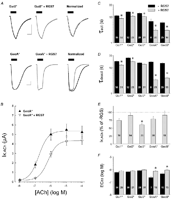
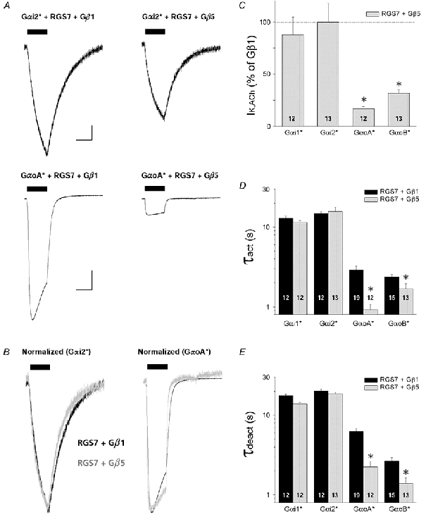
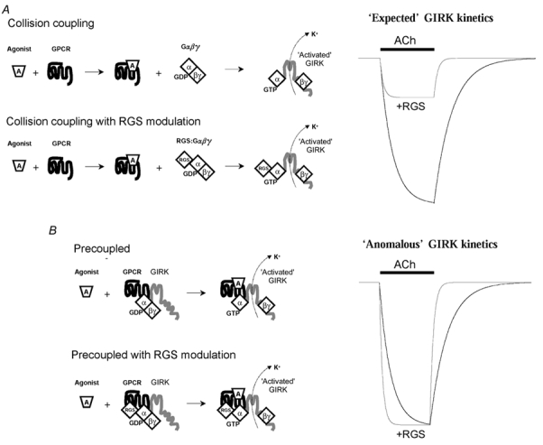
'Regulators of G protein Signalling' (RGSs) accelerate the activation and deactivation kinetics of G protein-gated inwardly rectifying K(+) (GIRK) channels. In an apparent paradox, RGSs do not reduce steady-state GIRK current amplitudes as expected from the accelerated rate of deactivation when reconstituted in Xenopus oocytes. We present evidence here that this kinetic anomaly is dependent on the degree of G protein-coupled receptor (GPCR) precoupling, which varies with different Galpha(i/o)-RGS complexes. The gating properties of GIRK channels (Kir3.1/Kir3.2a) activated by muscarinic m2 receptors at varying levels of G protein expression were examined with or without the co-expression of either RGS4 or RGS7 in Xenopus oocytes. Different levels of specific m2 receptor-Galpha coupling were established by uncoupling endogenous pertussis toxin (PTX)-sensitive Galpha(i/o) subunits with PTX, while expressing varying amounts of a single PTX-insensitive subunit (Galpha(i1(C351G)), Galpha(i2(C352G)), Galpha(i3(C351G)), Galpha(oA(C351G)), or Galpha(oB(C351G))). Co-expression of each of the PTX-insensitive Galpha(i/o) subunits rescued acetylcholine (ACh)-elicited GIRK currents (I(K,ACh)) in a concentration-dependent manner, with Galpha(o) isoforms being more effective than Galpha(i) isoforms. Receptor-independent 'basal' GIRK currents (I(K,basal)) were reduced with increasing expression of PTX-insensitive Galpha subunits and were accompanied by a parallel rise in I(K,ACh). These effects together are indicative of increased Gbetagamma scavenging by the expressed Galpha subunit and the subsequent formation of functionally coupled m2 receptor-G protein heterotrimers (Galpha((GDP))betagamma). Co-expression of RGS4 accelerated all the PTX-insensitive Galpha(i/o)-coupled GIRK currents to a similar extent, yet reduced I(K,ACh) amplitudes 60-90 % under conditions of low Galpha(i/o) coupling. Kinetic analysis indicated the RGS4-dependent reduction in steady-state GIRK current was fully explained by the accelerated deactivation rate. Thus kinetic inconsistencies associated with RGS4-accelerated GIRK currents occur at a critical threshold of G protein coupling. In contrast to RGS4, RGS7 selectively accelerated Galpha(o)-coupled GIRK currents. Co-expression of Gbeta5, in addition to enhancing the kinetic effects of RGS7, caused a significant reduction (70-85 %) in steady-state GIRK currents indicating RGS7-Gbeta5 complexes disrupt Galpha(o) coupling. Altogether these results provide further evidence for a GPCR-Galphabetagamma-GIRK signalling complex that is revealed by the modulatory affects of RGS proteins on GIRK channel gating. Our functional experiments demonstrate that the formation of this signalling complex is markedly dependent on the concentration and composition of G protein-RGS complexes.


Figures

Figure 1
Figure 1. Specific coupling of Gαi2(C352G) to m2 receptors and GIRK channel activation in Xenopus oocytes
A, typical ACh-evoked GIRK currents (IK,ACh) recorded from different oocytes from three separate experimental groups. Upper traces: IK,ACh from a ‘control’ oocyte expressing muscarinic m2 receptors and Kir3.1/Kir3.2a channel subunits, utilizing endogenous Gαi/o proteins for receptor activation. Middle trace: co-expression of PTX-S1 (1 ng cRNA/oocyte) effectively uncouples ACh-evoked GIRK currents utilizing oocyte Gαi/o proteins. Lower traces: expression of the PTX-insensitive Gαi2(C352G) subunit (5 ng cRNA) with PTX-S1 rescues m2 receptor-coupled GIRK currents. All GIRK currents were elicited by a 25 s application of different concentrations of ACh as indicated. Bottom traces: superimposed IK,ACh elicited by 1 μm ACh from the control oocyte (grey trace) and the Gαi2(C352G)-coupled oocyte (black trace). Peak amplitudes are normalized to illustrate the kinetic differences in the activation and deactivation time courses. B, ACh dose-response relations for GIRK activation via m2 receptors coupled to oocyte Gαi/o subunits (○, control) and Gαi2(C352G) at different levels of expression (▪ 1 ng, ▴ 5 ng, and • 10 ng cRNA/oocyte). C, ACh-dose-response curves from B normalized to maximal IK,ACh. D, comparison of receptor-independent basal GIRK currents (IK,basal) with varying levels of Gαi2(C352G) expression. IK,basal is expressed as the percentage change in the ‘control group’ mean value determined for each batch of oocytes. E, activation time constants (τact) and F, deactivation time constants (τdeact) for GIRK currents coupled to varying levels of Gαi2(C352G) expression and different concentrations of ACh. ○ control; ▴ 5 ng; and • 10 ng of Gαi2(C352G) cRNA/oocyte. Data in B-F represent the means ±s.e.m. from at least 3 batches of oocytes with the number of oocytes indicated. * P < 0.05.
Figure 2
Figure 2. Properties of ACh-evoked GIRK currents activated by m2 receptors coupled to five different Gαi/o subunits
A, typical ACh-evoked GIRK currents for each PTX-insensitive Gαi/o subunit examined (Gαi1(C351G), Gαi2(C352G), Gαi3(C351G), GαoA(C351G) and GαoB(C351G)). Currents were elicited by a 25 s application of 1 μm ACh. B, maximal amplitude of receptor-dependent GIRK currents (IK,ACh, grey bars) and receptor-independent GIRK currents (IK,basal, black bars) for each expressed PTX-insensitive Gα subunit (Gα*). Gα* expression was produced by 5 ng cRNA/oocyte. Maximal IK,ACh responses are to 10 μm ACh. C, EC50 values for control (open bar) and each PTX-insensitive Gα* subunit, derived from ACh dose-response relations for IK,ACh activation. D, activation (black bars) and deactivation time constants (grey bars) derived from exponential fits of the IK,ACh time course in response to rapid ACh application and washout. For B-D, data represent the means + s.e.m. from at least 3 batches of oocytes with the number of oocytes indicated (* P < 0.05). In B and D, statistical comparisons were with Gαi1(C351G), and in C comparisons were with oocyte Gαi/o coupling (open bar).
Figure 3
Figure 3. Biochemical analysis of PTX-insensitive Gαi/o protein levels in Xenopus oocytes
A, radiolabelling (upper panel) and Western blot analysis (lower panel) of oocytes injected with cRNAs encoding five different PTX-insensitive Gαi/o subunits (*) as described in Methods and Fig. 2. Oocytes were incubated for 3 days in OCM containing 0.5 mCi ml−1 [35S]Met/Cys. All groups were injected with cRNAs for the m2 receptor and GIRK channel subunits Kir3.1 and Kir3.2a (0.5 ng each/oocyte). PTX-insensitive Gαi/o cRNAs (5 ng/oocyte) were injected with PTX-S1 cRNA (1 ng/oocyte). Both endogenous and heterologously expressed Gα proteins were immunoprecipitated from the lysate equivalent of one oocyte using a ‘common’ Gα antibody, then separated by SDS-PAGE and transferred to a PVDF membrane for autoradiography and Western blotting. The 41 kDa band corresponding to Gαi/o proteins is indicated. B, quantitative analysis of Gαi/o proteins detected by radiolabelling and Western blot analysis. The 41 kDa band from the control group (no PTX-insensitive Gαi/o cRNA) served as an internal reference and was used to normalize the band intensity among the different experimental groups in each autoradiogram and Western blot. The Western blot results are the means + s.e.m. obtained from 3 independent experiments (separate batches of injected oocytes that were immunoprecipitated and immunostained as described in Methods). The radiolabelling data are the means + s.e.m. obtained from 2 of the Western blot experiments. * P < 0.05.
Figure 4
Figure 4. Activation kinetics of Gαo-coupled GIRK currents at different levels of GαoB(C352G) expression
A, kinetic scheme for GIRK channel activation via Gβγ binding to each of the four GIRK channel subunits. Four closed states are depicted with 0 to 3 bound Gβγ dimers. Channel opening (O) occurs with the binding of 4 Gβγ dimers. The level of free Gβγ dimers then determines the channel-bound state that precedes receptor activation. B, left panel: ACh-evoked GIRK currents from oocytes injected with either 1 or 5 ng GαoB(C351G) cRNA. Right panel: the activation phase for the 1 μm ACh-evoked GIRK currents are displayed at higher temporal resolution. Red lines represent non-linear fits to the data. A single exponential function (G1) best described the IK,ACh time course with low GαoB(C351G) expression (1 ng RNA/oocyte), having a time constant of 8.1 s. A third-order exponential function (G3) best described the activation time course at high GαoB(C351G) expression (5 ng RNA/oocyte), having a time constant of 4.4 s. C, ACh dose-response curves for m2 receptor-activated GIRK currents from control oocytes (○) and oocytes expressing GαoB(C351G) at two different levels (▪ 1 ng and ▴ 5 ng cRNA/oocyte). D, effects of GαoB(C351G) expression levels on receptor-independent GIRK channel activity (IK,basal). The open bar is from control oocyte responses, and black bars are from oocytes injected with varying amount of GαoB(C351G) cRNA and PTX-S1 cRNA (1 ng/oocyte). Data represent the means + s.e.m. from 4 batches of oocytes with the number of oocytes indicated.
Figure 5
Figure 5. Effects of RGS4 on GIRK currents coupled to specific PTX-insensitive Gαi/o subunits
All data are from oocytes injected with 5 ng Gα* cRNA/oocyte, with or without 10 ng RGS4 cRNA/oocyte. Endogenous PTX-sensitive Gαi/o subunits were inactivated by either PTX injection or PTX-S1 expression as described in Methods. Data are means and s.e.m.; * P < 0.05. A, representative ACh-evoked GIRK currents from oocytes expressing either Gαi2(C352G) (upper traces) or GαoA(C351G) (lower traces) with and without RGS4 coexpression. Scale bars indicate 1 μA and 10 s. B, ACh dose-response relations for m2 receptor-coupled GIRK currents from oocytes expressing Gαi2(C352G) in the absence (▴) or presence of RGS4 (▿). C, IK,ACh activation time constants (τact) from oocytes expressing PTX-insensitive Gαi/o subunits alone (black bars) or with co-expressed RGS4 (grey bars). Time constants were derived from a single exponential fit to the rising phase of IK,ACh elicited by 1 μm ACh. D, deactivation time constants (τdeact) derived from ACh-elicited GIRK currents recorded from oocytes expressing PTX-insensitive Gαi/o subunits alone (black bars) or with co-expressed RGS4 (grey bars). Time constants were derived from a single exponential fit to the decay of IK,ACh after rapid washout of 1 μm ACh. E, effects of RGS4 on maximal IK,ACh responses elicited by m2 receptors coupled to PTX-insensitive Gαi/o subunits. IK,ACh amplitudes (10 μm ACh) from RGS4-expressing oocytes are expressed as a percentage of the mean IK,ACh amplitude from oocytes expressing the PTX-insensitive Gαi/o subunit alone (RGS-). F, effects of RGS4 on the EC50 for ACh activation of GIRK currents coupled to m2 receptors and PTX-insensitive Gαi/o subunits. Black bars are without RGS4 expression (-RGS4), grey bars are with RGS4 co-expression (+RGS4).
Figure 6
Figure 6. Effects of RGS7 on GIRK currents coupled to specific PTX-insensitive Gαi/o subunits
All data are from oocytes injected with 5 ng Gα* cRNA/oocyte, with or without 10 ng RGS7 cRNA/oocyte. Endogenous PTX-sensitive Gαi/o subunits were inactivated by either PTX injection or PTX-S1 expression as described in Methods. Data are means and s.e.m.; * P < 0.05. A, representative ACh-evoked GIRK currents from oocytes expressing either Gαi2(C352G) (upper traces) or GαoA(C351G) (lower traces) with and without RGS7 coexpression. Scale bars indicate 1 μA and 10 s. B, ACh dose-response relations for m2 receptor-coupled GIRK currents from oocytes expressing Gαi2(C352G) in the absence (▴) or presence of RGS7 (▿). C, IK,ACh activation time constants (τact) from oocytes expressing PTX-insensitive Gαi/o subunits alone (black bars) or with co-expressed RGS7 (grey bars). Time constants are from GIRK currents evoked by 1 μm ACh. D, deactivation time constants (τdeact) from ACh-elicited GIRK currents recorded from oocytes expressing PTX-insensitive Gαi/o subunits alone (black bars) or with co-expressed RGS7 (grey bars). Time constants were derived after rapid washout of 1 μm ACh. E, effects of RGS7 on maximal IK,ACh responses elicited by m2 receptors coupled to PTX-insensitive Gαi/o subunits. IK,ACh amplitudes (10 μm ACh) from RGS4-expressing oocytes as a percentage of the mean IK,ACh amplitude from oocytes expressing the PTX-insensitive Gαi/o subunit alone (no RGS7). F, effects of RGS7 on the EC50 for ACh activation of GIRK currents coupled to m2 receptors and PTX-insensitive Gαi/o subunits. Black bars are without RGS7 expression, grey bars are with RGS7 co-expression.
Figure 7
Figure 7. Effects of Gβ5 on RGS7-accelerated GIRK currents coupled to specific PTX-insensitive Gαi/o subunits
All data are from oocytes injected with 5 ng Gα* cRNA/oocyte and 10 ng RGS7 cRNA/oocyte, with either 5 ng Gβ5 cRNA/oocyte or 5 ng Gβ1 cRNA/oocyte as a negative control. Oocytes also received Kir3.1 and Kir3.2a cRNA (0.5 ng/oocyte each), and m2 receptor cRNA (0.5 ng/oocyte), and endogenous PTX-sensitive Gαi/o subunits were inactivated by PTX-S1 expression (1.0 ng/oocyte). Data are means + s.e.m.; * P < 0.05. A, representative ACh-evoked GIRK currents from oocytes expressing RGS7 and Gαi2(C352G) with either Gβ1 or Gβ5 (upper traces), or GαoA(C351G) with either Gβ1 or Gβ5 (lower traces). Scale bars indicate 1 μA and 10 s. B, GIRK currents from A, normalized to peak amplitude and superimposed to highlight their temporal features (black traces, RGS7+Gβ1; grey traces, RGS7+Gβ5). C, Gβ5 selectively suppresses RGS7/ Gαo-coupled GIRK currents. IK,ACh amplitudes are expressed as a percentage of the mean IK,ACh amplitude from oocytes expressing the negative control Gβ1. D, GIRK activation time constants (τact) derived from oocytes expressing different PTX-insensitive Gαi/o subunits with either RGS7 + Gβ1 (black bars) or RGS7 + Gβ5 (grey bars). Time constants were derived from a single exponential fit to the rising phase of IK,ACh elicited by 1 μm ACh. E, deactivation time constants (τdeact) derived from ACh-elicited GIRK currents as in D. Time constants were derived from a single exponential fit to the decay of IK,ACh after rapid washout of 1 μm ACh.
Figure 8
Figure 8. RGS modulation of GPCR → GIRK signal transduction at varying levels of G protein coupling
A, collision coupling model (low Gα expression) and B precoupled model (high Gα expression). The precoupled model assumes a GPCR-G protein-RGS-GIRK channel complex displaying ‘anomalous’ kinetic behaviour that was characteristic of oocytes expressing Gαo subunits with either RGS4 or RGS7, together with co-expressed m2 receptors and GIRK channel Kir3.1/3.2a subunits.

Similar articles

Cited by

References

    1. Berman DM, Wilkie TM, Gilman AG. GAIP and RGS4 are GTPase-activating proteins for the Gi subfamily of G protein α subunits. Cell. 1996;86:445–452. - PubMed
    1. Blake BL, Wing MR, Zhou JY, Lei Q, Hillmann JR, Behe CI, Morris RA, Harden TK, Bayliss DA, Miller RJ, Siderovski DP. Gβ association and effector interaction selectivities of the divergent Gγ subunit Gγ13. Journal of Biological Chemistry. 2001;276:49267–49274. - PubMed
    1. Breitwieser GE, Szabo G. Mechanism of muscarinic receptor-induced K+ channel activation as revealed by hydrolysis-resistant GTP analogues. Journal of General Physiology. 1988;91:469–494. - PMC - PubMed
    1. Burgon PG, Lee WL, Nixon AB, Peralta EG, Casey PJ. Phosphorylation and nuclear translocation of a regulator of G protein signaling (RGS10) Journal of Biological Chemistry. 2001;276:32828–32834. - PubMed
    1. Chase DL, Patikoglou GA, Koelle MR. Two RGS proteins that inhibit Gαo and Gαq signaling in C. elegans neurons require a Gβ5-like subunit for function. Current Biology. 2001;11:222–231. - PubMed

Publication types

MeSH terms

LinkOut - more resources