. 2002 Dec 1;545(2):399-406.
doi: 10.1113/jphysiol.2002.021881.

Cytoskeletal disrupting agents prevent calmodulin kinase, IQ domain and voltage-dependent facilitation of L-type Ca2+ channels

Affiliations

Cytoskeletal disrupting agents prevent calmodulin kinase, IQ domain and voltage-dependent facilitation of L-type Ca2+ channels

Igor Dzhura et al. J Physiol. .

Erratum in

  • J Physiol. 2003 Feb 1;546(Pt 3):955

Retraction in

Abstract

A calmodulin (CaM) binding 'IQ' domain on the L-type Ca(2+) channel (LTCC) C terminus and calmodulin kinase II (CaMK) both signal increases in LTCC opening probability (P(o)) by shifting LTCCs into a gating mode (mode 2) with long openings through a process called facilitation. However, the mechanism whereby CaMK and the IQ domain are targeted to LTCCs is unknown. Endogenous CaMK is targeted to LTCCs in excised cell membrane patches because LTCC P(o) increased significantly in CaM-enriched (20 microM) bath solution and this effect was prevented by a specific CaMK inhibitory peptide, but not by an inactive control peptide. Pre-exposure of myocytes to the cytoskeletal disrupting agents nocodazole (microtubule specific) or cytochalasin D (microfilament specific) prevented the effects of CaM-dependent increases in P(o) of LTCCs in excised membrane patches. Neither cytochalasin D nor nocodazole altered the distribution of LTCC gating modes under basal conditions in on-cell mode or excised cell membrane patches, but each of these agents occluded the response of LTCCs to exogenous, constitutively active CaMK and to an IQ-mimetic peptide (IQmp). Cytochalasin D and nocodazole pretreatment also prevented LTCC facilitation that followed a cell membrane depolarizing prepulse. In contrast, cytochalasin D and nocodazole did not affect the increase in LTCC P(o) or prevent the shift to mode 2 gating in response to protein kinase A, indicating that cytoskeletal disruption specifically prevents prepulse, CaMK and IQ-dependent LTCC facilitation.


Figures

Figure 1
Figure 1. Cytoskeletal proteins are required for endogenous CaMK to facilitate L-type Ca2+ channels (LTCC) in excised cell membrane patches
A-C show LTCC opening probability (Po) diary plots for 500 depolarizing steps. Exposure to high CaM (20 μm) solution increases channel activity (B) compared with control conditions (5 μm CaM, A). C, the effect of increased CaM was blocked by adding AC3-I (10 μm), a specific CaMK inhibitory peptide. D, summary data for LTCC Po under control conditions (n = 6), 20 μm CaM (n = 5), 20 μm CaM with an inactive control peptide, AC3-C (10 μm, n = 4), 20 μm CaM and AC3-I (n = 4), and 20 μm CaM after treatment with cytochalasin D (Cyto-D, n = 7) or nocodazole (Noco, n = 8). *P < 0.05 compared with control.
Figure 2
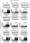
Figure 2. The effect of cytoskeletal disruption on CaMK-, IQmp- and PKA-mediated increases in LTCC Po
A-C show single channel records following exposure to exogenous, constitutively active CaMK (top) with superimposed ensemble-averaged currents before and after CaMK application. The effect of CaMK (indicated by the horizontal bar) on Po during an ensemble of 500 depolarizing steps is also shown (bottom). DMSO control, cytochalasin D and nocodazole treatment conditions are indicated above each panel. D-F are ordered as in A-C, but show single LTCC records following exposure to IQmp (10 μm), indicated by the horizontal bar. G-I are ordered as the previous panels, but show the effect of the catalytic subunit of PKA (1 μm, top) on Po, indicated by the horizontal bar.
Figure 3
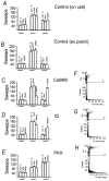
Figure 3. Cytochalasin D (Cyto-D) and nocodazole (Noco) prevent increased LTCC Po by CaMK and IQmp, but leave PKA signalling intact
Summary data for LTCC Po from on cell mode (A) and excised cell membrane patch recordings (B) following treatment with DMSO (control), cytochalasin D or nocodazole. Neither cytochalasin or nocodazole affect LTCC Po compared with control. However, both constitutively active CaMK (C) and IQmp (D) fail to increase LTCC Po after treatment with cytochalasin D or nocodazole. E, in contrast, PKA-induced increases in LTCC Po are not affected by cytochalasin D or nocodazole compared to control. Numerals (abscissa) indicate the number of LTCCs studied in each condition. *P < 0.05 compared with control.
Figure 4
Figure 4. Cytochalasin D and nocodazole prevent LTCC facilitation by CaMK and IQmp, while PKA-mediated LTCC facilitation is preserved
Summary data for the distribution of LTCC gating modes from on-cell mode (A) and excised cell membrane patch recordings (B) following treatment with cytochalasin D, nocodazole or DMSO (control). Neither cytochalasin D or nocodazole affect the distribution of gating modes compared with control. However, both constitutively active CaMK (C) and IQmp (D) fail to increase the proportion of sweeps with prolonged channel openings (mode 2) after treatment with cytochalasin D or nocodazole, compared to control. E, in contrast, cytochalasin D or nocodazole do not affect PKA-mediated increases in mode 2 gating sweeps. *P < 0.05 compared with cytochalasin D and nocodazole treatment. Analysis of LTCC gating modes (Yue et al. 1990) for cells treated with CaMK (F), IQmp (G) and PKA (H) after treatment with cytochalasin D. Only PKA was capable of increasing the probability of LTCCs entering into a gating mode with prolonged openings (mode 2) after treatment with cytoskeletal disrupting agents. Numerals indicate gating modes 0, 1 and 2 (see Methods for details). All analyses were performed on n = 3 LTCCs.
Figure 5
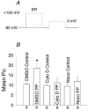
Figure 5. Cytoskeletal disrupting agents prevent prepulse (PP) LTCC facilitation
A, schematic representation of the voltage clamp protocol for eliciting PP facilitation. Control measurements omitted the PP and had a constant holding potential of −80 mV (dashed line). LTCC Po was measured during the depolarizing step to 0 mV. B, mean LTCC Po for cells treated with DMSO control vehicle, cytochalasin D (Cyto D) and nocodazole (noco). The number of LTCCs studied is indicated on the abscissa. *P + 0.003 compared with other groups.

Comment in

  • Findings of research misconduct.
    [No authors listed] [No authors listed] NIH Guide Grants Contracts (Bethesda). 2014 Dec 5:NOT-OD-15-031. NIH Guide Grants Contracts (Bethesda). 2014. PMID: 25528784 Free PMC article. No abstract available.

References

    1. Anderson ME, Braun AP, Schulman H, Premack BA. Multifunctional Ca2+/calmodulin-dependent protein kinase mediates Ca(2+)-induced enhancement of the L-type Ca2+ current in rabbit ventricular myocytes. Circulation Research. 1994;75:854–861. - PubMed
    1. Armstrong DL, Rossier MF, Shcherbatko AD, White RE. Enzymatic gating of voltage-activated calcium channels. Annals of the New York Academy of Sciences. 1991;635:26–34. - PubMed
    1. Bayer KU, De Koninck P, Leonard AS, Hell JW, Schulman H. Interaction with the NMDA receptor locks CaMKII in an active conformation. Nature. 2001;411:801–805. - PubMed
    1. Bayer KU, Harbers K, Schulman H. AlphaKAP is an anchoring protein for a novel CaM kinase II isoform in skeletal muscle. EMBO Journal. 1998;17:5598–5605. - PMC - PubMed
    1. Bers DM, Patton CW, Nuccitelli R. A practical guide to the preparation of Ca2+ buffers. Methods in Cell Biology. 1994;40:3–29. - PubMed

Publication types

MeSH terms

LinkOut - more resources