. 2002 Dec 1;545(2):441-51.
doi: 10.1113/jphysiol.2002.029561.

Voltage-dependent priming of rat vanilloid receptor: effects of agonist and protein kinase C activation

Affiliations

Voltage-dependent priming of rat vanilloid receptor: effects of agonist and protein kinase C activation

Gerard P Ahern et al. J Physiol. .

Abstract

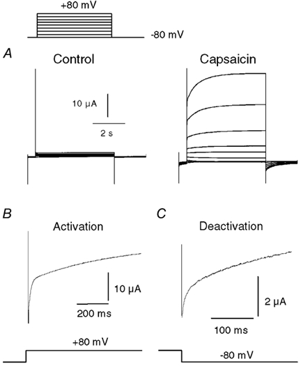
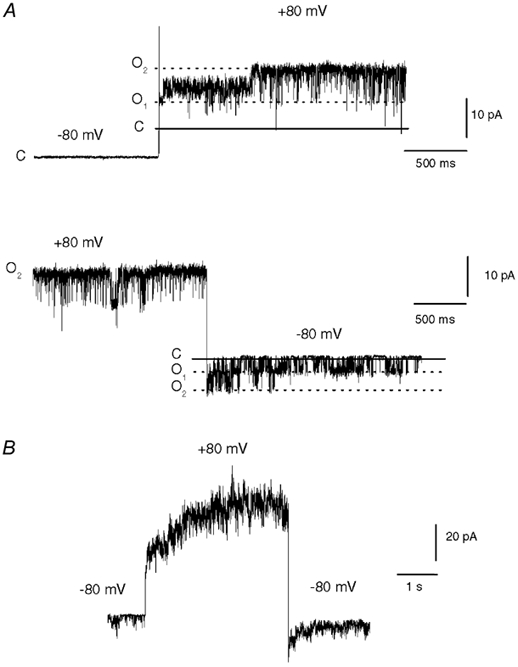
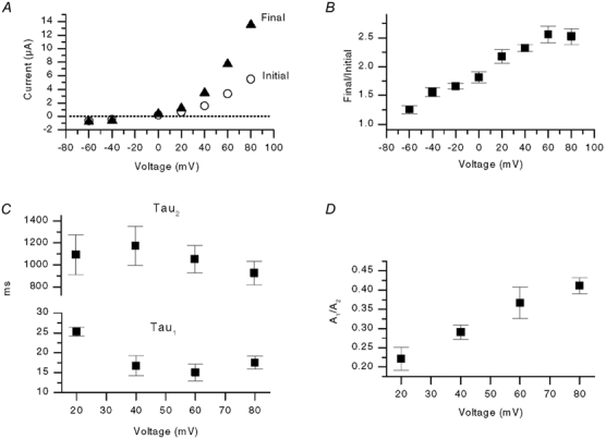
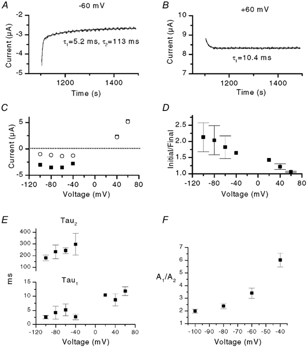
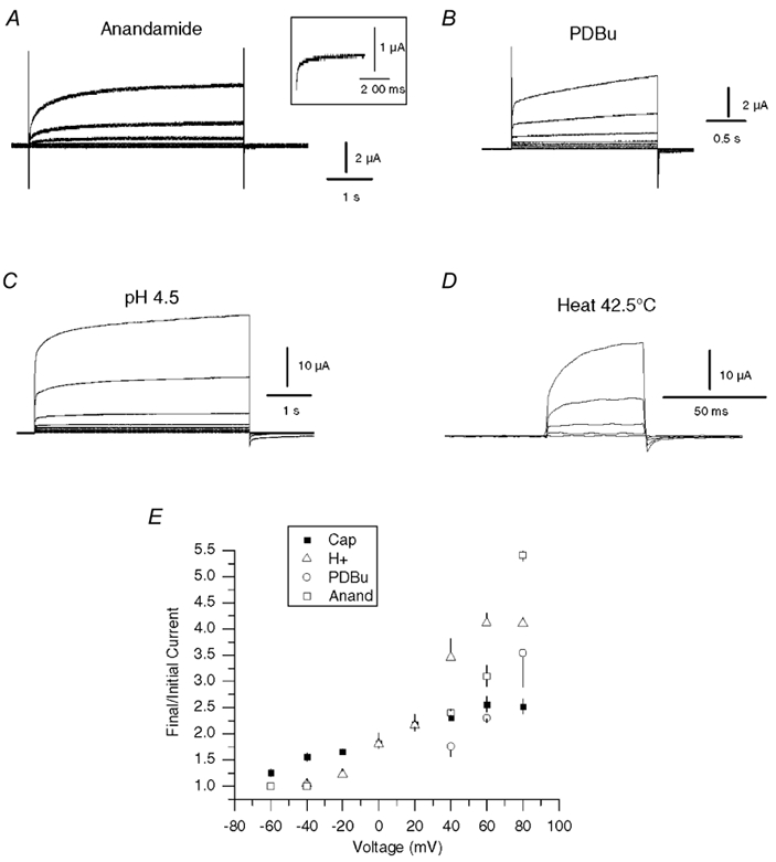
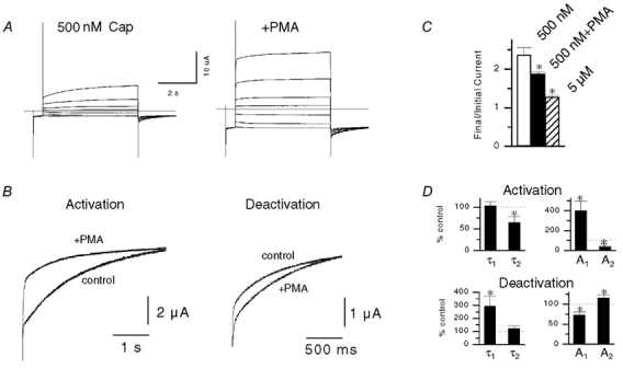
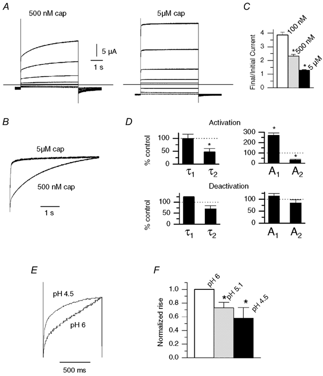
The responses of vanilloid receptor (VR) channels to changing membrane potential were studied in Xenopus oocytes and rat dorsal root ganglion (DRG) neurons. In oocytes, capsaicin-evoked VR currents increased instantaneously upon a step depolarization and thereafter rose biexponentially with time constants of approximately 20 and 1000 ms. Similarly, upon repolarization the current abruptly decreased, followed by a biexponential decay with time constants of approximately 4 and 200 ms. Qualitatively similar effects were observed in single channel recordings of native VR channels from DRG neurons and with endogenous VR activators, including heat (43 degrees C), H(+), anandamide and protein kinase C (PKC). The magnitude of the time-dependent current rise increased with membrane depolarization. This effect was accompanied by an increase in the relative proportion of the fast kinetic component, A(1). In contrast, the time constants of the activation and deactivation processes were not strongly voltage dependent. Increasing the agonist concentration both reduced the magnitude of the current rise and increased its overall rate, without significantly altering the deactivation rate. In contrast, PKC both speeded the current rise and slowed its decay. These results suggest that voltage interacts with agonists in a synergistic manner to augment VR current and this mechanism will be enhanced under conditions of inflammation when VRs are likely to be phosphorylated.


Figures

Figure 1
Figure 1. Voltage modulation of capsaicin-activated currents in whole oocytes
A, family of current traces showing the response of a VR1-expressing oocyte to a series of voltage steps (in 20 mV increments) from −80 to +80 mV, either in the absence or in the presence of 500 nm capsaicin. B, expanded trace from the data in A showing the rising phase of current after the voltage step to +80 mV. C, expanded trace showing the tail current recorded at −80 mV after a 5 s step to +80 mV.
Figure 2
Figure 2. Voltage modulation of single VR channels in DRG neurons
A, current traces from a cell-attached patch from a DRG neuron showing the response of two VR channels to voltage steps between −80 and +80 mV. The continuous and dashed lines indicate the closed (C) and two open current levels (O1 and O2), respectively. Note that one channel initially opens to a subconductance level after the depolarization step (top trace). The pipette solution contained 500 nm capsaicin and the bath solution contained a high [K+] to eliminate the cell membrane potential. B, an outside-out patch from a DRG neuron containing four to five VR channels activated by 500 nm capsaicin during a step from −80 to +80 mV. A double exponential fit to the rising current gave time constants of 16 and 1465 ms.
Figure 3
Figure 3. Voltage dependence and kinetics of VR current rise in oocytes
A, plot of instantaneous current and final current (5 s later) versus voltage from one experiment. B, mean normalized rise (final current/initial current) versus voltage from five oocytes activated by 500 nm capsaicin. C, plot of the time constants, τ1 and τ2, versus voltage (n = 7). The rising phase of current was fitted with a double exponential function described in the Methods. D, plot of the ratio of the amplitude of the fast component of the rising phase (A1) and the slower component (A2) versus voltage (n = 6).
Figure 4
Figure 4. Voltage dependence and kinetics of VR current deactivation in oocytes
A and B, traces showing tail currents at −60 and +60 mV, respectively, after a 5 s pre-pulse to +80 mV. The continuous lines show double and single exponential fits, respectively, with time constants as indicated. Note that only a single exponential function is required to fit the data at +60 mV. C, the initial or peak tail current (▪) and the final current (500 ms later) (○) as a function of voltage from a single experiment. D, plot of initial/final tail current versus voltage from three oocytes. E, plot of the time constants, τ1 and τ2, versus voltage. The decaying phase of current was fitted with either a double or single exponential function as shown in A. Difference in values for τ1 between +60 mV and −100 to −40 mV were significant at P < 0.05. F, plot of the ratio of the amplitudes of the fast and slow components of the tail current (A1/A2) versus voltage (three oocytes).
Figure 5
Figure 5. Voltage modulation of VR1 currents activated by anandamide, H+, heat and PKC
A-D, current traces from oocytes activated with anandamide (10 μm), PKC (pretreatment with 1 μm PDBu), H+ (pH 4.5) and heat (42.5 °C). Oocytes were depolarized with 20 mV incremental voltage steps from −80 to +80 mV, except for heat traces, which were depolarized to voltages between 0 and +80 mV, from a holding potential of −80 mV. Control traces in the absence of agonist were subtracted from the test data. For heat, traces obtained at 37 °C were subtracted from the 42.5 °C data. Tail currents with anandamide are not clearly seen. The inset in A shows expanded tail current from a separate recording with greater VR1 expression. E, the mean normalized rise (final current/initial current) versus voltage for oocytes activated with 500 nm capsaicin (n = 5), pH 4.5 (n = 4), after 1 μm PDBu (n = 4) and 10 μm anandamide (n = 3).
Figure 7
Figure 7. Regulation of voltage modulation by PKC
A, response of an oocyte to 5 s depolarizing voltage steps (-80 to +60 mV, 20 mV steps) in the presence of 500 nm capsaicin before and after treatment with 500 nm PMA. B, normalized traces of the rising (+60 mV) and decaying components (-80 mV) before and after PMA. C, plot of relative current increase (final/initial current) during depolarization to +80 mV with 500 nm capsaicin before and after PMA treatment (n = 3, P < 0.05, paired t test). Data for 5 μm capsaicin (from Fig. 6) are shown for comparison. D, mean changes in the amplitude and time course of double exponential fits to currents at ±80 mV (n = 4 activation, n = 3 deactivation; * P < 0.05, paired t test).
Figure 6
Figure 6. Influence of capsaicin and H+ concentration on voltage modulation
A, response of an oocyte to depolarizing voltage steps in the presence of 500 nm capsaicin and 5 μm capsaicin. B, scaled traces from data in A (+80 mV) show a faster rise with 5 μm capsaicin. C, plot of relative current increase (final/initial current) during 5 s depolarization to +80 mV with 100 nm (n = 3), 500 nm (n = 9) and 5 μm capsaicin (n = 6). Differences between all groups were significant (P < 0.01, two-sample t test) D, changes in time constants and amplitudes for double exponential fits to the current activation at +80 mV and to the current deactivation at −80 mV, after switching from 500 nm to 5 μm capsaicin (n = 4 for both groups, * P < 0.05, paired t test). E, current traces from oocytes during 1 s depolarizations to +80 mV with pH 6 and pH 4.5. F, the final/initial current (normalized to pH 6) with pH 4.5-6 (n = 4, * P < 0.05 paired t test versus pH 6).

Similar articles

Cited by

References

    1. Abdulla FA, Smith PA. Axotomy- and autotomy-induced changes in the excitability of rat dorsal root ganglion neurons. Journal of Neurophysiology. 2001;85:630–643. - PubMed
    1. Ahern GP, Kessler M, Premkumar LS. Voltage-dependence of the capsaicin receptor channel (VR1) Society for Neuroscience Abstracts. 2000;26:39.
    1. Caterina MJ, Julius D. The vanilloid receptor: a molecular gateway to the pain pathway. Annual Review of Neuroscience. 2001;24:487–517. - PubMed
    1. Caterina MJ, Schumacher MA, Tominaga M, Rosen TA, Levine JD, Julius D. The capsaicin receptor: a heat-activated ion channel in the pain pathway. Nature. 1997;389:816–824. - PubMed
    1. Cesare P, Dekker LV, Sardini A, Parker PJ, McNaughton PA. Specific involvement of PKC-epsilon in sensitization of the neuronal response to painful heat. Neuron. 1999;23:617–624. - PubMed

MeSH terms

LinkOut - more resources