

cAMP-independent responses of olfactory neurons in Xenopus laevis tadpoles and their projection onto olfactory bulb neurons
- PMID: 12456827
- PMCID: PMC2290687
- DOI: 10.1113/jphysiol.2002.031914
cAMP-independent responses of olfactory neurons in Xenopus laevis tadpoles and their projection onto olfactory bulb neurons
Abstract
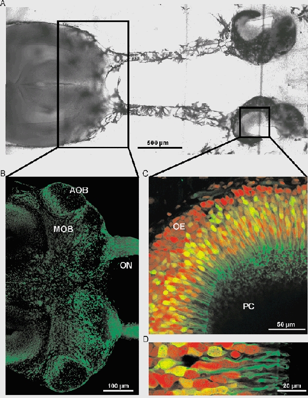
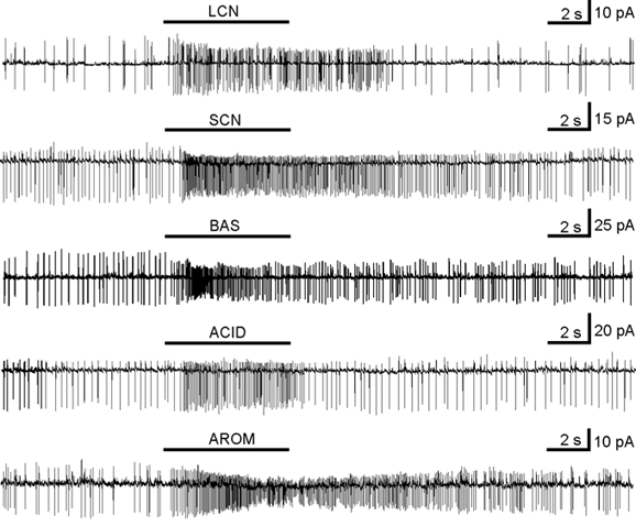
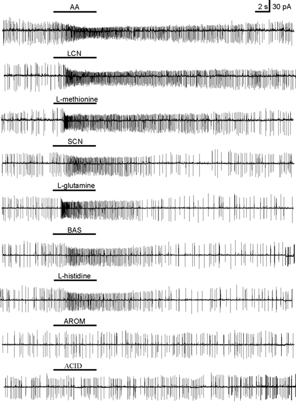
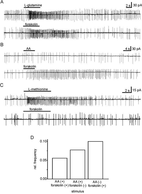
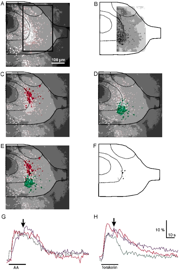
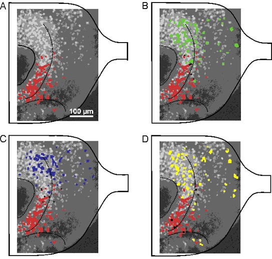
We report on responses of olfactory receptor neurons (ORNs) upon application of amino acids and forskolin using a novel slice preparation of the olfactory epithelium of Xenopus laevis tadpoles. Responses were measured using the patch-clamp technique. Both amino acids and forskolin proved to be potent stimuli. Interestingly, a number of ORNs that responded to amino acids did not respond to forskolin. This suggests that some amino acids activate transduction pathways other than the well-known cAMP-mediated one. The differential processing of cAMP-mediated stimuli on the one hand and amino acid stimuli on the other was further elucidated by calcium-imaging of olfactory bulb neurons using a novel nose-olfactory bulb preparation of Xenopus laevis tadpoles. The projection pattern of amino acid-sensitive ORNs to olfactory bulb neurons differed markedly from the projection pattern of forskolin-sensitive ORNs. Olfactory bulb neurons activated by amino acids were located laterally compared to those activated by forskolin, and only a small proportion responded to both stimuli. The ensemble of neurons activated by forskolin was also activated by the phosphodiesterase inhibitor 3-isobutyl-1-methylxanthine (IBMX) and the membrane-permeant cAMP analogue 8-(4-chlorophenylthio)adenosine 3',5'-cyclic monophosphate (pCPT-cAMP). We therefore conclude that sensory transduction of a number of amino acids is cAMP independent, and amino acid- and cAMP-mediated responses are processed differentially at the level of the olfactory bulb.
Figures
Similar articles
-
cAMP-independent olfactory transduction of amino acids in Xenopus laevis tadpoles.J Physiol. 2003 Aug 15;551(Pt 1):115-23. doi: 10.1113/jphysiol.2003.043059. Epub 2003 Jun 24. J Physiol. 2003. PMID: 12824450 Free PMC article.
-
ATP activates both receptor and sustentacular supporting cells in the olfactory epithelium of Xenopus laevis tadpoles.Eur J Neurosci. 2006 Jan;23(1):119-28. doi: 10.1111/j.1460-9568.2005.04533.x. Eur J Neurosci. 2006. PMID: 16420422
-
One Special Glomerulus in the Olfactory Bulb of Xenopus laevis Tadpoles Integrates a Broad Range of Amino Acids and Mechanical Stimuli.J Neurosci. 2016 Oct 26;36(43):10978-10989. doi: 10.1523/JNEUROSCI.4631-15.2016. J Neurosci. 2016. PMID: 27798179 Free PMC article.
-
Information coding in the vertebrate olfactory system.Annu Rev Neurosci. 1996;19:517-44. doi: 10.1146/annurev.ne.19.030196.002505. Annu Rev Neurosci. 1996. PMID: 8833453 Review.
-
Making scents out of how olfactory neurons are ordered in space.Nat Neurosci. 2009 Feb;12(2):103-4. doi: 10.1038/nn0209-103. Nat Neurosci. 2009. PMID: 19172161 Review. No abstract available.
Cited by
-
Classes and narrowing selectivity of olfactory receptor neurons of Xenopus laevis tadpoles.J Gen Physiol. 2004 Feb;123(2):99-107. doi: 10.1085/jgp.200308970. J Gen Physiol. 2004. PMID: 14744986 Free PMC article.
-
Correlation between olfactory receptor cell type and function in the channel catfish.J Neurosci. 2003 Oct 15;23(28):9328-39. doi: 10.1523/JNEUROSCI.23-28-09328.2003. J Neurosci. 2003. PMID: 14561860 Free PMC article.
-
Purinergic receptor-mediated Ca signaling in the olfactory bulb and the neurogenic area of the lateral ventricles.Purinergic Signal. 2010 Dec;6(4):429-45. doi: 10.1007/s11302-010-9207-6. Epub 2010 Dec 1. Purinergic Signal. 2010. PMID: 21437013 Free PMC article.
-
Amino acid- vs. peptide-odorants: responses of individual olfactory receptor neurons in an aquatic species.PLoS One. 2012;7(12):e53097. doi: 10.1371/journal.pone.0053097. Epub 2012 Dec 27. PLoS One. 2012. PMID: 23300867 Free PMC article.
-
Response profiles to amino acid odorants of olfactory glomeruli in larval Xenopus laevis.J Physiol. 2007 Jun 1;581(Pt 2):567-79. doi: 10.1113/jphysiol.2007.130518. Epub 2007 Mar 8. J Physiol. 2007. PMID: 17347262 Free PMC article.
References
-
- Buck L, Axel R. A novel multigene family may encode odorant receptors: a molecular basis for odor recognition. Cell. 1991;65:175–187. - PubMed
-
- Edwards FA, Konnerth A, Sakmann B, Takahashi T. A thin slice preparation for patch-clamp recordings from neurones of the mammalian central nervous system. Pflügers Archiv. 1989;414:600–612. - PubMed
MeSH terms
Substances
LinkOut - more resources
Full Text Sources