. 2002 Dec 1;545(2):485-99.
doi: 10.1113/jphysiol.2002.031161.

Single-cell epithelial defects close rapidly by an actinomyosin purse string mechanism with functional tight junctions

Affiliations

Single-cell epithelial defects close rapidly by an actinomyosin purse string mechanism with functional tight junctions

P Florian et al. J Physiol. .

Abstract

Restitution of single-cell defects, a frequent event in epithelia with high turnover, is poorly understood. Morphological and functional changes were recorded, using intravital time-lapse video microscopy, confocal fluorescence microscopy, and conductance scanning techniques. After artificial single-cell loss from an HT-29/B6 colonic cell monolayer, the basal ends of adjacent cells extended. Concurrently, the local conductive leak associated with the defect sealed with an exponential time course (from 0.48 +/- 0.05 microS 2 min post lesion to 0.17 +/- 0.02 microS 8 min post lesion, n = 17). Between 3 and 10 min post lesion, a band of actin arose around the gap, which colocalized with a ring of ZO-1 and occludin. Hence, tight junction proteins bound to the actin band facing the gap, and competent tight junctions assembled in the adjoining cell membranes. Closure and sealing were inhibited when actin polymerization was blocked by cytochalasin D, delayed following decrease of myosin-ATPase activity by butanedione monoxime, and blocked after myosin light chain kinase inhibition by ML-7. The Rho-associated protein kinase inhibitor Y-27632 did not affect restitution. After loosening of intercellular contacts in low Ca(2+) Ringer solution, the time course of restitution was not significantly altered. Albeit epithelial conductivity was 12-fold higher in low Ca(2+) Ringer solution than in controls, under both conditions the repaired epithelium assumed the same conductivity as distant intact epithelium. In conclusion, epithelial restitution of single-cell defects comprises rapid closure by an actinomyosin 'purse-string' mechanism and simultaneous formation of a functional barrier from tight junction proteins also associated with the purse string.


Figures

Figure 1
Figure 1. Illustration of the conductance scanning technique (not drawn to scale)
gleak, the conductance of the focal leak associated with a single-cell lesion, was determined as follows. Electric current (AC, 24 Hz) was imposed across the epithelium of HT-29/B6 cells. A probe, consisting of a mechanically coupled pair of microelectrodes, was moved (at a distance z0 to the surface) along the x axis (parallel to the surface), starting directly above the lesion. The potential difference at the probe (amplified with synchronous demodulation) is a measure of the local current density. Spatial integration and division by the transepithelial voltage yielded the conductance of the leak (gleak).
Figure 2
Figure 2. Time-lapse video microscopy of the closure of a single-cell defect
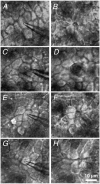
The intact HT-29/B6 monolayer immediately before setting the lesion (A, C, E and G) and about 10 min after it (B, D, F and H), as seen in the conductance scanning set-up. A and B, under control conditions; C and D, after incubation with cytochalasin D; E and F, with 2,3-butanedione monoxime; and G and H, in low free [Ca2+]o Ringer solution. Closure of the defect started below the surface (defined by the apical cell membranes). The difficulty in focusing the level of minimal defect area using conventional light microscopy was overcome by applying confocal fluorescence microscopy.
Figure 3
Figure 3. Confocal immunofluorescence microscopy of single-cell defects in HT-29/B6. Staining of tight junction elements (ZO-1 and occludin)
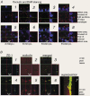
A, staining with occludin antibody (red) and DAPI (blue) at different times: images 1 and 5, 3 min post lesion (p.l.); 2 and 6, 6 min p.l.; 3 and 7, 10 min p.l.; 4 and 8, 15 min p.l. The quadratic images show x-y scans parallel to the epithelium. At the positions indicated by the green and red lines, x-z and y-z projections were computed (rectangular images on top and to the right of the quadratic image). In x-z and y-z planes, the blue lines indicate the level of the x-y plane. Images are focused on either the plane of tight junctions in the intact epithelium (1-4) or a parallel plane below, where the defect appeared minimal (5-8). In images 1 and 5, the confocal scanning range of the z axis was slightly smaller than in the others. See Movie 1 in Supplementary material. B, staining with ZO-1 and occludin antibodies, 3 (1-3) or 10 min (4-7) post lesion. In pictures 1-6, the arrangement of x-y, x-z and y-z planes is the same as above. 1 and 4 show ZO-1 (green), 2 and 5 show occludin (red), 3 and 6 show the merged images (yellow indicates colocalization). Images are focused on the plane where the defect appeared minimal. Image 7 shows a superposition of x scans in the y-z plane (with ZO-1 and occludin merged) for the defect 10 min p.l. The tight junction proteins delineate the funnel-like shape of the defect. For 3D-reconstruction see Movies 2-4 in Supplementary material.
Figure 4
Figure 4. Immunofluorescence staining of ZO-1 and F-actin
Cells were analysed with ZO-1 antibody (green) and Alexa Fluor 594-phalloidin (red) for F-actin at 3 min (1-3) and 10 min (4-6) p.l. Images 1 and 4, ZO-1; 2 and 5, F-actin; 3 and 6 merged images of ZO-1 and F-actin staining. The arrangement of images (x-y, x-z, y-z planes) is the same as in Fig. 3. The scale bar (10 μm) applies to all images. (m: damage caused by marks indicating the position of the defect investigated. The mark consisted of lines in a right-angled pattern that were scratched into the monolayer with a microelectrode.)
Figure 7
Figure 7. Conductance of the leak, gleak, as a function of time associated with single-cell defects
Time course of restitution in HT-29/B6 cells (28th-31st passage), under control conditions (A), after incubation with cytochalasin D (B), with 2,3-butanedione monoxime (C), and in low free [Ca2+]o Ringer solution (D). Under control conditions (A), repair resulted in exponential decline of gleak, which allowed extrapolation (dashed line) of the initial (t + 0) leak at 0.69 ± 0.06 μS. Sealing of the leak (described by gleak) was blocked after inhibition of actin polymerization (B), and was slower after impairment of myosin ATPases (C), but proceeded at the same speed in low Ca2+ Ringer solution (D).
Figure 5
Figure 5. Conductivity of HT-29/B6 monolayers measured in a conventional Ussing chamber as a function of time
A, the time course was recorded under control conditions (n = 19) and the effects of cytoskeleton modulators, cytochalasin D (n = 10) or butanedione monoxime (n = 10), and low free extracellular calcium (250 nmol l−1, n = 8) were studied. With both inhibitors and under low free calcium, the increase of GUssing was reversible upon washout with regular Ringer solution. B, cells from a different passage (32nd-33rd) were analysed under control conditions (n = 14) and the effects of the myosin light chain kinase inhibitor ML-7 (n = 7), and the Rho-associated kinase inhibitor Y-27632 (n = 6) were studied. With the inhibitor ML-7, the increase of GUssing was reversible upon washout with regular Ringer solution, but the effect of Y-27632 was not.
Figure 6
Figure 6. Distribution of the recorded signal, expressed as apparent conductivity GA, as a function of space (horizontal distance from lesion) and time (post lesion)
A typical experiment under control conditions is shown. The apparent conductivity (GA) is a normalized measure of the local current density. After positioning the probe above the lesion (produced at t + 0), the probe was repeatedly moved along the x axis, and measurements were taken at certain points of the x axis, with a constant distance z0 to the surface. By spatial integration of GA(x, t), the conductance of the leak, gleak, was determined as a function of time.
Figure 8
Figure 8. Conductance of the leak, gleak, as a function of time associated with single-cell defects
Time course of restitution in HT-29/B6 cells (32nd-33rd passage), under control conditions (A), after incubation with ML-7 (B) and with Y-27632 treatment (C). Under control conditions (A), repair resulted in exponential decline of gleak (n = 7). Sealing of the leak (described by gleak) was dramatically delayed after inhibition of myosin light chain kinase activity (n = 8), but proceeded at the same speed as controls after Y-27632 treatment (n = 8).
Figure 9
Figure 9. Geometrical model of tight junction distribution during the repair of a single-cell lesion
The sum of cell circumferences per epithelial area in an epithelium of hexagonal symmetry (A) does not change when a single cell is removed (B), because the loss of cellular junctions is compensated for by the extensions of the adjoining cells closing the defect, adding an equal length of cellular junctions.
Figure 10
Figure 10. Restitution of single-cell defects in an epithelial monolayer
In undisturbed intestinal epithelium, an intracellular perijunctional actin band is located adjacent to the apical junctional complex, which includes the tight junction. This actin, together with myosin II, is assumed to control the tight junction and thus paracellular conductivity (Turner, 2000). After the loss of a single cell, the cell membranes facing the gap lack these structures (A). However, the neighbouring cells flatten and their basal ends extend into the gap. A circumferential actinomyosin band forms that pulls taut the suture and serves as a base for new tight junction proteins (B). In addition, new tight junctions assemble rapidly, like a zipper being closed, between the extensions of the neighbouring cells closing in (cf. Fig. 9).

Similar articles

Cited by

References

    1. Albers TM, Lomakina I, Moore RP. Structural and functional roles of cytoskeletal proteins during repair of native guinea pig intestinal epithelium. Cell Biology International. 1996;20:821–830. - PubMed
    1. Balda MS, Gonzalez-Mariscal L, Matter K, Cereijido M, Anderson JM. Assembly of the tight junction: the role of diacylglycerol. Journal of Cell Biology. 1993;123:293–302. - PMC - PubMed
    1. Bement WM, Forscher P, Mooseker MS. A novel cytoskeletal structure involved in purse string wound closure and cell polarity maintenance. Journal of Cell Biology. 1993;121:565–578. - PMC - PubMed
    1. Bement WM, Mandato CA, Kirsch MN. Wound-induced assembly and closure of an actomyosin purse string in Xenopus oocytes. Current Biology. 1999;9:579–587. - PubMed
    1. Brock J, Midwinter K, Lewis J, Martin P. Healing of incisional wounds in the embryonic chick wing bud: characterization of the actin purse-string and demonstration of a requirement for Rho activation. Journal of Cell Biology. 1996;135:1097–1107. - PMC - PubMed

Publication types

MeSH terms