. 2002 Dec 1;545(2):521-35.
doi: 10.1113/jphysiol.2002.022103.

The contribution of intracellular calcium stores to mEPSCs recorded in layer II neurones of rat barrel cortex

Affiliations

The contribution of intracellular calcium stores to mEPSCs recorded in layer II neurones of rat barrel cortex

Christopher R L Simkus et al. J Physiol. .

Abstract

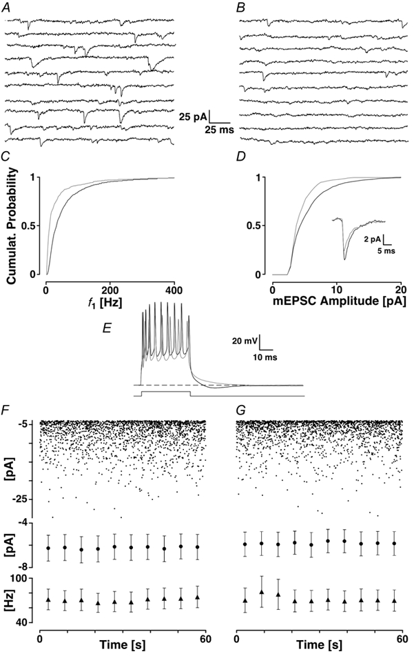
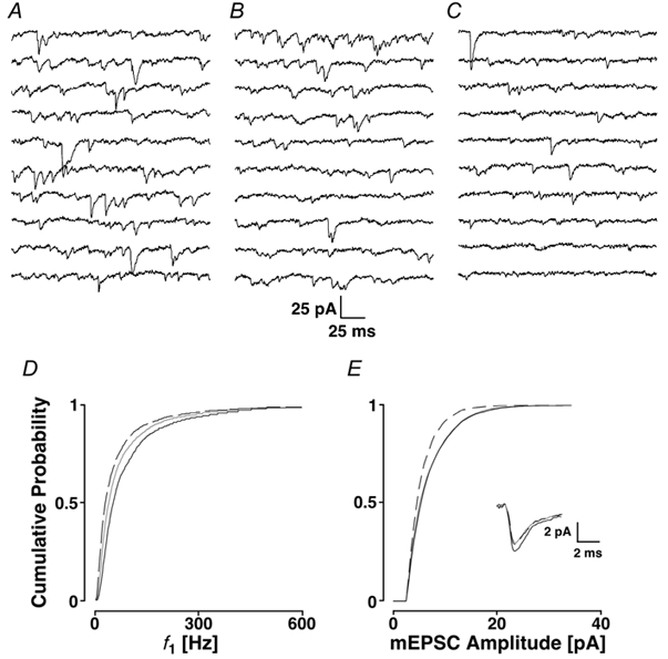
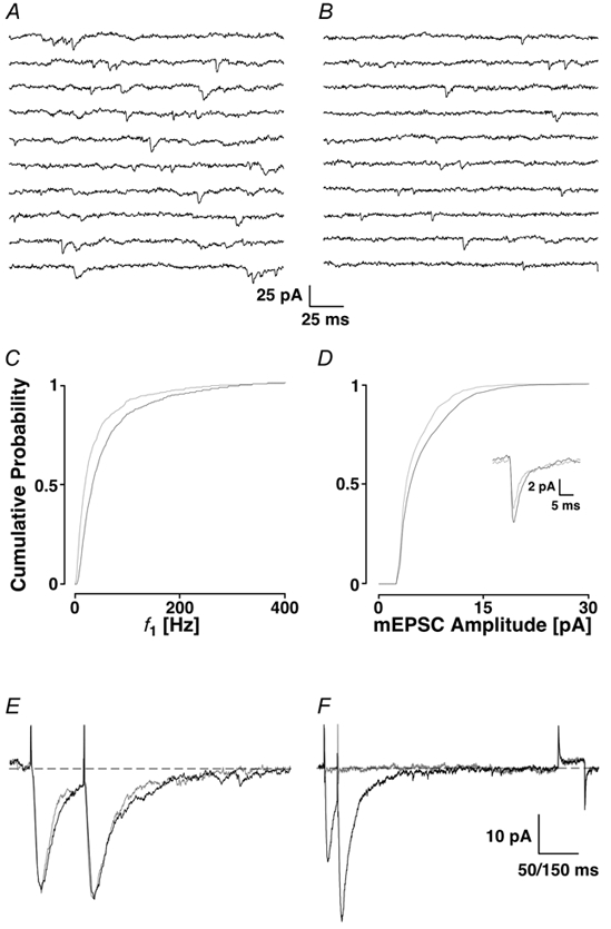
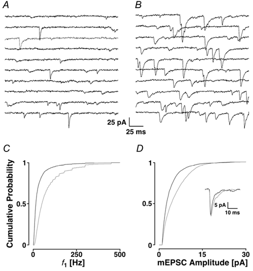
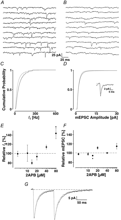
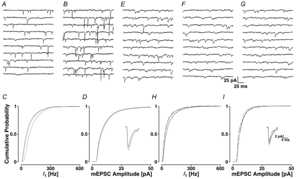
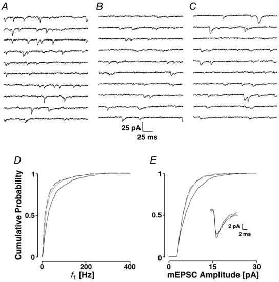
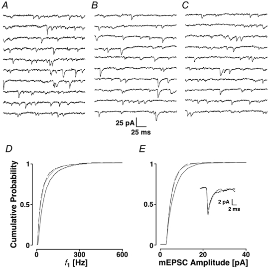
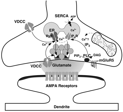
Loading slices of rat barrel cortex with 50 microM BAPTA-AM while recording from pyramidal cells in layer II induces a marked reduction in both the frequency and amplitudes of mEPSCs. These changes are due to a presynaptic action. Blocking the refilling of Ca(2+) stores with 20 microM cyclopiazonic acid (CPA), a SERCA pump inhibitor, in conjunction with neuronal depolarisation to activate Ca(2+) stores, results in a similar reduction of mEPSCs to that observed with BAPTA-AM, indicating that the source for intracellular Ca(2+) is the endoplasmic reticulum. Block or activation of ryanodine receptors by 20 microM ryanodine or 10 mM caffeine, respectively, shows that a significant proportion of mEPSCs are caused by Ca(2+) release from ryanodine stores. Blocking IP(3) receptors with 14 microM 2-aminoethoxydiphenylborane (2APB) also reduces the frequency and amplitude of mEPSCs, indicating the involvement of IP(3) stores in the generation of mEPSCs. Activation of group I metabotropic receptors with 20 microM (RS)-3,5-dihydroxyphenylglycine (DHPG) results in a significant increase in the frequency of mEPSCs, further supporting the role of IP(3) receptors and indicating a role of group I metabotropic receptors in causing transmitter release. Statistical evidence is presented for Ca(2+)-induced Ca(2+) release (CICR) from ryanodine stores after the spontaneous opening of IP(3) stores.


Figures

Figure 1
Figure 1. Loading the slice with 50 μm BAPTA-AM
The first 2 s of recording taken before (A) and after the addition of BAPTA-AM and waiting for > 20 min (B). C, the cPDF of f1 before (black line) and after the addition of BAPTA-AM (grey line). D, the cPDF of the mEPSC amplitudes before (black line) and following the addition of BAPTA-AM (grey line). The average mEPSC time courses are given in the inset before (black line) and after (grey line) the addition of BAPTA-AM. E, bursts of action potentials elicited by an 80 ms current pulse of 0.3 nA before (black line) and after loading (grey line). The grey trace no longer shows the sAHP. The dashed line indicates resting membrane potential. F and G, assessing the stability of mEPSCs illustrated with a 1 min recording in control (left) and after 20 min (right). The peak amplitudes are plotted in time. There is no difference in either frequency or amplitude. Below are pooled data of 5 experiments with average values taken every 6 s for the peak amplitudes (•) and f1 (▴). There was no difference between control and a recording 20 min later.
Figure 2
Figure 2. Preventing the loading of ER stores with the SERCA pump inhibitor CPA (20 μm)
A, control sequence; B, after the addition of CPA; and C, in the presence of CPA after a K+ depolarisation. Below are the cPDFs of the three conditions illustrated for f1 (D) and amplitudes (E). The black line corresponds to the control condition, the grey line to CPA alone and the dashed line to CPA following a K+ depolarisation. The inset in E shows the time courses of the average mEPSCs under the three conditions.
Figure 3
Figure 3. Block of ryanodine receptors using 20 μm ryanodine
A recording sequence of 2 s before (A) and after (B) the block of ryanodine receptors with the corresponding cPDFs shown in C and D for f1 and amplitudes (black lines control; grey lines, ryanodine receptors blocked). E, EPSC-like currents evoked by AMPA iontophoresis on the apical dendrite recorded from a pyramidal cell. Inter-pulse interval is 50 ms. The black line was obtained during control and the grey line when ryanodine with a K+ depolarisation was washed in. F, in an experiment with different time courses, 10 μm NBQX abolished the EPSC-like currents (grey line), illustrating that the iontophoresis activated AMPA receptors.
Figure 4
Figure 4. Activation of ryanodine receptors using 10 mm caffeine
A, a control period is shown and B, after the addition of caffeine. The cPDFs are illustrated for f1 (C) and amplitudes (D). Control conditions are in black and responses after the activation of ryanodine receptors are in grey. The inset refers to the time course of the mEPSCs before and after the addition of caffeine.
Figure 5
Figure 5. Block of the IP3 receptors using 14 μm 2APB
Two seconds of recording are illustrated in control (A) and after the addition of 2APB (B) with the appropriate cPDFs illustrated for f1 (C) and for the amplitudes (D) with the time course information shown in the inset. The results in 2APB are shown in grey. Concentration-response curves for average frequency (f0) are shown in E and amplitude in F. An asterisk marks significant deviations from control. Note the minimum at 14 μm 2APB. G, EPSC-like currents evoked by AMPA iontophoresis on the apical dendrite. The black line was obtained during control and the grey line when 2APB was present.
Figure 6
Figure 6. Activation of presynaptic metabotropic receptors using 20 μm DHPG
The control is illustrated in A and the response to DHPG in B. The cPDFs are shown for f1 and amplitudes in C and D, respectively, and the time courses are illustrated in the inset. Grey lines represent the data after the addition of DHPG. Activation of mGluRs by DHPG fails to increase mEPSC frequency after IP3 receptor blockade. E, control period, F, after the addition of 2APB and G, after the further addition of DHPG. In H and I, the cPDFs are shown for f1 and amplitudes, respectively. Black lines, control; grey lines, 2APB; dashed lines, subsequent addition of DHPG in the presence of 2APB. The inset shows the time courses of the mEPSCs.
Figure 7
Figure 7. Interaction of ryanodine stores with IP3 stores
A, control period; B, after the addition of ryanodine (with K+ depolarisation); and C, after the subsequent addition of 2APB. In D and E, the cPDFs are shown for f1 and amplitudes, respectively. Black lines, control; grey lines, ryanodine; and dashed lines, subsequent addition of 2APB. Inset refers to the time courses.
Figure 8
Figure 8. Interaction of IP3 stores with ryanodine stores
A, control period; B, after the addition of 2APB; and C, after the subsequent addition of ryanodine. In D and E, the cPDFs are shown for f1 and amplitudes, respectively. Black lines, control; grey lines, 2APB; and dashed lines, subsequent addition of ryanodine. The inset refers to the time courses.
Figure 9
Figure 9. Schematic drawing illustrating the generation of mEPSCs with the sites of actions of the drugs used indicated
Abbreviations: RyR, ryanodine receptor; IP3R, IP3 receptor; PLC, phospholipase C; DAG, diacylglycerol; and PiP2, phosphatidylinositol bisphosphate. Pharmacologically, ryanodine receptors are activated by caffeine and blocked by ryanodine; IP3 receptors are blocked by 2APB; DHPG activates mGluR5 receptors; SERCA pumps are blocked by CPA.

Similar articles

Cited by

References

    1. Bird MM. Presynaptic and postsynaptic organelles of synapses formed in cultures of previously dissociated mouse spinal cord. Cell and Tissue Research. 1978;194:503–511. - PubMed
    1. Capogna M, Gähwiler BH, Thompson SM. Presynaptic enhancement of inhibitory synaptic transmission by protein kinases A and C in the rat hippocampus in vitro. Journal of Neuroscience. 1995;15:1249–1260. - PMC - PubMed
    1. Carter AG, Vogt KE, Foster KA, Regehr WG. Assessing the role of calcium-induced calcium release in short-term presynaptic plasticity at excitatory central synapses. Journal of Neuroscience. 2002;22:21–28. - PMC - PubMed
    1. Chavez-Noriega LE, Stevens CF. Increased transmitter release at excitatory synapses produced by direct activation of adenylate cyclase in rat hippocampal slices. Journal of Neuroscience. 1994;14:310–317. - PMC - PubMed
    1. Del Castillo J, Katz B. Quantal components of the end-plate potential. Journal of Physiology. 1954;124:560–573. - PMC - PubMed

Publication types

MeSH terms

Substances

LinkOut - more resources