. 2002 Dec 1;545(2):595-614.
doi: 10.1113/jphysiol.2002.024331.

A cold- and menthol-activated current in rat dorsal root ganglion neurones: properties and role in cold transduction

Affiliations

A cold- and menthol-activated current in rat dorsal root ganglion neurones: properties and role in cold transduction

Gordon Reid et al. J Physiol. .

Abstract

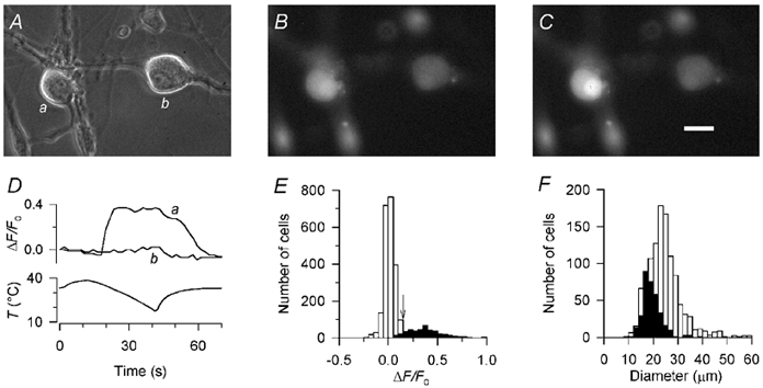
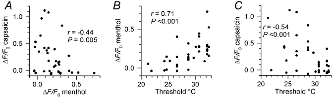
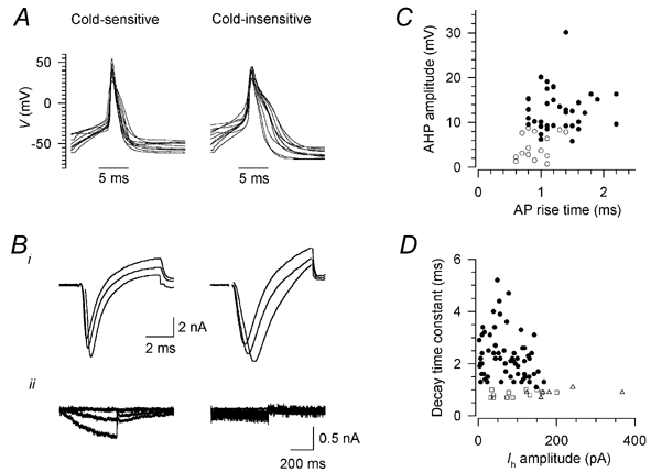
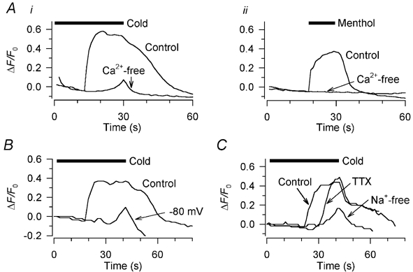
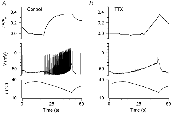
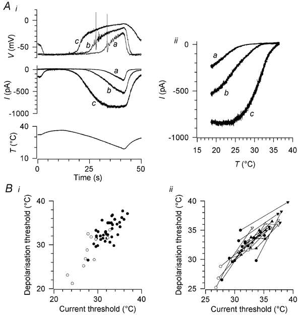
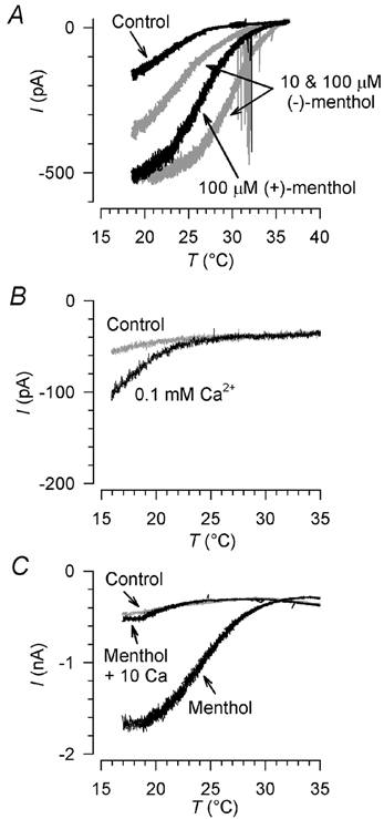
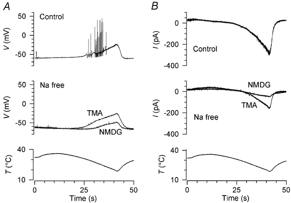
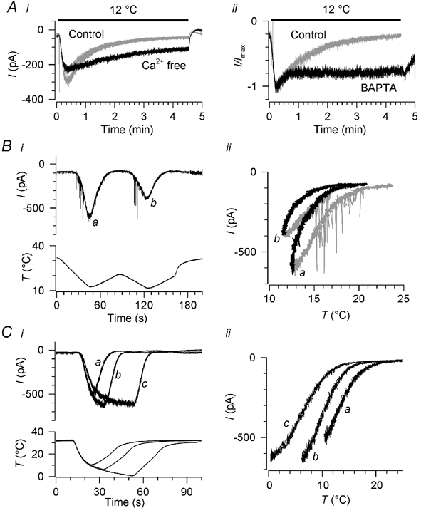
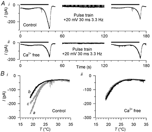
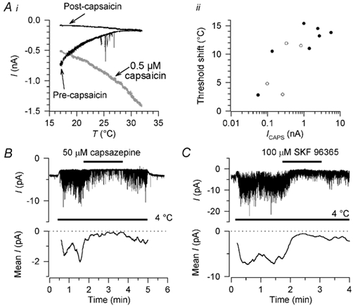
Skin temperature is sensed by peripheral thermoreceptors. Using the neuronal soma in primary culture as a model of the receptor terminal, we have investigated the mechanisms of cold transduction in thermoreceptive neurones from rat dorsal root ganglia. Cold-sensitive neurones were pre-selected by screening for an increase in [Ca(2+)](i) on cooling; 49 % of them were also excited by 0.5 microM capsaicin. Action potentials and voltage-gated currents of cold-sensitive neurones were clearly distinct from those of cold-insensitive neurones. All cold-sensitive neurones expressed an inward current activated by cold and sensitised by (-)-menthol, which was absent from cold-insensitive neurones. This current was carried mainly by Na(+) ions and caused a depolarisation on cooling accompanied by action potentials, inducing voltage-gated Ca(2+) entry; a minor fraction of Ca(2+) entry was voltage-independent. Application of (-)-menthol shifted the threshold temperatures of the cold-induced depolarisation and the inward current to the same extent, indicating that the cold- and menthol-activated current normally sets the threshold temperature for depolarisation during cooling. The action of menthol was stereospecific, with the (+)-isomer being a less effective agonist than the (-)-isomer. Extracellular Ca(2+) modulated the cold- and menthol-activated current in a similar way to its action on intact cold receptors: lowered [Ca(2+)](o) sensitised the current, while raised [Ca(2+)](o) antagonised the menthol-induced sensitisation. During long cooling pulses the current showed adaptation, which depended on extracellular Ca(2+) and was mediated by a rise in [Ca(2+)](i). This adaptation consisted of a shift in the temperature sensitivity of the channel. In capsaicin-sensitive neurones, capsaicin application caused a profound depression of the cold-activated current. Inclusion of nerve growth factor in the culture medium shifted the threshold of the cold-activated current towards warmer temperatures. The current was blocked by 50 microM capsazepine and 100 microM SKF 96365. We conclude that the cold- and menthol-activated current is the major mechanism responsible for cold-induced depolarisation in DRG neurones, and largely accounts for the known transduction properties of intact cold receptors.


Figures

Figure 1
Figure 1. Responses of rat dorsal root ganglion neurones to cooling
A, phase contrast image of two DRG neurones after 2 days in culture. B, epifluoresence image of the same two neurones, loaded with Calcium Green-1, just before applying the cooling stimulus. C, the same neurones at the peak of the cold stimulus. Scale bar 20 μm. D, ratio of the fluorescence change during the cold stimulus (ΔF) to the fluorescence intensity before the stimulus (F0) for the two neurones shown in A-C. Neurone a was defined as cold-sensitive according to the criteria described in Results, while neurone b was defined as insensitive to cold. In all cases, fluorescence intensity was averaged over the cytoplasmic area of the neurone excluding the nucleus, in order to measure only the change in cytoplasmic [Ca2+]i. E, histogram of ΔF/F0 in 2664 DRG neurones, 421 from the ‘threshold’ group (▪) and 2243 from the ‘no-threshold’ group (□; see text for definitions). The cutoff point for defining a neurone as cold-sensitive (ΔF/F0 ≥ 0.15) is arrowed; neurones in the ‘threshold’ group with peak ΔF/F0 ≥ 0.15 were considered as cold-sensitive (see Results). F, histogram of the diameters of 331 cold-sensitive neurones (▪) and 1106 cold-insensitive neurones (□); cold-sensitive neurones were among the smallest in DRG cultures.
Figure 2
Figure 2. The relationship between cold, menthol and capsaicin sensitivity
A, correlation between the amplitudes of the increases in [Ca2+]i (expressed as ΔF/F0) induced by 100 μm (-)-menthol and 0.5 μm capsaicin in cold-sensitive neurones. B and C, correlations between the rise in [Ca2+]i induced by menthol (B) and capsaicin (C) and the threshold temperature at which [Ca2+]i began to rise during cooling. Spearman rank correlation coefficients (r) are shown on each plot, along with the statistical significance (P) of the correlations.
Figure 3
Figure 3. Action potentials and current signatures in cold-sensitive and cold-insensitive neurones
A, action potentials in 10 randomly selected cold-sensitive neurones (left) and 10 cold-insensitive neurones (right) in response to 5 ms depolarising stimuli, aligned so that the peak occurs at the same time. Resting potentials were between −40 and −70 mV; in some cases the beginning of the stimulus is beyond the left edge of the trace. Bi, transient inward currents in one cold-sensitive (left) and one cold-insensitive neurone (right); from a holding potential of −60 mV, after a 150 ms prepulse to −80 mV, the potential was stepped to −10, 0 and +10 mV for 6 ms. The decay time constant was measured for the most positive pulse. Bii, hyperpolarisation-activated current Ih in one cold-sensitive (left) and one cold-insensitive neurone (right); from a holding potential of −60 mV, the potential was stepped to −70, −90 and −110 mV for 500 ms. Current amplitude was measured for the most negative pulse. Whole-cell (not perforated-patch) recordings, K2SO4 pipette solution; recordings were corrected offline for leak and capacitive transients, and any residual capacitive transients are blanked. C, relation between action potential rise time and the amplitude of the after-hyperpolarisation (AHP) in cold-sensitive (○) and cold-insensitive (•) neurones. D, relation between Ih amplitude and decay time constant of the transient inward current (see B) in cold-insensitive neurones (•) and cold-sensitive neurones sensitive (□) and insensitive (▵) to capsaicin.
Figure 4
Figure 4. Source of the cold- and menthol-induced rise in [Ca2+]i in rat DRG neurones
Ai, the cold-induced increase in [Ca2+]i was abolished in Ca2+-free extracellular solution containing 1 mm EGTA; the cold stimulus here (horizontal bar) was a ramp from 32 to 18 °C without the usual pre-warming step (see Methods). Aii, in the same neurone, the increase in [Ca2+]i elicited by 10 μm (-)-menthol was also abolished by removing extracellular Ca2+. B, in a different neurone, the cold-induced rise in [Ca2+]i was strongly reduced by voltage clamping at −80 mV. The cold stimulus here (horizontal bar) and in all other figures except Fig. 4A was the standard one shown in Fig. 1D. C, in a third neurone, the effects on the cold-induced [Ca2+]i increase of 1 μm tetrodotoxin (TTX) and of a Na+-free extracellular solution (N-methyl-d-glucamine used as Na+ substitute) are shown. Three successive cold stimuli were applied, 5 min apart.
Figure 5
Figure 5. Cold-induced depolarisation and its relation to the [Ca2+]i signal
A, simultaneous recording of the cold-induced [Ca2+]i signal (top trace) and membrane potential (middle trace) during the temperature stimulus shown in the bottom trace. The threshold temperatures for action potential activity and the increase in [Ca2+]i were the same. B, recording in the same neurone 5 min later during application of 100 nm TTX, which completely abolished action potential activity. The [Ca2+]i increase was delayed and threshold was shifted to a lower temperature than in A. Unusually, this neurone was firing action potentials at the base temperature of 32 °C before TTX application, and the resting [Ca2+]i was also relatively high, falling rapidly as the action potentials were shut off by warming (see A). For this reason the baseline fluorescence intensity (F0) for the calculation of ΔF/F0 in A was measured 5 s after the start of the stimulus instead of just before the stimulus as was usual.
Figure 6
Figure 6. Cold-induced depolarisation and inward current, and threshold shift caused by menthol
Ai, top, cold-induced depolarisation in the presence of 100 nm TTX (a), and its sensitization by 10 μm (b) and 100 μm (c) (-)-menthol. Middle, cold- and menthol-induced currents in the same neurone at a holding potential of −60 mV; the letters a-c have the same meanings as in the top panel. Note that 100 μm (-)-menthol depolarised the neurone and activated a current at the base temperature of 32 °C; these effects were reversed by warming to 37 °C. Bottom, the thermal stimulus. Aii, the currents in Ai during the falling phase of the temperature ramp (10-42 s), plotted against stimulus temperature. Bi, threshold temperatures of the cold-induced depolarisation and inward current in 38 neurones cultured in the presence of 50 ng ml−1 nerve growth factor (NGF) (•) and in 11 neurones cultured without NGF (○). Bii, shift in current and depolarisation thresholds from control values (•, ○) induced by 10 μm (▴, ▵) and 100 μm (-)-menthol (▾, ▿) in 16 neurones cultured with NGF (filled symbols) and five cultured without NGF (open symbols).
Figure 7
Figure 7. Stereospecificity of menthol action and modulation of the cold- and menthol-activated current by extracellular Ca2+
A, current-temperature relation of cold- and menthol-activated currents in standard extracellular solution and 100 μm (+)-menthol. Currents in the same neurone in 10 μm and 100 μm (-)-menthol are shown in grey for comparison. Holding potential −60 mV. Action potentials in cell processes are apparent during menthol application. B, potentiation of the current by lowered extracellular [Ca2+]: the current in 2 mm Ca2+, shown in grey, was increased about twofold by lowering extracellular [Ca2+] to 0.1 mm. C, antagonism of the sensitizing effect of (-)-menthol by raised extracellular [Ca2+]: the control current, shown in grey, was sensitized by 10 μm (-)-menthol; this sensitization was abolished by raising extracellular [Ca2+] to 10 mm. Holding potential in B and C was −80 mV.
Figure 8
Figure 8. Dependence of the cold-induced depolarisation and inward current on external Na+
A, effects of two Na+ substitutes on cold-induced depolarisation. Three cold stimuli were applied at intervals of 4.5 min, in the following order: control (top trace), N-methyl-d-glucamine (middle trace, NMDG), tetramethylammonium (middle trace, TMA). B, in the same neurone, replacement of Na+ by NMDG strongly inhibited the cold-activated inward current while a substantial current was present in TMA. Holding potential was −60 mV.
Figure 9
Figure 9. Adaptation of the cold- and menthol-activated current: time course, Ca2+ dependence and shift in temperature sensitivity
Ai, time course of adaptation on cooling and the effect of removing extracellular Ca2+. Three pulses to 12 °C were applied, separated by 5 min, the second one (black) being in Ca2+-free extracellular solution and the first and third (grey) in normal [Ca2+]. Extracellular [Mg2+] was raised to 10 mm throughout. Aii, the effect on adaptation time course of including 10 mm BAPTA in the pipette solution, recorded in two different neurones in standard extracellular solution. The control recording was in the perforated-patch configuration and was chosen because its adaptation time constant (69 s) was close to the mean for all perforated-patch recordings (71.5 s). Currents are normalised; peak currents were ≈200 pA (BAPTA) and ≈500 pA (control). B, shift in the temperature sensitivity of the cold-activated current during successive falling and rising temperature ramps, in standard extracellular solution; i, raw currents and temperature stimulus, and ii, current-temperature relationship. Falling temperature ramps are shown in grey in ii, and rising ramps in black; the two pairs of ramps are marked a and b for easy identification. C, effect of cooling pulses of duration 10, 20 and 40 s on the temperature dependence of the cold-activated current, measured during rising ramps (a-c) following each cold pulse, in standard extracellular solution. i, raw currents; ii, current-temperature relationship, showing only currents during the rising ramps a-c. Holding potential was −60 mV in A-C.
Figure 10
Figure 10. Induction of adaptation by raised [Ca2+]i
A, induction of adaptation by raising [Ca2+]i with trains of depolarising pulses. Ai, a cooling stimulus of the standard type (horizontal bar; see Fig. 1D) was followed immediately by a 1 min train of depolarising pulses (to +20 mV for 30 ms at 300 ms intervals) and then another cooling stimulus (horizontal bar); the cold-activated current was reduced after the pulse train. Aii, in the same neurone in Ca2+-free extracellular solution, the cold-activated current was unchanged by the pulse train. Extracellular [Mg2+] was raised to 10 mm throughout. B, current-temperature relationship of the currents in A. Cold-activated currents before the pulse train are shown in grey and marked a in i; currents immediately after the pulse train are in black and marked b in i. In addition, currents 5 min after the pulse train are plotted; these are in grey and marked c in i. Holding potential was −60 mV.
Figure 11
Figure 11. Cross-adaptation caused by capsaicin-induced current and block by capsazepine and SKF 96365
Ai, application of 0.5 μm capsaicin for 1 min induced adaptation of the cold-activated current in a capsaicin-sensitive neurone; current activated by capsaicin is shown in grey. Holding potential was −60 mV. ii, correlation between the shift in threshold after capsaicin application and the amplitude of the capsaicin-induced current (ICAPS) in 11 cold-sensitive neurones, seven recorded in perforated-patch (•) and four in conventional whole-cell configuration (○). Capsaicin-induced current amplitude is shown on a logarithmic scale because of the wide range of values. In neurones like that in A, where an 18 °C stimulus no longer elicited a cold-activated current, the threshold was assumed to be 18 °C for the purposes of calculating the threshold shift shown here. B, block of cold-activated channel activity by 50 μm capsazepine in a multi-channel outside-out patch, and recovery on washout. C, block of cold-activated channel activity by 100 μm SKF 96365 in another multi-channel outside-out patch, with slow and partial recovery on washout. Holding potential in B and C was −80 mV. Mean current traces (lower traces) are moving averages over 10 s of activity, corrected for leak at 4 °C; only the periods at 4 °C are shown in the mean current traces.

References

    1. Assad JA, Hacohen N, Corey DP. Voltage dependence of adaptation and active bundle movement in bullfrog saccular hair cells. Proceedings of the National Academy of Sciences of the USA. 1989;86:2918–2922. - PMC - PubMed
    1. Babes A, Amuzescu B, Krause U, Scholz A, Flonta M-L, Reid G. Cooling inhibits capsaicin-induced currents in cultured rat dorsal root ganglion neurones. Neuroscience Letters. 2002;317:131–134. - PubMed
    1. Baccaglini PI, Hogan PG. Some rat sensory neurons in culture express characteristics of differentiated pain sensory cells. Proceedings of the National Academy of Sciences of the USA. 1983;80:594–598. - PMC - PubMed
    1. Bessou P, Perl ER. Response of cutaneous sensory units with unmyelinated fibers to noxious stimuli. Journal of Neurophysiology. 1969;32:1025–1043. - PubMed
    1. Bevan S, Hothi S, Hughes G, James IF, Rang HP, Shah K, Walpole CS, Yeats JC. Capsazepine: a competitive antagonist of the sensory neurone excitant capsaicin. British Journal of Pharmacology. 1992;107:544–552. - PMC - PubMed

Publication types

MeSH terms

LinkOut - more resources