Differential activation of ion channels by inositol 1,4,5-trisphosphate (IP3)- and ryanodine-sensitive calcium stores in rat basilar artery vasomotion
- PMID: 12456838
- PMCID: PMC2290697
- DOI: 10.1113/jphysiol.2002.027904
Differential activation of ion channels by inositol 1,4,5-trisphosphate (IP3)- and ryanodine-sensitive calcium stores in rat basilar artery vasomotion
Abstract
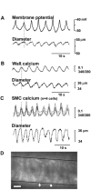
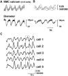
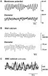
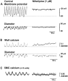
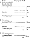
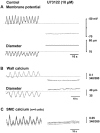
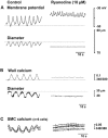
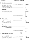
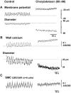
Spontaneous, rhythmical contractions, or vasomotion, can be recorded from cerebral vessels under both normal physiological and pathophysiological conditions. Using electrophysiology to study changes in membrane potential, the ratiometric calcium indicator Fura-2 AM to study changes in [Ca(2+)](i) in both the arterial wall and in individual smooth muscle cells (SMCs), and video microscopy to study changes in vessel diameter, we have investigated the cellular mechanisms underlying vasomotion in the juvenile rat basilar artery. During vasomotion, rhythmical oscillations in both membrane potential and [Ca(2+)](i) were found to precede rhythmical contractions. Nifedipine depolarized SMCs and abolished rhythmical contractions and depolarizations. [Ca(2+)](i) oscillations in the arterial wall became reduced and irregular, while [Ca(2+)](i) oscillations in adjacent SMCs were no longer synchronized. BAPTA-AM, thapsigargin and U73122 hyperpolarized SMCs, relaxed the vessel, decreased basal calcium levels and abolished vasomotion. Chloride substitution abolished rhythmical activity, depolarized SMCs, increased basal calcium levels and constricted the vessel, while niflumic acid and DIDS abolished vasomotion. Ryanodine, charybdotoxin and TRAM-34, but not iberiotoxin, 4-aminopyridine or apamin, each depolarized SMCs and increased the frequency of rhythmical depolarizations and [Ca(2+)](i) oscillations. We conclude that vasomotion in the basilar artery depends on the release of intracellular calcium from IP(3) (inositol 1,4,5,-trisphosphate)-sensitive stores which activates calcium-dependent chloride channels to depolarize SMCs. Depolarization in turn activates voltage-dependent calcium channels, synchronizing contractions of adjacent cells through influx of extracellular calcium. Subsequent calcium-induced calcium release from ryanodine-sensitive stores activates an intermediate conductance potassium channel, hyperpolarizing the SMCs and providing a negative feedback pathway for regeneration of the contractile cycle.
Figures
Similar articles
-
Differential effect of calcium-activated potassium and chloride channels on rat basilar artery vasomotion.J Huazhong Univ Sci Technolog Med Sci. 2014 Aug;34(4):482-490. doi: 10.1007/s11596-014-1303-3. Epub 2014 Aug 19. J Huazhong Univ Sci Technolog Med Sci. 2014. PMID: 25135715
-
Voltage independence of vasomotion in isolated irideal arterioles of the rat.J Physiol. 2002 Apr 1;540(Pt 1):219-29. doi: 10.1113/jphysiol.2001.013698. J Physiol. 2002. PMID: 11927681 Free PMC article.
-
Intracellular Ca(2+) release from endoplasmic reticulum regulates slow wave currents and pacemaker activity of interstitial cells of Cajal.Am J Physiol Cell Physiol. 2015 Apr 15;308(8):C608-20. doi: 10.1152/ajpcell.00360.2014. Epub 2015 Jan 28. Am J Physiol Cell Physiol. 2015. PMID: 25631870 Free PMC article.
-
Mechanisms of cellular synchronization in the vascular wall. Mechanisms of vasomotion.Dan Med Bull. 2010 Oct;57(10):B4191. Dan Med Bull. 2010. PMID: 21040688 Review.
-
Venous Vasomotion.Adv Exp Med Biol. 2019;1124:313-328. doi: 10.1007/978-981-13-5895-1_13. Adv Exp Med Biol. 2019. PMID: 31183833 Review.
Cited by
-
Role of voltage-dependent modulation of store Ca2+ release in synchronization of Ca2+ oscillations.Biophys J. 2006 Jan 1;90(1):1-23. doi: 10.1529/biophysj.104.058743. Epub 2005 Jul 22. Biophys J. 2006. PMID: 16040741 Free PMC article.
-
Synchrony of spontaneous Ca2+ activity in microvascular mural cells.J Smooth Muscle Res. 2020;56(0):1-18. doi: 10.1540/jsmr.56.1. J Smooth Muscle Res. 2020. PMID: 32249242 Free PMC article. Review.
-
Mechanism of asynchronous Ca(2+) waves underlying agonist-induced contraction in the rat basilar artery.Br J Pharmacol. 2009 Feb;156(4):587-600. doi: 10.1111/j.1476-5381.2008.00063.x. Epub 2009 Jan 16. Br J Pharmacol. 2009. PMID: 19154440 Free PMC article.
-
Properties of synchronous spontaneous Ca2+ transients in the mural cells of rat rectal arterioles.Pflugers Arch. 2017 Sep;469(9):1189-1202. doi: 10.1007/s00424-017-1978-y. Epub 2017 Apr 21. Pflugers Arch. 2017. PMID: 28429070
-
Cathepsin B is secreted apically from Xenopus 2F3 cells and cleaves the epithelial sodium channel (ENaC) to increase its activity.J Biol Chem. 2012 Aug 31;287(36):30073-83. doi: 10.1074/jbc.M111.338574. Epub 2012 Jul 10. J Biol Chem. 2012. PMID: 22782900 Free PMC article.
References
-
- Auer LM, Gallfhoffer B. Rhythmic activity of cat pial vessels in vivo. European Neurology. 1981;20:448–468. - PubMed
-
- Bertuglia S, Colantuoni A, Inaglietta M. Effects of l-NMMA and indomethacin on arteriolar vasomotion in skeletal muscle microcirculation of conscious and anaethetized hamsters. Microvasular Research. 1994;48:68–84. - PubMed
-
- Bonev AD, Jaggar JH, Rubart M, Nelson MT. Activators of protein kinase C decrease Ca2+ spark frequency in smooth muscle cells from cerebral arteries. American Journal of Physiology. 1997;273:C2090–2095. - PubMed
-
- Bouskela E, Grampp W. Spontaneous vasomotion in hamster cheek pouch arterioles in varying experimental conditions. American Journal of Physiology. 1992;262:H478–485. - PubMed
MeSH terms
Substances
LinkOut - more resources
Full Text Sources
Miscellaneous

