

Excitability of human muscle afferents studied using threshold tracking of the H reflex
- PMID: 12456841
- PMCID: PMC2290676
- DOI: 10.1113/jphysiol.2002.026526
Excitability of human muscle afferents studied using threshold tracking of the H reflex
Abstract
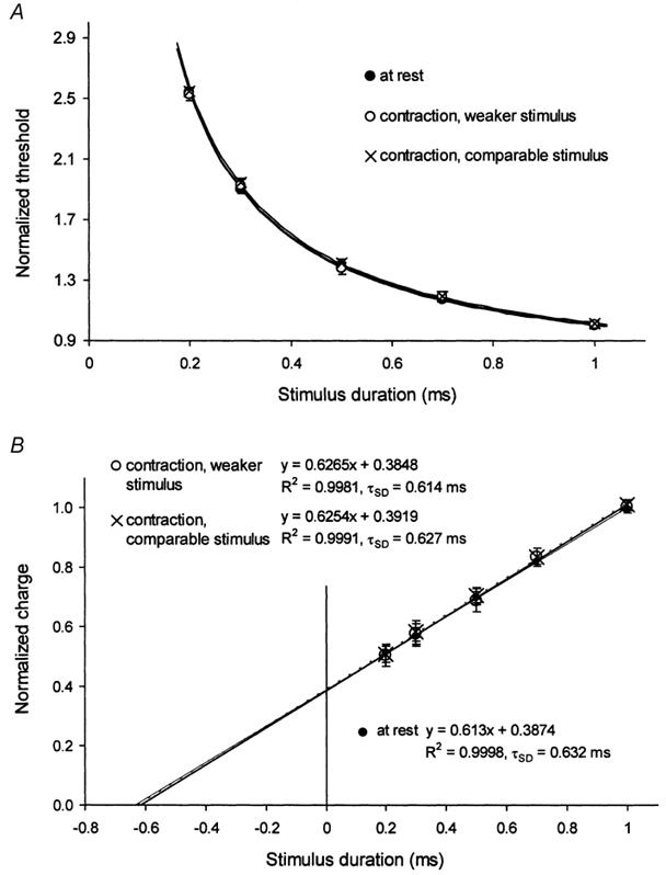
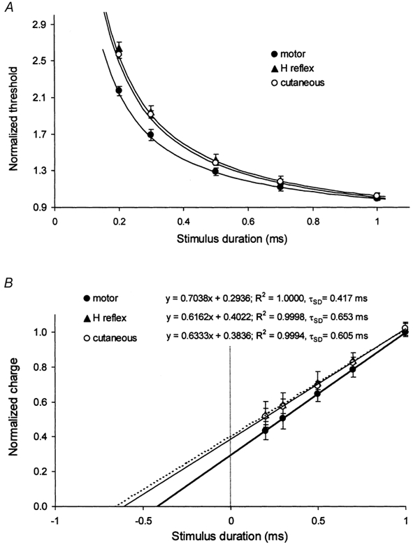
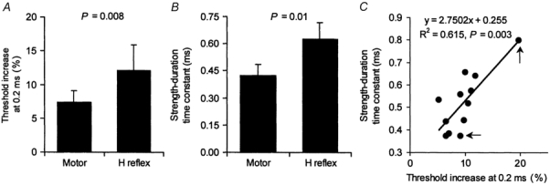
In human peripheral nerves, physiological evidence has been presented for a number of biophysical differences between cutaneous afferents and alpha motor axons. The differences in strength-duration properties for cutaneous afferents and motor axons in the median nerve have been attributed to greater expression of a persistent Na(+) conductance (I(Na,P)) on cutaneous afferents. However, it is unclear whether the biophysical properties of human group Ia afferents differ from those of cutaneous afferents. The present studies were undertaken to determine whether the properties of human group Ia afferents can be studied indirectly using 'threshold tracking' to measure the excitability changes in the H reflex, and to determine whether the excitability of group Ia afferents differs from that of cutaneous afferents. The strength-duration properties of the soleus H reflex and soleus motor axons were measured at rest and during sustained voluntary contractions. Similar experiments were performed on the median nerve at the wrist to study the strength-duration properties of cutaneous afferents, alpha motor axons and H reflex of the thenar muscles. In addition, the technique of 'latent addition' was used to determine whether there was a difference in a low-threshold conductance on soleus Ia afferent and motor axons. The present findings indicate that the strength-duration time constant (tau(SD)) for the H reflex is longer than that for alpha motor axons, but similar to that for cutaneous afferents. There were no differences in tau(SD) for the soleus H reflex at rest and during contractions, suggesting that tau(SD) for the H reflex is largely unaffected by changes in synaptic or motoneurone properties. Finally, the difference in latent addition suggests that the longer tau(SD) of the soleus H reflex may indeed be due to greater activity of a persistent Na(+) conductance on Ia afferents than on soleus alpha motor axons.
Figures

Similar articles
-
Modulation of transmission in the corticospinal and group Ia afferent pathways to soleus motoneurons during bicycling.J Neurophysiol. 2003 Jan;89(1):304-14. doi: 10.1152/jn.00386.2002. J Neurophysiol. 2003. PMID: 12522181
-
Effects of hip joint angle changes on intersegmental spinal coupling in human spinal cord injury.Exp Brain Res. 2005 Dec;167(3):381-93. doi: 10.1007/s00221-005-0046-6. Epub 2005 Jul 30. Exp Brain Res. 2005. PMID: 16059682 Free PMC article. Clinical Trial.
-
Differences in activity-dependent hyperpolarization in human sensory and motor axons.J Physiol. 2004 Jul 1;558(Pt 1):341-9. doi: 10.1113/jphysiol.2004.063966. Epub 2004 May 14. J Physiol. 2004. PMID: 15146048 Free PMC article.
-
Contributions to the understanding of gait control.Dan Med J. 2014 Apr;61(4):B4823. Dan Med J. 2014. PMID: 24814597 Review.
-
Revisiting the use of Hoffmann reflex in motor control research on humans.Eur J Appl Physiol. 2023 Apr;123(4):695-710. doi: 10.1007/s00421-022-05119-7. Epub 2022 Dec 26. Eur J Appl Physiol. 2023. PMID: 36571622 Review.
Cited by
-
Modulation of motor unit discharge rate and H-reflex amplitude during submaximal fatigue of the human soleus muscle.Exp Brain Res. 2004 Oct;158(3):345-55. doi: 10.1007/s00221-004-1907-0. Epub 2004 May 14. Exp Brain Res. 2004. PMID: 15146306
-
Clinical uses of H reflexes of upper and lower limb muscles.Clin Neurophysiol Pract. 2016 Apr 7;1:9-17. doi: 10.1016/j.cnp.2016.02.003. eCollection 2016. Clin Neurophysiol Pract. 2016. PMID: 30214954 Free PMC article. Review.
-
Stretch reflexes in human abdominal muscles.Exp Brain Res. 2004 Nov;159(2):206-13. doi: 10.1007/s00221-004-1948-4. Epub 2004 Jul 17. Exp Brain Res. 2004. PMID: 15258713
-
Prolonged infrapatellar tendon vibration does not influence quadriceps maximal or explosive isometric force production in man.Eur J Appl Physiol. 2014 Aug;114(8):1757-66. doi: 10.1007/s00421-014-2904-z. Epub 2014 May 21. Eur J Appl Physiol. 2014. PMID: 24846679
-
Gene expression in muscle in response to exercise.J Muscle Res Cell Motil. 2003;24(2-3):121-6. doi: 10.1023/a:1026041228041. J Muscle Res Cell Motil. 2003. PMID: 14609023 Review.
References
-
- Baker MD, Bostock H. Low-threshold, persistent sodium current in rat large dorsal root ganglion neurons in culture. Journal of Neurophysiology. 1997;77:1503–1513. - PubMed
-
- Bostock H, Baker M. Evidence for two types of potassium channel in human motor axons in vivo. Brain Research. 1988;462:354–358. - PubMed
-
- Bostock H, Burke D, Hales JP. Differences in behaviour of sensory and motor axons following release of ischaemia. Brain. 1994;117:225–234. - PubMed
-
- Bostock H, Cikurel K, Burke D. Threshold tracking techniques in the study of human peripheral nerve. Muscle and Nerve. 1998;21:137–158. - PubMed
Publication types
MeSH terms
LinkOut - more resources
Full Text Sources