Guiding neuronal growth with light
- PMID: 12456879
- PMCID: PMC138558
- DOI: 10.1073/pnas.252631899
Guiding neuronal growth with light
Abstract
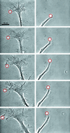
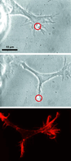
Control over neuronal growth is a fundamental objective in neuroscience, cell biology, developmental biology, biophysics, and biomedicine and is particularly important for the formation of neural circuits in vitro, as well as nerve regeneration in vivo [Zeck, G. & Fromherz, P. (2001) Proc. Natl. Acad. Sci. USA 98, 10457-10462]. We have shown experimentally that we can use weak optical forces to guide the direction taken by the leading edge, or growth cone, of a nerve cell. In actively extending growth cones, a laser spot is placed in front of a specific area of the nerve's leading edge, enhancing growth into the beam focus and resulting in guided neuronal turns as well as enhanced growth. The power of our laser is chosen so that the resulting gradient forces are sufficiently powerful to bias the actin polymerization-driven lamellipodia extension, but too weak to hold and move the growth cone. We are therefore using light to control a natural biological process, in sharp contrast to the established technique of optical tweezers [Ashkin, A. (1970) Phys. Rev. Lett. 24, 156-159; Ashkin, A. & Dziedzic, J. M. (1987) Science 235, 1517-1520], which uses large optical forces to manipulate entire structures. Our results therefore open an avenue to controlling neuronal growth in vitro and in vivo with a simple, noncontact technique.
Figures


Similar articles
-
Optical neuronal guidance.Methods Cell Biol. 2007;83:495-520. doi: 10.1016/S0091-679X(07)83021-4. Methods Cell Biol. 2007. PMID: 17613322
-
Controlled induction, enhancement, and guidance of neuronal growth cones by use of line optical tweezers.Opt Lett. 2005 Oct 1;30(19):2596-8. doi: 10.1364/ol.30.002596. Opt Lett. 2005. PMID: 16208911
-
Optical neuronal guidance in three-dimensional matrices.J Neurosci Methods. 2009 May 15;179(2):278-83. doi: 10.1016/j.jneumeth.2009.02.004. Epub 2009 Feb 21. J Neurosci Methods. 2009. PMID: 19428538 Free PMC article.
-
Actin dynamics in growth cone motility and navigation.J Neurochem. 2014 Apr;129(2):221-34. doi: 10.1111/jnc.12506. Epub 2013 Nov 17. J Neurochem. 2014. PMID: 24164353 Free PMC article. Review.
-
Micro-scale chromophore-assisted laser inactivation of nerve growth cone proteins.Microsc Res Tech. 2000 Jan 15;48(2):97-106. doi: 10.1002/(SICI)1097-0029(20000115)48:2<97::AID-JEMT5>3.0.CO;2-G. Microsc Res Tech. 2000. PMID: 10649510 Review.
Cited by
-
In-situ guidance of individual neuronal processes by wet femtosecond-laser processing of self-assembled monolayers.Appl Phys Lett. 2011 Oct 17;99(16):163701. doi: 10.1063/1.3651291. Appl Phys Lett. 2011. PMID: 27703280 Free PMC article.
-
Influence of distant femtosecond laser pulses on growth cone fillopodia.Cytotechnology. 2008 Oct;58(2):103-11. doi: 10.1007/s10616-008-9178-4. Epub 2008 Dec 16. Cytotechnology. 2008. PMID: 19085066 Free PMC article.
-
Visible light regulates neurite outgrowth of nerve cells.Cytotechnology. 2007 Jul;54(3):181-8. doi: 10.1007/s10616-007-9087-y. Epub 2007 Aug 15. Cytotechnology. 2007. PMID: 19003010 Free PMC article.
-
Optically guided neuronal growth at near infrared wavelengths.Opt Express. 2006 Oct 16;14(21):9786-93. doi: 10.1364/oe.14.009786. Opt Express. 2006. PMID: 19529370 Free PMC article.
-
Water-filtered infrared-A (wIRA) in acute and chronic wounds.GMS Krankenhhyg Interdiszip. 2009 Dec 16;4(2):Doc12. doi: 10.3205/dgkh000137. GMS Krankenhhyg Interdiszip. 2009. PMID: 20204090 Free PMC article.
References
-
- Bomze H. M., Bulsara, K. R., Iskandar, B. J., Caroni, P. & Skene, J. H. (2001) Nat. Neurosci. 4, 38-43. - PubMed
-
- Tessier-Lavigne M. & Goodman, C. S. (1996) Science 274, 1123-1133. - PubMed
-
- Mueller B. K. (1999) Annu. Rev. Neurosci. 22, 351-388. - PubMed
-
- Hong K., Nishiyama, M., Henley, J., Tessier-Lavigne, M. & Poo, M. (2000) Nature 403, 93-98. - PubMed
-
- Pantaloni D., Le Clainche, C. & Carlier, M.-F. (2001) Science 292, 1502-1506. - PubMed
Publication types
MeSH terms
Substances
LinkOut - more resources
Full Text Sources
Other Literature Sources

