. 2002 Dec 10;99(25):16255-60.
doi: 10.1073/pnas.252630899. Epub 2002 Nov 27.

Human cytomegalovirus gene expression during infection of primary hematopoietic progenitor cells: a model for latency

Affiliations

Human cytomegalovirus gene expression during infection of primary hematopoietic progenitor cells: a model for latency

Felicia D Goodrum et al. Proc Natl Acad Sci U S A. .

Abstract

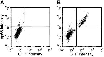
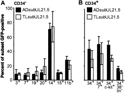
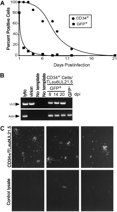
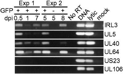
Human cytomegalovirus (HCMV) resides latently in hematopoietic cells of the bone marrow. Although viral genomes can be found in CD14+ monocytes and CD34+ progenitor cells, the primary reservoir for latent cytomegalovirus is unknown. We analyzed human hematopoietic subpopulations infected in vitro with a recombinant virus that expresses a green fluorescent protein marker gene. Although many hematopoietic cell subsets were infected in vitro, CD14+ monocytes and various CD34+ subpopulations were infected with the greatest efficiency. We have developed an in vitro system in which to study HCMV infection and latency in CD34+ cells cultured with irradiated stromal cells. Marker gene expression was substantially reduced by 4 days postinfection, and infectious virus was not made during the culture period. However, viral DNA sequences were maintained in infected CD34+ cells for >20 days in culture, and, importantly, virus replication could be reactivated by coculture with human fibroblasts. Using an HCMV gene array, we examined HCMV gene expression in CD34+ cells. The pattern of viral gene expression was distinct from that observed during productive or nonproductive infections. Some of these expressed viral genes may function in latency and are targets for further analysis. Altered gene expression in hematopoietic progenitors may be indicative of the nature and outcome of HCMV infection.


Figures

Fig 1.
Fig 1.
GFP is a reliable marker for TLsubUL21.5 infection of hematopoietic cells. Mononuclear cells enriched for CD34+ cells (≈60%) were mock-infected (A) or infected with TLsubUL21.5 at a multiplicity of 5 plaque-forming units (pfu) per cell (B). At 10 h postinfection, cells were analyzed for expression of GFP and staining for pp65 by flow cytometry. Data from a representative experiment are shown.
Fig 2.
Fig 2.
Immunophenotypes of hematopoietic cells infected with ADsubUL21.5 or TLsubUL21.5. Mononuclear cells were infected at a multiplicity of 5 pfu per cell. At 16–18 h postinfection, cells were labeled with fluorescently conjugated antibodies to distinguish hematopoietic subsets. The percentage of each subset infected (GFP+) was determined by flow cytometry. (A) Analysis of CD34-depleted cells. CD3 and CD7 cell surface markers identify T lymphocytes; CD19 and CD20 identify B lymphocytes; CD14 identifies monocytes; CD15 identifies neutrophils; and CD11b identifies predominantly granulocytes and monocytes. (B) Analysis of CD34-enriched cells containing progenitor cells. CD34+/CD7+ cells and CD34+/c-kit+ cells are subsets of progenitor cells. CD34+/CD38−/lineage-negative cells represent a rare progenitor subset containing stem cells. Each bar represents an average of three to five independent experiments. Standard errors are indicated.
Fig 3.
Fig 3.
In vitro model for HCMV latency. Mononuclear cells were infected with TLsubUL21.5 at a multiplicity of 5 pfu per cell. Infected CD34+ cells were isolated at 16–20 h postinfection and put into long-term bone marrow culture. (A) Time course of CD34 and GFP expression during long-term bone marrow culture. (B) Detection of UL5 and actin sequences from infected cells during long-term bone marrow culture by PCR. (C) Reactivation of HCMV replication in infected hematopoietic cells after coculture with fibroblasts. Fibroblasts were cocultured with infected CD34+ cells from long-term bone marrow cultures (Upper) or with a lysate prepared from an equivalent number of cells (Lower), and reactivation was monitored by GFP fluorescence. Representative data are shown from three independent experiments.
Fig 4.
Fig 4.
Gene arrays reveal cell-type-specific differences in HCMV RNA expression. Representative array segments with cDNAs corresponding to HCMV genes UL1 through UL79 are displayed. Fibroblasts were infected at a multiplicity of 5 pfu per cell, and viral RNAs were analyzed at 72 h postinfection (Upper). CD34+ cells were infected at the 10 pfu per cell and RNAs were analyzed at 5 days postinfection (Lower).
Fig 5.
Fig 5.
HCMV gene expression in CD34+ cells was confirmed by RT-PCR. cDNA prepared from two independent experiments was analyzed for the expression of IRL3, UL5, UL40, UL64, UL106, and US23. IRL3, UL5, UL40, and UL64 were detected in CD34+ cells by array analysis. UL106 and US23 were not detected by array analysis. Controls included GFP− CD34+ cells in experiment 2, an RNA sample that received no reverse transcriptase during cDNA amplification, DNA from a lytic infection, and cDNA from a lytic and mock infection.

References

    1. Britt W. J. & Alford, C. A. (1996) in Fields Virology, eds. Fields, B. N., Knipe, D. M. & Howley, P. M. (Lippincott–Raven, Philadelphia), Vol. 2, pp. 2493–2524.
    1. Sinzger C., Grefte, A., Plachter, B., Gouw, A. S., The, T. H. & Jahn, G. (1995) J. Gen. Virol. 76, 741-750. - PubMed
    1. Soderberg C., Larsson, S., Bergstedt-Lindqvist, S. & Moller, E. (1993) J. Virol. 67, 3166-3175. - PMC - PubMed
    1. Schrier R. D., Nelson, J. A. & Oldstone, M. B. (1985) Science 230, 1048-1051. - PubMed
    1. Boeckh M., Hoy, C. & Torok-Storb, B. (1998) Clin. Infect. Dis. 26, 209-210. - PubMed

Publication types

MeSH terms