

Crystal structure of a DNA decamer containing a cis-syn thymine dimer
- PMID: 12456887
- PMCID: PMC138548
- DOI: 10.1073/pnas.242422699
Crystal structure of a DNA decamer containing a cis-syn thymine dimer
Abstract
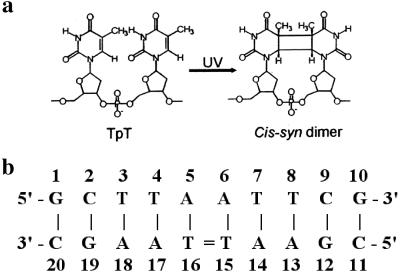
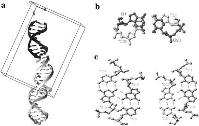
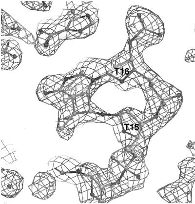
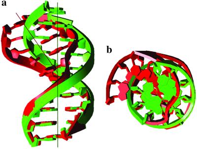
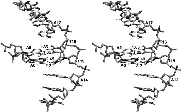
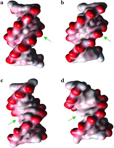
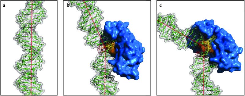
It is well known that exposure to UV induces DNA damage, which is the first step in mutagenesis and a major cause of skin cancer. Among a variety of photoproducts, cyclobutane-type pyrimidine photodimers (CPD) are the most abundant primary lesion. Despite its biological importance, the precise relationship between the structure and properties of DNA containing CPD has remained to be elucidated. Here, we report the free (unbound) crystal structure of duplex DNA containing a CPD lesion at a resolution of 2.0 A. Our crystal structure shows that the overall helical axis bends approximately 30 degrees toward the major groove and unwinds approximately 9 degrees, in remarkable agreement with some previous theoretical and experimental studies. There are also significant differences in local structure compared with standard B-DNA, including pinching of the minor groove at the 3' side of the CPD lesion, a severe change of the base pair parameter in the 5' side, and serious widening of both minor and major groves both 3' and 5' of the CPD. Overall, the structure of the damaged DNA differs from undamaged DNA to an extent that DNA repair proteins may recognize this conformation, and the various components of the replicational and transcriptional machinery may be interfered with due to the perturbed local and global structure.
Figures
References
-
- Cadet J. & Vigny, P., (1990) The Photochemistry of Nucleic Acids (Wiley, New York).
-
- Cleaver J. E. & Crowley, E. (2002) Front. Biosci. 7, 1024-1043. - PubMed
-
- Beukers R., Ylstra, J. & Berends, W. (1960) Rec. Trav. Chim. 79, 101-104.
-
- Wacker A., Dellweg, H. & Weinblum, D. (1960) Naturwissenschaften 47, 477.
-
- Setlow R. B. & Carrier, W. L. (1966) J. Mol. Biol. 17, 237-254. - PubMed
Publication types
MeSH terms
Substances
Associated data
- Actions
- Actions
Grants and funding
LinkOut - more resources
Full Text Sources
Miscellaneous
