. 2002 Dec 10;99(25):15908-13.
doi: 10.1073/pnas.212628899. Epub 2002 Nov 27.

An mRNA structure that controls gene expression by binding FMN

Affiliations

An mRNA structure that controls gene expression by binding FMN

Wade C Winkler et al. Proc Natl Acad Sci U S A. .

Abstract

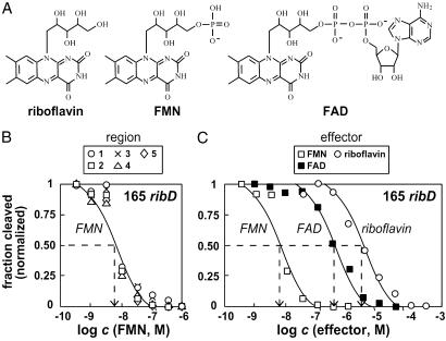
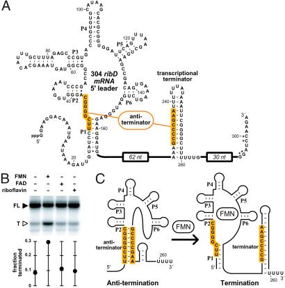
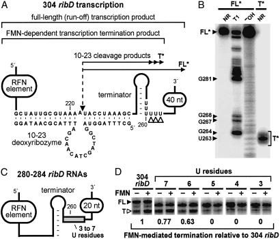
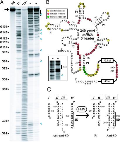
The RFN element is a highly conserved domain that is found frequently in the 5'-untranslated regions of prokaryotic mRNAs that encode for flavin mononucleotide (FMN) biosynthesis and transport proteins. We report that this domain serves as the receptor for a metabolite-dependent riboswitch that directly binds FMN in the absence of proteins. Our results also indicate that in Bacillus subtilis, the riboswitch most likely controls gene expression by causing premature transcription termination of the ribDEAHT operon and precluding access to the ribosome-binding site of ypaA mRNA. Sequence and structural analyses indicate that the RFN element is a natural FMN-binding aptamer, the allosteric character of which is harnessed to control gene expression.


Figures

Fig 1.
Fig 1.
FMN-induced structure modulation of an RFN element. (A) Structural model and conserved sequences of the RFN element derived from a phylogenetic analysis of prokaryote mRNA sequences. Nucleotides defined by letters are present in >90% of the RNAs examined. The letters R, Y, K, and N represent purine, pyrimidine, G or U, and any nucleotide identity, respectively. The six stem–loop structures are labeled P1–P6. The original model and the sequence data used to create this adaptation are described in ref. . (B) Structural probing of the 165 ribD RNA (arrow) in the presence (+) and absence (−) of 10 μM FMN. Also, 5′ 32P-labeled ribD RNA precursors were not reacted (NR) or were subjected to partial digest with RNase T1 (T1) or alkali (-OH) as indicated. Blue arrowheads indicate regions of FMN-dependent modulation of spontaneous cleavage. Regions 1 (nucleotide 136), 2 (nucleotides 113 and 114), 3 (nucleotides 89–91), 4 (nucleotide 56), and 5 (nucleotides 33 and 34) were subsequently used to establish an apparent KD value for FMN binding (Fig. 2). See Materials and Methods for experimental details. (C) Secondary-structure model of the 165 ribD RNA and the nucleotide positions of spontaneous cleavage in the presence (yellow) and absence (yellow and red) of FMN as identified from B. Cleavage occurs 3′ relative to the nucleotides highlighted in color.
Fig 2.
Fig 2.
Affinities and specificity of the 165 ribD RNA. (A) Chemical structures of riboflavin, FMN, and FAD. (B) Determination of the apparent KD value for FMN binding to the 165 ribD RNA. The extent of spontaneous RNA cleavage (normalized relative to the highest and lowest cleavage values measured for each region) was plotted for five regions (Fig. 1B) that exhibit FMN-dependent modulation. The dashed line identifies the apparent KD value or the concentration of FMN required for conversion of half the RNAs into the ligand-bound form. RNAs were subjected to in-line probing as described for Fig. 1 by using various concentrations of ligand as indicated. (C) The normalized fraction of spontaneous cleavage at region two for FMN (from B) and for the related compounds FAD and riboflavin as indicated. Details are as described for B.
Fig 3.
Fig 3.
FMN causes transcription termination of the ribD RNA in vitro. (A) Sequence and secondary structure of the 304 ribD RNA. Shaded regions identify nucleotides that are complementary and might serve as an antiterminator structure. Nucleotides denoted with an asterisk have been altered from the wild-type sequence to generate a restriction site. The nucleotide sequence comprising the 62- and 30-nt regions (not shown) are presented in refs. and . (B) In vitro transcription termination assays. In vitro transcriptions were conducted by using T7 RNA polymerase and a double-stranded DNA construct that serves as a template for the synthesis of 304 ribD RNA. Reactions were incubated in the absence (−) or presence (+) of 100 μM of the compounds as indicated for each lane. FL and T denote full-length and terminated RNA transcripts, respectively. The fraction of total transcripts that are termination products is plotted in the graph. See Materials and Methods for experimental details. (C) Model for the FMN-dependent riboswitch. Shaded regions identify the putative antiterminator structure that is disrupted after binding of FMN and formation of the P1 structure.
Fig 4.
Fig 4.
Mapping of the transcription termination site and the importance of the U-rich domain. (A) The 304 ribD construct (see also Fig. 3A) and processed fragments used to map the transcription termination site. The nucleotides presented depict the interaction with a 10–23 deoxyribozyme and the U-rich region where transcription termination was expected to occur. The dashed arrow identifies the location of deoxyribozyme-mediated cleavage. The portions of the construct that correspond to the full-length product, the FMN-induced transcription product and the deoxyribozyme digestion products FL* and T* that are derived from the full-length and terminated RNAs, respectively, are also identified. The open arrowheads identify the site of FMN-modulated transcription termination. (B) PAGE analysis of the 5′ 32P-labeled FL* and T* RNAs. RNAs were subjected to no additional reaction (NR) or were subjected to partial digestion with RNase T1 (T1) or alkali (-OH) as indicated for each lane. Bands corresponding to the FL* and T* RNAs, along with several T1 digestion products, are identified. The T* RNA corresponds to termination at nucleotides U261–U263, the mobility of which differs from the markers by 1 nt equivalent due to the absence of a 2′,3′-cyclic phosphate (unpublished data). (C) Schematic representation of ribD RNAs ranging from 280 to 284 nucleotides that carry three through seven U residues, respectively, in the U-rich domain (shaded box). Other than the U insertion or deletions, the RNA is identical in sequence to that of nucleotide 1–284 of the 304 ribD RNA. (D) Transcription termination assays with templates that encode the 304 ribD RNA and for 280–284 ribD variants that carry three through seven U residues, respectively, as indicated for each lane. Transcription reactions were conducted as described for Fig. 3B and incubated in the absence (−) or presence (+) of 100 μM FMN. FL and T denote full-length and terminated RNA transcripts, respectively. The numbers below each gel image reflect the relative level of FMN-induced termination compared with the nonmutated 304 ribD construct.
Fig 5.
Fig 5.
FMN-induced structure modulation of the ypaA riboswitch. (A) Structural probing of the 349 ypaA RNA (arrow) in the presence (+) and absence (−) of 100 μM FMN. The bar identifies the region of cleavage products that is expanded in B Inset. Additional details are as described for Fig. 1B. (B) Secondary-structure model of the 349 ypaA RNA and the nucleotide positions of spontaneous cleavage in the presence (yellow and green) and absence (yellow and red) of FMN as identified from A. Nucleotides marked with an asterisk have been altered from the wild-type sequence to generate a restriction site. Sequences not depicted in the 133- and 45-nt domains can be obtained from the B. subtilis genome sequence (23). (Inset) An image of an extended PAGE separation of the region encompassing the SD element. (C) Proposed mechanism for the FMN-modulated formation of structures within the expression platform that control translation. RNA domains ii and iii form in the absence of FMN, thus exposing the SD element for ribosome binding. In contrast, FMN binding requires the formation of P1, which occupies RNA domain ii and permits the sequestration of the SD element by RNA domain iii.

References

    1. Lundrigan M. D., Köster, W. & Kadner, R. J. (1991) Proc. Natl. Acad. Sci. USA 88, 1479-1483. - PMC - PubMed
    1. Nahvi A., Sudarsan, N., Ebert, M. S., Zou, X., Brown, K. L. & Breaker, R. R. (2002) Chem. Biol. 9, 1043-1049. - PubMed
    1. Miranda-Rios J., Navarro, M. & Soberón, M. (2001) Proc. Natl. Acad. Sci. USA 98, 9736-9741. - PMC - PubMed
    1. Winkler W., Nahvi, A. & Breaker, R. R. (2002) Nature 419, 952-956. - PubMed
    1. Henkin T. M. (2000) Curr. Opin. Microbiol. 3, 149-153. - PubMed

Publication types

MeSH terms

LinkOut - more resources