. 2002 Nov;27(6):991-1002.
doi: 10.1053/jhsu.2002.35878.

Biomechanical evaluation of ligamentous stabilizers of the scaphoid and lunate

Affiliations

Biomechanical evaluation of ligamentous stabilizers of the scaphoid and lunate

Walter H Short et al. J Hand Surg Am. 2002 Nov.

Abstract

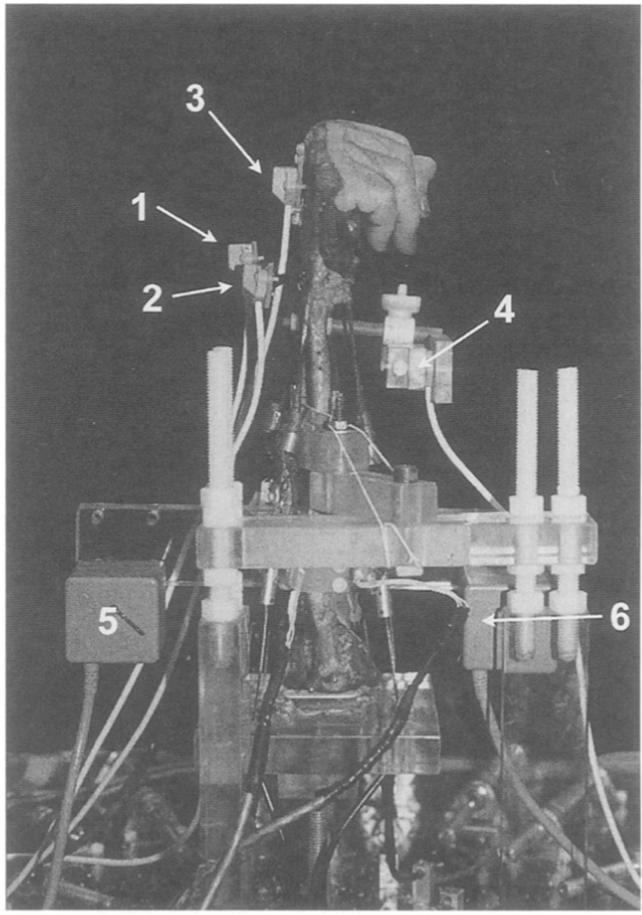
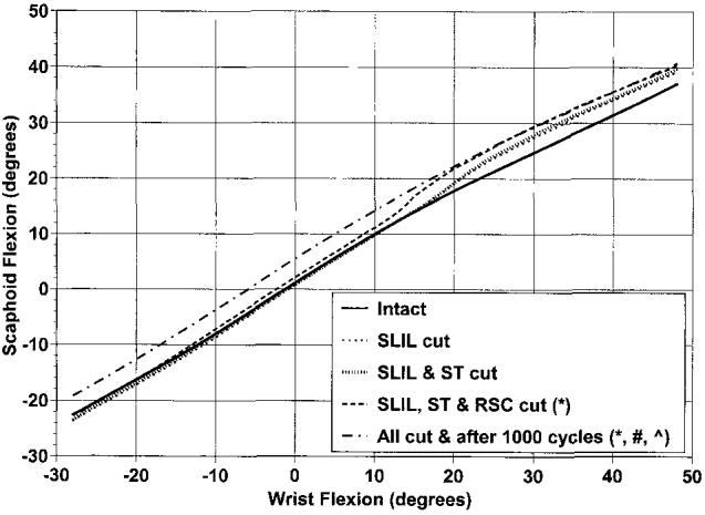
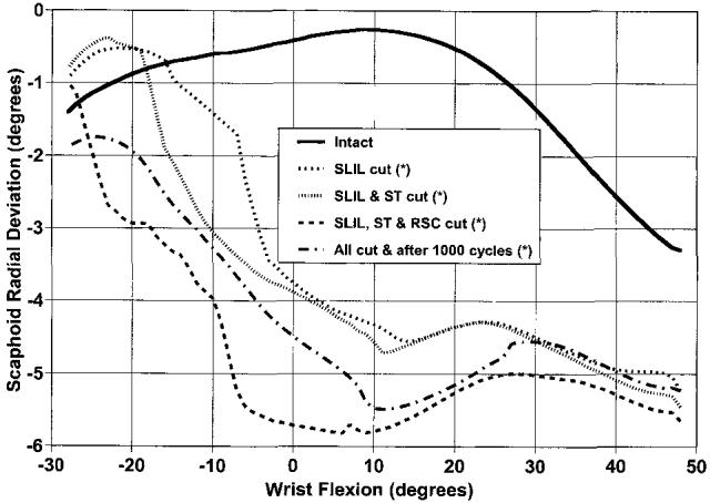
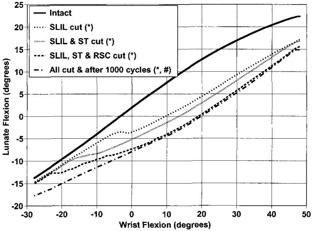
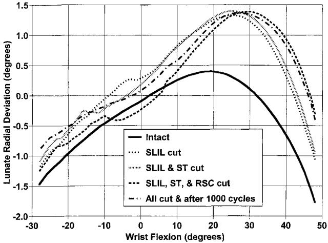
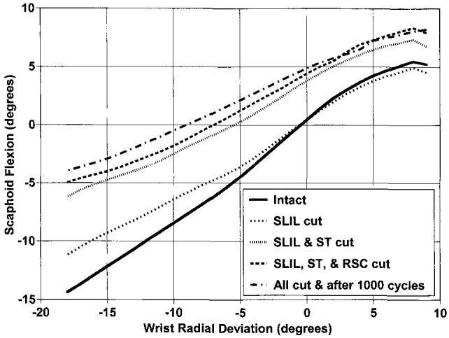
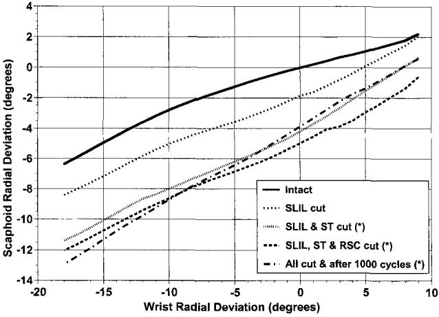
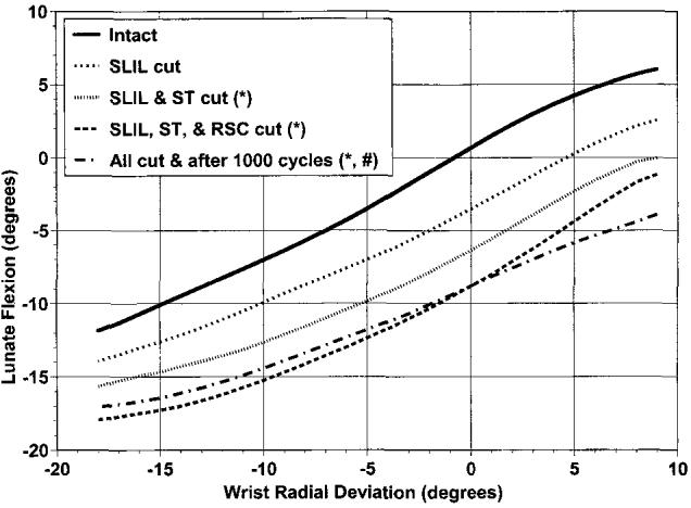
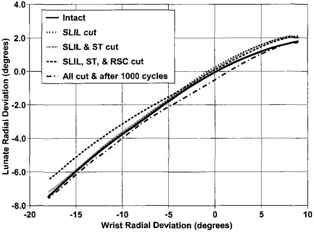
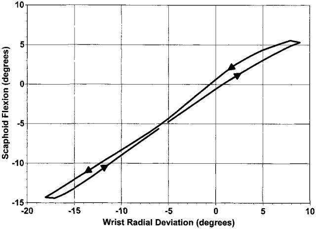
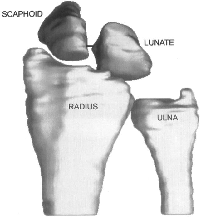
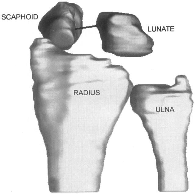
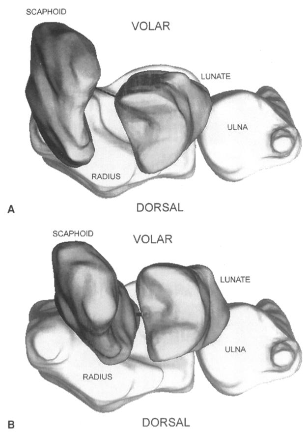
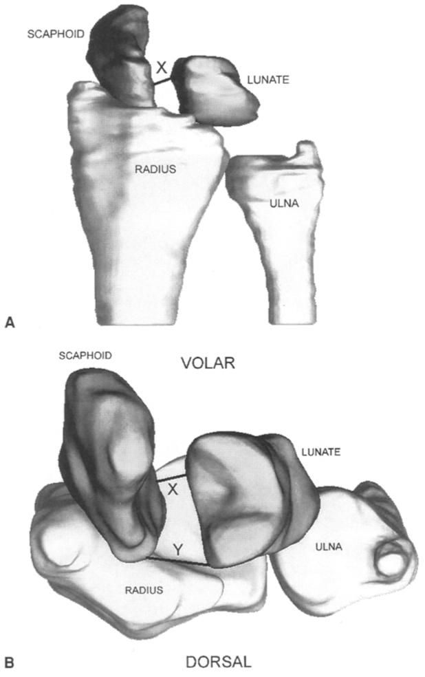
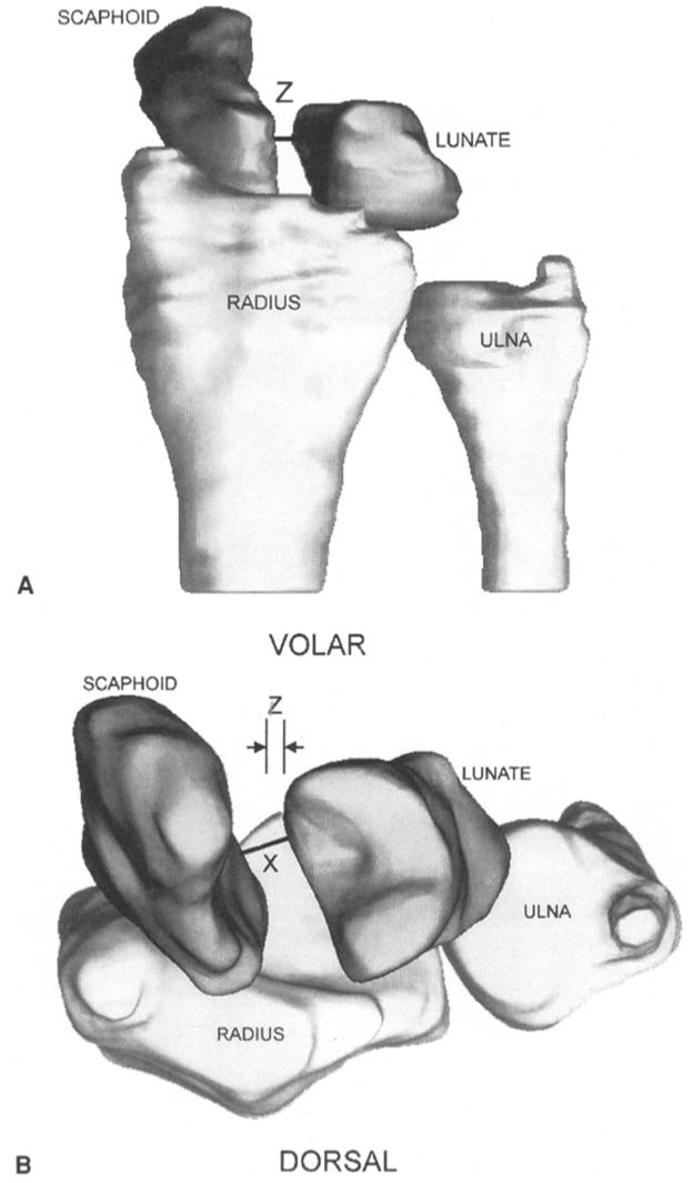
This study evaluated the effects of sectioning the scapholunate interosseous ligament, radioscaphocapitate ligament, and scaphotrapezial ligament on the kinematics of the scaphoid and lunate. Eight cadaver upper extremities were placed in a wrist joint simulator and moved in continuous cycles of flexion-extension and radial-ulnar deviation. Positional data of the scaphoid and lunate were obtained in the intact state, after the scapholunate ligament was cut; after the scapholunate and scaphotrapezial ligaments were cut; after the scapholunate, scaphotrapezial, and radioscaphocapitate ligaments were cut; and after all 3 ligaments were cut and the specimen was placed through an additional 1,000 cycles of flexion-extension. Cutting the scapholunate ligament caused changes in scaphoid and lunate motion during flexion-extension, but not radial-ulnar deviation. Additional sectioning of the scaphotrapezial ligament followed by the radioscaphocapitate ligament caused further kinematic changes in these carpal bones. One thousand cycles of motion after all 3 ligaments were sectioned caused additional kinematic changes in the scaphoid and lunate. The scapholunate ligament appears to be the primary stabilizer between the scaphoid and lunate. The radioscaphocapitate and scaphotrapezial ligaments are secondary restraints. Repetitive cyclic motion after ligament sectioning appears to have additional deleterious effects on carpal kinematics.


Figures

Figure 1
Figure 1
Experimental setup. Wrist joint motion simulator with Fastrak sensors mounted on scaphoid (1), lunate (2), third metacarpal (3), and distal radius (4). The Fastrak electromagnetic sources are mounted on the ulna dorsally (5) for use with the third metacarpal sensor and volarly (6) for use with the scaphoid, lunate, and radial sensors.
Figure 2
Figure 2
Average scaphoid flexion and extension as a function of wrist flexion and extension in the intact wrist and after sequential ligamentous sectioning. The graph represents the motion as the wrist moves from wrist extension to flexion. Flexion is positive. *Scaphoid position is statistically different from intact specimen as the specimen is moving from maximum flexion to neutral. #Scaphoid position is statistically different from the intact specimen as the wrist was going into and coming out of flexion. ^Scaphoid position is statistically different when compared with SLIL cut specimen when the wrist was going into and out of extension.
Figure 3
Figure 3
Average scaphoid radial and ulnar deviation as a function of wrist flexion and extension in the intact wrist and after sequential ligamentous sectioning. The graph represents the motion as the wrist moves from wrist flexion to extension. Flexion is positive, radial deviation is positive. *Scaphoid position is statistically different from intact specimen when the wrist is moving from flexion to extension.
Figure 4
Figure 4
Average lunate flexion and extension as a function of wrist flexion and extension in the intact wrist and after sequential ligamentous sectioning. The graph represents the motion as the wrist moves from wrist flexion to extension. Flexion is positive. *Lunate position is statistically different from intact specimen during wrist flexion. #Lunate position is statistically different from intact specimen when the wrist is extended.
Figure 5
Figure 5
Average lunate radial and ulnar deviation as a function of wrist flexion and extension in the intact wrist and after sequential ligamentous sectioning. The graph represents the motion as the wrist moves from wrist flexion to extension. Flexion is positive, radial deviation is positive.
Figure 6
Figure 6
Average scaphoid flexion and extension as a function of wrist radial and ulnar deviation in the intact wrist and after sequential ligamentous sectioning. The graph represents the motion as the wrist moves from radial to ulnar deviation. Flexion is positive, radial deviation is positive.
Figure 7
Figure 7
Average scaphoid radial and ulnar deviation as a function of wrist radial and ulnar deviation in the intact wrist and after sequential ligamentous sectioning. The graph represents the motion as the wrist moves from radial to ulnar deviation. Radial deviation is positive. *Scaphoid position is statistically different when compared with intact or SLIL-sectioned specimen as the wrist is going into ulnar deviation.
Figure 8
Figure 8
Average lunate flexion and extension as a function of wrist radial and ulnar deviation in the intact wrist and after sequential ligamentous sectioning. The graph represents the motion as the wrist moves from radial to ulnar deviation. Flexion is positive, radial deviation is positive. *Lunate position is statistically different when compared with intact or SLIL-sectioned specimen throughout the entire radial-ulnar motion. #Further statistical difference when compared with the SLIL-sectioned specimen during nearly all of wrist radial and ulnar deviation.
Figure 9
Figure 9
Average lunate radial and ulnar deviation as a function of wrist radial and ulnar deviation. The graph represents the motion as the wrist moves from radial to ulnar deviation. Radial deviation is positive.
Figure 10
Figure 10
Average scaphoid flexion and extension as a function of wrist radial and ulnar deviation in the intact wrist. The direction of wrist motion is shown with the arrowhead.
Figure 11
Figure 11
Dorsal view of the distal radius, ulna, scaphoid, and lunate for one intact wrist in 50° of flexion. The line connecting the scaphoid and lunate connects the points on the bones where the minimum distance (1.7 mm) of bones occurs. In some views part of the minimum distance line may be hidden by the scaphoid or lunate.
Figure 12
Figure 12
Dorsal view of the distal radius, ulna, scaphoid, and lunate after the SLIL, ST, and RSC ligaments have been sectioned. This is the same wrist as in Fig. 11 and is also in 50° of flexion. The line connecting the scaphoid and lunate indicates the minimum distance (6.0 mm) between the bones. In some views part of the minimum distance line may be hidden by the scaphoid or lunate.
Figure 13
Figure 13
Visualization of the scaphoid clunk after all 3 ligaments have been sectioned (A) Three-dimensional transverse view of one wrist in neutral wrist flexion-extension as the wrist is going from flexion into extension. The scaphoid has risen up onto the dorsal rim of the radius. The minimum distance between the bones is 3.8 mm as shown by the line connecting the bones. In some views part of the minimum distance line may be hidden by the scaphoid or lunate. (B) Three-dimensional transverse view of the same wrist but with the wrist in 20° of wrist extension as the wrist is moving from maximum extension toward neutral. Here the scaphoid has jumped back into the radioscaphoid fossa. The minimum distance between the bones is 1.0 mm as shown by the line connecting the bones.
Figure 14
Figure 14
Variation in gap measurement between the scaphoid and lunate depends on where it is measured. (A) In a simulated typical radiographic measurement of the gap between the scaphoid and lunate in a wrist, in neutral flexion-extension, with the SLIL, ST, and RSC ligaments sectioned, a gap of 3.8 mm and labeled as “X,” would be measured. (B) When the motion of these bones is viewed 3-dimensionally from a transverse view, the gap between the scaphoid and lunate can be seen to be opening up, and is labeled as “Y.” In some views part of the minimum distance line may be hidden by the scaphoid or lunate. Therefore the calculated minimum distance between the bones may not completely represent changes in the scaphoid and lunate positions. Furthermore the gap measurement is dependent on which points are chosen on the scaphoid and lunate.
Figure 15
Figure 15
Effect of radiographic arm position on gap measurement between scaphoid and lunate. (A) An apparently small gap could be measured from a radiograph with the forearm slightly rotated so that the radiograph is not a true posteroanterior x-ray. (A and B) Here a simulated whole arm rotation of 10° would suggest a gap of 2.4 mm, labeled as “Z” on the figure, whereas the true minimum distance is 3.8 mm as seen in B as “X” and in Fig. 14A as “X.” In some views part of the minimum distance line may be hidden by the scaphoid or lunate.

References

    1. Berger RA. The gross and histologic anatomy of the scapholunate interosseous ligament. J Hand Surg. 1996;21A:170–178. - PubMed
    1. Berger RA, Landsmeer JMF. The palmar radiocarpal ligaments: a study of adult and fetal human wrist joints. J Hand Surg. 1990;15A:847–854. - PubMed
    1. Berger RA. The ligaments of the wrist. A current overview of anatomy with considerations of their potential functions. Hand Clin. 1997;13:63–82. - PubMed
    1. Moritomo H, Viegas SF, Nakamura K, DaSilva MF, Patterson RM. The scaphotrapezio-trapezoidal joint. Part 1: an anatomic and radiographic study. J Hand Surg. 2000;25A:899–910. - PubMed
    1. Drewniany JJ, Palmer AK, Flatt AE. The scaphotrapezial ligament complex: an anatomic and biomechanical study. J Hand Surg. 1985;10A:492–498. - PubMed

Publication types

LinkOut - more resources