

Substance P up-regulates macrophage inflammatory protein-1beta expression in human T lymphocytes
- PMID: 12458047
- PMCID: PMC4009682
- DOI: 10.1016/s0165-5728(02)00277-1
Substance P up-regulates macrophage inflammatory protein-1beta expression in human T lymphocytes
Abstract
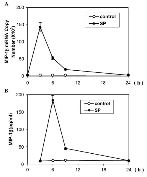
Substance P (SP) is an important modulator of neuroimmunoregulation. We have demonstrated that human T lymphocytes express SP and neurokinin-1 receptor (NK-1R), a primary SP receptor. In the present study, we investigated whether SP stimulates synthesis of macrophage inflammatory protein-1beta (MIP-1beta) in human T lymphocytes. SP significantly enhanced MIP-1beta expression at both the mRNA and protein level in a human T cell line (Jurkat) containing the SP receptor gene (J-SPR) as determined by real-time PCR and ELISA assays. SP-induced MIP-1beta expression is abrogated by the specific NK-1R antagonist (CP-96,345). The supernatants from SP-stimulated J-SPR T cell cultures enhanced T lymphocyte chemotaxis in vitro, indicating functional activity of SP-induced MIP-1beta. In addition, SP augmented secretion of MIP-1beta from primary cultures of peripheral blood lymphocytes (PBL) isolated from some of the donors. This donor variability was due to differential expression of the primary SP receptor (NK-1R) on PBL from different donors. PBL from two of seven donors that did not respond to SP stimulation had undetectable NK-1R expression. Our mechanistic studies showed that SP activated NF-kappaB promoter-directed luciferase activity, which may be responsible for its effect on MIP-1beta expression in human T cells. Our data provide a potential mechanism by which SP selectively influences cellular immune responses such as beta-chemokine expression in human T lymphocytes through NK-1R, which may have an important in vivo implication in inflammatory diseases.
Figures


Similar articles
-
Interleukin-1beta upregulates functional expression of neurokinin-1 receptor (NK-1R) via NF-kappaB in astrocytes.Glia. 2004 Nov 15;48(3):259-66. doi: 10.1002/glia.20079. Glia. 2004. PMID: 15390113 Free PMC article.
-
Neuropeptide substance P upregulates chemokine and chemokine receptor expression in primary mouse neutrophils.Am J Physiol Cell Physiol. 2007 Aug;293(2):C696-704. doi: 10.1152/ajpcell.00060.2007. Epub 2007 May 9. Am J Physiol Cell Physiol. 2007. PMID: 17494633
-
Human neuronal cells (NT2-N) express functional substance P and neurokinin-1 receptor coupled to MIP-1 beta expression.J Neurosci Res. 2003 Feb 15;71(4):559-66. doi: 10.1002/jnr.10504. J Neurosci Res. 2003. PMID: 12548712 Free PMC article.
-
A non-peptide substance P antagonist (CP-96,345) inhibits morphine-induced NF-kappa B promoter activation in human NT2-N neurons.J Neurosci Res. 2004 Feb 15;75(4):544-53. doi: 10.1002/jnr.10873. J Neurosci Res. 2004. PMID: 14743438
-
Substance P and fibrotic diseases.Neuropeptides. 2019 Aug;76:101941. doi: 10.1016/j.npep.2019.101941. Epub 2019 Jun 24. Neuropeptides. 2019. PMID: 31256921 Review.
Cited by
-
Interleukin-1beta upregulates functional expression of neurokinin-1 receptor (NK-1R) via NF-kappaB in astrocytes.Glia. 2004 Nov 15;48(3):259-66. doi: 10.1002/glia.20079. Glia. 2004. PMID: 15390113 Free PMC article.
-
Gastrointestinal neuroendocrine peptides/amines in inflammatory bowel disease.World J Gastroenterol. 2017 Jul 28;23(28):5068-5085. doi: 10.3748/wjg.v23.i28.5068. World J Gastroenterol. 2017. PMID: 28811704 Free PMC article. Review.
-
The neuroscience of cancer: Focus on neuropeptidergic systems.Acta Pharm Sin B. 2025 May;15(5):2323-2350. doi: 10.1016/j.apsb.2025.03.025. Epub 2025 Mar 13. Acta Pharm Sin B. 2025. PMID: 40487638 Free PMC article. Review.
-
Mucosal neuroimmune mechanisms in gastro-oesophageal reflux disease (GORD) pathogenesis.J Gastroenterol. 2024 Mar;59(3):165-178. doi: 10.1007/s00535-023-02065-9. Epub 2024 Jan 14. J Gastroenterol. 2024. PMID: 38221552 Free PMC article. Review.
-
Neurokinin receptors and their implications in various autoimmune diseases.Curr Res Immunol. 2021 Jul 1;2:66-78. doi: 10.1016/j.crimmu.2021.06.001. eCollection 2021. Curr Res Immunol. 2021. PMID: 35492389 Free PMC article. Review.
References
-
- Bozic CR, Lu B, Hopken UE, Gerard C, Gerard NP. Neurogenic amplification of immune complex inflammation. Science. 1996;273:1722–1725. - PubMed
-
- Carolan EJ, Casale TB. Effects of neuropeptides on neutrophil migration through noncellular and endothelial barriers. J. Allergy Clin. Immunol. 1993;92:589–598. - PubMed
-
- Dunzendorfer S, Kaser A, Meierhofer C, Tilg H, Wiedermann CJ. Cutting edge: peripheral neuropeptides attract immature and arrest mature blood-derived dendritic cells. J. Immunol. 2001;166:2167–2172. - PubMed
-
- Hassan NF, Campbell DE, Douglas SD. Purification of human monocytes on gelatin-coated surfaces. J. Immunol. Methods. 1986;95:273–276. - PubMed
-
- Ho WZ, Cnaan A, Li YH, Zhao H, Lee HR, Song L, Douglas SD. Substance P modulates human immunodeficiency virus replication in human peripheral blood monocyte-derived macrophages. AIDS Res. Hum. Retrovir. 1996a;12:195–198. - PubMed
Publication types
MeSH terms
Substances
Grants and funding
LinkOut - more resources
Full Text Sources
Miscellaneous
