. 2002 Dec;107(4):472-9.
doi: 10.1046/j.1365-2567.2002.01538.x.

Induction of 4-1BB (CD137) expression by DNA damaging agents in human T lymphocytes

Affiliations

Induction of 4-1BB (CD137) expression by DNA damaging agents in human T lymphocytes

Kwang-Mi Kim et al. Immunology. 2002 Dec.

Abstract

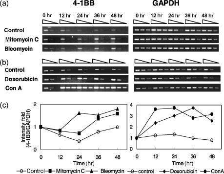
4-1BB(CD137) is a member of the tumour necrosis factor receptor superfamily and is expressed on activated T cells, monocytes and natural killer (NK) cells. The interaction of 4-1BB and 4-1BB ligand provides a costimulatory signal leading to T-cell activation. The expression of 4-1BB has been known to be activation dependent. Interestingly, we found that expression of 4-1BB increased in human peripheral blood mononuclear cells after exposure to mitomycin C. Thus, we tested whether the treatment with other DNA-damaging agents, such as doxorubicin, bleomycin, and gamma-irradiation, could induce 4-1BB expression. The data indicated that 4-1BB expression increased dose-dependently by these agents reaching maximum at 2-3 days after the exposure. We found that the major 4-1BB-expressing population was CD3+ T cells, although a moderate number of CD14+ cells and a few NKB1+ cells also expressed 4-1BB. The levels of 4-1BB expression induced by anticancer drugs, were relatively lower than that induced by CD3 ligation. Interestingly, at subcytotoxic concentrations, doxorubicin and bleomycin considerably enhanced 4-1BB expression induced by CD3 ligation in CEM cells. The ligation of the damage-induced 4-1BB by monoclonal antibody enhanced the viability and proliferating capacity of the cells. In conclusion, the expression of 4-1BB might be one of the cellular responses of the immune cells against various genotoxic stresses.


Figures

Figure 5
Figure 5
4-1BB mRNA expression is increased by anticancer drugs. Purified peripheral blood T cells from two healthy donors were cultured with bleomycin (60 µg/ml) or mitomycin C (1000 ng/ml) (a), doxorubicin (200 ng/ml) or Con A (10 µg/ml) (b). The cells were harvested at 0, 12, 24, 36, and 48 hr. The total RNA was extracted and analysed for relative levels of 4-1BB mRNA by RT–PCR as described in Materials and Methods. GAPDH was used to normalize for template quantity. Three-fold serial dilutions of cDNA at each time were subjected to PCR and equal amount of product for 4-1BB (368 bp) and GAPDH (500 bp) were resolved on 1·5% agarose gels stained with ethidium bromide. Data shown were the representative results from 15 independent experiments with the samples from five donors. (c) The first band intensity of each time point was divided by the corresponding GAPDH band intensity and the results were represented as intensity fold when 0 hr value in each reagent group was designated as 1.
Figure 1
Figure 1
4-1BB induction by anticancer drugs in normal human PBMC. (a) Fresh human PBMC obtained from healthy volunteers were incubated in presence of indicated concentrations of mitomycin C for 2 days. Then the cells were harvested, stained with 4B4 (filled) or control mAb (unfilled), and analysed by flow cytometry to detect 4-1BB expression. The percentage of 4-1BB-expressing cells increased according to the dose of mitomycin C. (b) Human PBMC were incubated in presence of indicated concentrations of doxorubicin, bleomycin, or mitomycin C. At days 1, 2, 3, and 4, the cells were harvested and analysed by flow cytometry to detect 4-1BB expression. The data were presented as the percentage of the 4-1BB-expressing cells in whole PBMC population, calculated as percentage 4B4-binding cells − percentage control mAb-binding cells. 4-1BB molecules were induced by doxorubicin, bleomycin, mitomycin, and the percentage of 4-1BB-expressing cells peaked at days 2–3 showing dose-dependent tendency, and decreased at day 4. These data show the representative results from six independent experiments. Each sample was determined in duplicate and the mean values were presented.
Figure 2
Figure 2
The predominant 4-1BB-expressing population is CD3+ cells and CD14+ cells. Fresh human PBMC were incubated in presence of 200 ng/ml doxorubicin, 60 µg/ml bleomycin, or 1000 ng/ml mitomycin C for 48 hr, then were double stained with one of CD3, CD14, or NKB1-specific antibodies together with an anti 4-1BB mAb or a control mAb. The numerical values in bold style represent the percentage of the 4-1BB-positive cells, and the values in parenthesis represent the background staining with isotype control mAb. The percentage of 4-1BB-expressing cells was highest in CD3+ cells, although some CD14+ cells and a few NKB1+ cells also expressed 4-1BB. Data shown were the representative results from four independent experiments.
Figure 3
Figure 3
Direct 4-1BB expression by anticancer drugs in purified T cells. Peripheral blood T cells were isolated from PBMC by two rounds of rosetting with sheep red blood cells. The purity of T cells was verified by flow cytometry (CD3+ T cell > 95%, data not shown). The purified T cells were cultured with anticancer drugs and harvested at indicated time points, then analysed by flow cytometry to detect 4-1BB expression. The percentages of the cells expressing 4-1BB were presented as a function of treatment time and concentration of each anticancer drug. Each sample was determined in duplicate and the mean values were presented. Anticancer drugs directly induced 4-1BB expression in the purified T cells in the absence of APC or other cells. These data show the representative results from three independent experiments.
Figure 4
Figure 4
4-1BB molecules are induced by γ-irradiation both in CD4+ and CD8+ cells. Freshly isolated PBMC were irradiated with single doses at indicated dosed by caesium irradiator. The irradiated cells were cultured and harvested at indicated time points and analysed by flow cytometry to detect 4-1BB expression. Cells were double stained with CD4- (a and c) or CD8-specific (b and d) antibodies together with an anti 4-1BB mAb or a control mAb. Each sample was determined in duplicate and the mean values were presented as a function of time and irradiation dose (a and b), or the representative results were presented in flow cytometry histograms (c and d). Data shows that γ-irradiation induced considerable expression of 4-1BB in both CD4+ and CD8+ cells.
Figure 6
Figure 6
Comparative analysis of 4-1BB expression by DNA damaging agents and by TCR stimulation. CEM cells were treated with various concentrations of anticancer drugs for 2 hr at 37°, then were transferred to 48-well plates coated with various concentrations of anti-CD3 mAb or control mAb. After 24 hr, The CEM cells were harvested and the 4-1BB expression was measured by flow cytometry. The levels of 4-1BB expression induced by anticancer drugs were relatively lower than that induced by CD3 ligation. At the lower concentrations, doxorubicin and bleomycin considerably enhanced 4-1BB expression induced by CD3 ligation, while mitomycin C did not. Data shown were the representative results from three independent experiments. Each sample was determined in duplicate and the mean values were presented.
Figure 7
Figure 7
The ligation of DNA damage-induced 4-1BB enhances the viability and proliferating capacity of the cells treated with anticancer drugs. Freshly isolated peripheral blood T cells were cultured with doxorubicin (200 ng/ml) or bleomycin (60 µg/ml) or mitomycin C (1000 ng/ml) for 2 days. At day 2, only the viable cells were isolated by Ficoll density gradient centrifugation and 3 × 10⁵ cells were plated in each well of 96-well plates coated with an anti-4-1BB mAb or an isotype control mAb, then cultured for 4 days. At the end of culture, the viability of the cells was measured by MTT assay (a). At the same time, the abilities of the cells to proliferate in response to mitogenic signal (10 µg/ml Con A) were evaluated by [methyl-3H]thymidine uptake (b). Data shown were the representative results from four independent experiments. Each sample was determined in triplicate wells and the data were represented as mean value ± SD. The differences between control mAb (MAG56) and 4B4 mAb treatment samples were analysed by Student's t-test. *P < 0·05.

Similar articles

Cited by

References

    1. Kwon BS, Weissman SM. cDNA sequences of two inducible T-cell genes. Proc Natl Acad Sci USA. 1989;86:1963–7. - PMC - PubMed
    1. Mallett S, Barclay AN. A new superfamily of cell surface proteins related to the nerve growth factor receptor. Immunol Today. 1991;12:220–2. - PubMed
    1. Smith CA, Farrah T, Goodwin RG. The TNF receptor superfamily of cellular and viral proteins. Activation, Costimulation, Death Cell. 1994;76:959–62. - PubMed
    1. Armitage RJ. Tumor necrosis factor receptor superfamily members and their ligands. Curr Opin Immunol. 1994;6:407–13. - PubMed
    1. Pollok KE, Kim YH, Zhou Z, Hurtado J, Kim KK, Pickard RT, Kwon BS. Inducible T-cell antigen 4–1BB. An analysis of expression and function. J Immunol. 1993;150:771–81. - PubMed

Publication types

MeSH terms