. 2002 Dec 3;106(23):2961-6.
doi: 10.1161/01.cir.0000038705.69871.d9.

Expression and regulation of ST2, an interleukin-1 receptor family member, in cardiomyocytes and myocardial infarction

Affiliations

Expression and regulation of ST2, an interleukin-1 receptor family member, in cardiomyocytes and myocardial infarction

Ellen O Weinberg et al. Circulation. .

Abstract

Background: We identified an interleukin-1 receptor family member, ST2, as a gene markedly induced by mechanical strain in cardiac myocytes and hypothesized that ST2 participates in the acute myocardial response to stress and injury.

Methods and results: ST2 mRNA was induced in cardiac myocytes by mechanical strain (4.7+/-0.9-fold) and interleukin-1beta (2.0+/-0.2-fold). Promoter analysis revealed that the proximal and not the distal promoter of ST2 is responsible for transcriptional activation in cardiac myocytes by strain and interleukin-1beta. In mice subjected to coronary artery ligation, serum ST2 was transiently increased compared with unoperated controls (20.8+/-4.4 versus 0.8+/-0.8 ng/mL, P<0.05). Soluble ST2 levels were increased in the serum of human patients (N=69) 1 day after myocardial infarction and correlated positively with creatine kinase (r=0.41, P<0.001) and negatively with ejection fraction (P=0.02).

Conclusions: These data identify ST2 release in response to myocardial infarction and suggest a role for this innate immune receptor in myocardial injury.


Figures

Figure 1
Figure 1
Induction of soluble ST2 mRNA in cardiac myocytes. a, Induction of soluble ST2 mRNA by mechanical strain in NRCM. Northern analyses showing early (left) and later (right) time course. Maximal induction occurs at 2 hours, is sustained for 9 hours, and declines by 15 hours. b, Induction of soluble ST2 mRNA by mechanical strain, interleukin-1β (IL-1, 10 ng/mL), combined strain and interleukin-1β, and phorbol ester (PMA, 200 nmol/L) at 1 and 3 hours. Top, ST2 mRNA. Bottom, Ethidium bromide staining of 18S ribosomal RNA. No Str indicates no strain.
Figure 2
Figure 2
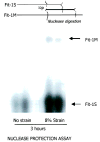
Nuclease protection assay demonstrating mRNA induction of soluble (Fit-1S) and membrane (Fit-1M) forms of ST2 in cardiac myocytes by mechanical strain. The expression of soluble ST2 mRNA is more abundant than membrane ST2 mRNA.
Figure 3
Figure 3
Activation of human ST2 promoter-luciferase constructs. a, Schematic representation of proximal and distal ST2 promoter region usage that gives rise to soluble and membrane ST2. Promoter usage is cell-type specific to produce a predominant transcript (indicated by thick lines) and a minor transcript (thin lines). b, 3.4-kb DNA flanking ST2 exon 1b (proximal promoter region) fused to luciferase reporter was responsive to interleukin-1β, mechanical strain, and phorbol ester. 2.1-kb DNA flanking ST2 exon 1a (distal promoter region) fused to luciferase reporter was slightly repressed by interleukin-1β, nonresponsive to mechanical strain, and weakly responsive to phorbol ester. *P < 0.05.
Figure 4
Figure 4
Serum ST2 levels in mice after MI. Serum ST2 levels were significantly increased 1 day after MI compared with sham-operated mice. Three days after MI, serum ST2 levels were similar among unoperated, sham-operated, and MI mice.
Figure 5
Figure 5
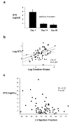
ST2 protein levels in the circulation of human patients after MI. Serial blood samples from 69 patients from the HEART study were analyzed for ST2 by ELISA. a, ST2 was significantly increased 1 day after MI compared with day 14 and day 90. b, Linear regression analysis demonstrating a significant positive relationship (P < 0.001) between circulating ST2 and creatine kinase 1 day after MI. Log ST2(ng/mL)=0.454[log CK(U/L)]−1.07. c, Linear regression analysis demonstrating a significant negative relationship (P = 0.02) between circulating ST2 and LV ejection fraction 1 day after MI.

Similar articles

Cited by

References

    1. Iwahana H, Yanagisawa K, Ito-Kosaka A, et al. Different promoter usage and multiple transcription initiation sites of the interleukin-1 receptor-related human ST2 gene in UT-7 and TM12 cells. Eur J Biochem. 1999;264:397–406. - PubMed
    1. Bergers G, Reikerstorfer A, Braselmann S, et al. Alternative promoter usage of the fos-responsive gene Fit-1 generates RNA isoforms coding for either secreted or membrane-bound proteins related to the IL-1 receptor. EMBO J. 1994;13:1176–1188. - PMC - PubMed
    1. Tominaga S. A putative protein of a growth specific cDNA from BALB/c-3T3 cells is highly similar to the extracellular portion of mouse interleukin-1 receptor. FEBS Lett. 1989;258:301–304. - PubMed
    1. Lohning M, Stroehmann A, Coyle AJ, et al. T1/ST2 is preferentially expressed on murine Th2 cells, independent of interleukin 4, interleukin 5, and interleukin 10, and important for Th2 effector function. Proc Natl Acad Sci U S A. 1998;95:6930–6935. - PMC - PubMed
    1. Coyle AJ, Lloyd C, Tian J, et al. Crucial role of the interleukin 1 receptor family member T1/ST2 in T helper cell type 2-mediated lung mucosal immune responses. J Exp Med. 1999;190:895–902. - PMC - PubMed

Publication types

MeSH terms