. 2002 Dec 9;159(5):821-31.
doi: 10.1083/jcb.200112131. Epub 2002 Dec 2.

Brain-derived neurotrophic factor can act as a pronecrotic factor through transcriptional and translational activation of NADPH oxidase

Affiliations

Brain-derived neurotrophic factor can act as a pronecrotic factor through transcriptional and translational activation of NADPH oxidase

Sun H Kim et al. J Cell Biol. .

Abstract

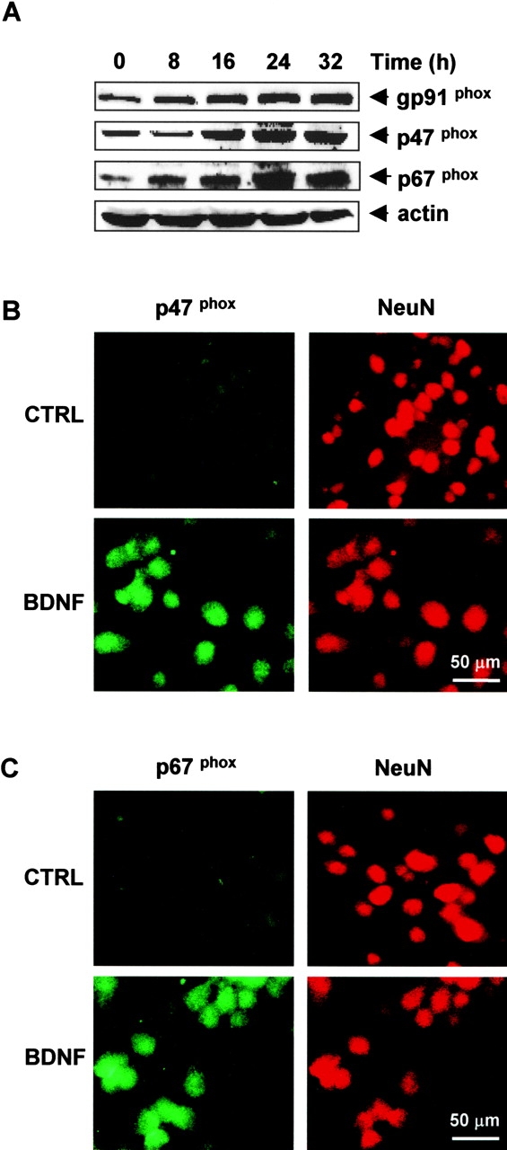
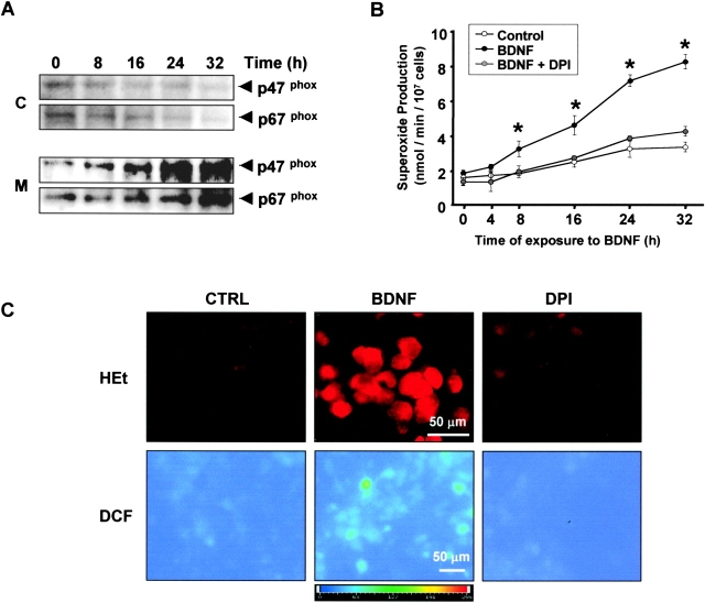
Several lines of evidence suggest that neurotrophins (NTs) potentiate or cause neuronal injury under various pathological conditions. Since NTs enhance survival and differentiation of cultured neurons in serum or defined media containing antioxidants, we set out experiments to delineate the patterns and underlying mechanisms of brain-derived neurotrophic factor (BDNF)-induced neuronal injury in mixed cortical cell cultures containing glia and neurons in serum-free media without antioxidants, where the three major routes of neuronal cell death, oxidative stress, excitotoxicity, and apoptosis, have been extensively studied. Rat cortical cell cultures, after prolonged exposure to NTs, underwent widespread neuronal necrosis. BDNF-induced neuronal necrosis was accompanied by reactive oxygen species (ROS) production and was dependent on the macromolecular synthesis. cDNA microarray analysis revealed that BDNF increased the expression of cytochrome b558, the plasma membrane-spanning subunit of NADPH oxidase. The expression and activation of NADPH oxidase were increased after exposure to BDNF. The selective inhibitors of NADPH oxidase prevented BDNF-induced ROS production and neuronal death without blocking antiapoptosis action of BDNF. The present study suggests that BDNF-induced expression and activation of NADPH oxidase cause oxidative neuronal necrosis and that the neurotrophic effects of NTs can be maximized under blockade of the pronecrotic action.


Figures

Figure 1.
Figure 1.
Prolonged exposure to NTs produces neuronal cell necrosis in cortical cell cultures. (A) Mixed cortical cell cultures (DIV 12–15) were continuously exposed to 10, 30, and 100 ng/ml of NGF, BDNF, or NT-3 for the indicated points of time. Neuronal cell death was assessed by measurement of LDH efflux to the bathing medium, mean ± SEM (n = 16 culture wells per condition). *Significant difference from the relevant control (sham washed control) at P < 0.05 using analysis of variance and Student-Neuman-Keuls test. (B) Brain sections were stained with hematoxylin-eosin at 2 d after intrastriatal injections of 5 μl of saline or 5 μg BDNF. Bright field photomicrogrphs showing a representative striatal area 1 mm lateral to the injection site of saline (a) or BDNF (b). Note the pyknotic neurons (arrows) in BDNF-treated striatal area. Striatal lesion was analyzed by measuring the injured areas (c), mean ± SEM (n = 5 rats per each condition). *Significant difference from the relevant control (saline injected) at P < 0.05 using Independent-Samples t test. (C) Phase–contrast (top) and electron (bottom) photomicrographs of cortical neurons 32 h after a sham wash (a and c) or continuous exposure to 100 ng/ml BDNF (b and d). Note that BDNF treatment induces swelling of neuronal cell body (arrow), scattering condensation of nuclear chromatin (arrowhead), and fenestration of plasma membrane characteristic of necrosis. (D) Patterns of BDNF-induced neuronal death were analyzed at 32 h after exposure of cortical cell cultures to 100 ng/ml BDNF. Approximately 200 neurons from control and BDNF-treated cultures were randomly selected and observed under transmission electron microscope. Degenerating neurons were defined as normal, necrosis (see above), or apoptosis (shrinkage of cytoplasm and nuclear membrane rupture with intact plasma membrane). (E) Cortical cell cultures (DIV 12–15) were continuously exposed to 100 ng/ml BDNF, alone or with 100 μg/ml anti-BDNF blocking antibody, 150 nM K252a, 10 μM MK-801, 50 μM CNQX, 10 μM MK 801 plus 50 μM CNQX, 100 μM trolox, or 1 μg/ml cycloheximide (CHX). Neuronal death was analyzed 36 h later by measurement of LDH efflux into the bathing medium, mean ± SEM (n = 16 culture wells per condition). *Significant difference from the relevant control (BDNF alone) at P < 0.05 using analysis of variance and Student-Neuman-Keuls test.
Figure 2.
Figure 2.
BDNF produces ROS in cortical neurons: involvement of protein synthesis. (A) Cortical cell cultures (DIV 12–15) were exposed to a sham wash (•) or 100 ng/ml BDNF (○). Levels of ROS in neurons were analyzed at indicated times by measuring fluorescence intensity of oxidized DCDHF-DA (DCF), mean ± SEM (n = 30–35 neurons randomly chosen from four culture wells per condition). *Significant difference from the relevant control (sham wash) at P < 0.05 using analysis of variance and Student-Neuman-Keuls test. (B and C) Fluorescence photomicrographs (B) and quantitation (C) of DCF in cortical neurons after 32 h exposure of cortical cell cultures (DIV 12–15) to a sham wash (CTRL) or 100 ng/ml BDNF, alone (BDNF) or in the presence of 1 μg/ml cycloheximide (CHX) or 100 μM trolox (TROLOX). *Significant difference from the control (CTRL); #significant difference from the BDNF control (BDNF alone) at P < 0.05 using analysis of variance and Student-Neuman-Keuls test.
Figure 3.
Figure 3.
BDNF increases mRNA levels of NADPH oxidase subunits in cortical cultures. RT-PCR analysis of NADPH oxidase subunits (p22-phox, gp91-phox, and p47-phox) and GAPDH mRNA expression in cortical cell cultures (DIV 12) exposed to 100 ng/ml BDNF for indicated times. The mRNA level of each NADPH oxidase subunit was normalized to the relevant control value (t = 0). Similar results were observed from three separate experiments.
Figure 4.
Figure 4.
BDNF increases expression of NADPH oxidase subunits in cortical neurons. (A) Western blot analysis of NADPH oxidase subunits (gp91-phox, p47-phox, and p67-phox) and actin in cortical cell cultures after exposure to 100 ng/ml BDNF for indicated times. Similar results were obtained from three separate experiments. (B) Fluorescence photomicrograph of cortical cell cultures immunolabeled with NeuN (right; anti–mouse immunoglobulin G conjugated with Texas red) and anti-p47-phox (left; anti–goat immunoglobulin G conjugated with FITC) after exposure to a sham wash (top) and 100 ng/ml BDNF (bottom) for 32 h. (C) Same as B except that cultures were immunolabeled with anti–p67-phox antibody (anti–goat immunoglobulin G conjugated with FITC) instead of anti–p47-phox. Note that levels of p47-phox and p67-phox were increased selectively in neurons after exposure of mixed cortical cell cultures to BDNF.
Figure 5.
Figure 5.
Translocation and activation of NADPH oxidase by BDNF. (A) Western blot analysis of the cytosolic fraction (C) and the membrane fraction (M) using anti–p47-phox and anti–p67-phox antibodies that were obtained from cortical cell cultures (DIV 12) after exposure to 100 ng/ml BDNF for the indicated times. (B) Superoxide production was analyzed by measuring reduction of cytochrome c in cortical cultures exposed to a sham wash (○) or 100 ng/ml BDNF with (gray colored circles) or without 3 nM DPI (•) for indicated times, mean ± SEM (n = 12 culture wells per condition). *Significant difference from the sham control at P < 0.05 using analysis of variance and Student-Neuman-Keuls test. (C) Fluorescence photomicrograph of the oxidized hydroethydine (HEt, top) and DCF (bottom) in cortical neurons after exposure to a sham operation (CTRL), 100 ng/ml BDNF (BDNF), or 100 ng/ml BDNF plus 3 nM DPI (DPI) for 32 h.
Figure 6.
Figure 6.
Activation of NADPH oxidase mediates BDNF-induced neuronal death. Cortical cell cultures (DIV 12–15) were exposed to 100 ng/ml BDNF or 30 μM Fe2+, alone or in the presence of 3 nM DPI or 10 μM AEBSF. Neuronal death was analyzed 36 h later by measurement of LDH efflux into the bathing medium, mean ± SEM (n = 16 culture wells per condition). *Significant difference from the relevant control (BDNF or Fe2+ alone) at P < 0.05 by Student-Neuman-Keuls test.
Figure 7.
Figure 7.
DPI or trolox enhances the neuroprotective effect of BDNF against serum deprivation. Neuron-rich cortical cell cultures (DIV 7) were deprived of serum, alone (serum deprivation) or in the presence of 100 ng/ml BDNF, 100 ng/ml BDNF plus 3 nM DPI, 3 nM DPI, 100 ng/ml BDNF plus 100 μM trolox, or 100 μM trolox. Neuronal death was assessed 24 and 48 h later by counting viable neurons, mean ± SEM (n = 16 fields randomly chosen from four culture wells per condition). *Significant difference from the relevant control (serum deprivation alone) at P < 0.05 using analysis of variance and Student-Neuman-Keuls test.

Similar articles

Cited by

References

    1. Alcantara S., J. Frisen, J.A. del Rio, E. Soriano, M. Barbacid, and I. Silos-Santiago. 1997. TrkB signaling is required for postnatal survival of CNS neurons and protects hippocampal and motor neurons from axotomy-induced cell death. J. Neurosci. 17:3623–3633. - PMC - PubMed
    1. Alderson, R.F., A.L. Alterman, Y.A. Barde, and R.M. Lindsay. 1990. Brain-derived neurotrophic factor increases survival and differentiated functions of rat septal cholinergic neurons in culture. Neuron. 5:297–306. - PubMed
    1. Atwal, J.K., B. Massie, F.D. Miller, and D.R. Kaplan. 2000. The TrkB-Shc site signals neuronal survival and local axon growth via MEK and P13-kinase. Neuron. 27:265–277. - PubMed
    1. Bannister, J.V., P. Bellavite, A. Davoli, P.J. Thornalley, and F. Rossi. 1982. The generation of hydroxyl radicals following superoxide production by neutrophil NADPH oxidase. FEBS Lett. 150:300–302. - PubMed
    1. Barde, Y.A. 1994. Neurotrophins: a family of proteins supporting the survival of neurons. Prog. Clin. Biol. Res. 390:45–56. - PubMed

Publication types

MeSH terms