. 2002 Dec 2;196(11):1447-59.
doi: 10.1084/jem.20020436.

GRP94 (gp96) and GRP94 N-terminal geldanamycin binding domain elicit tissue nonrestricted tumor suppression

Affiliations

GRP94 (gp96) and GRP94 N-terminal geldanamycin binding domain elicit tissue nonrestricted tumor suppression

Julie C Baker-LePain et al. J Exp Med. .

Abstract

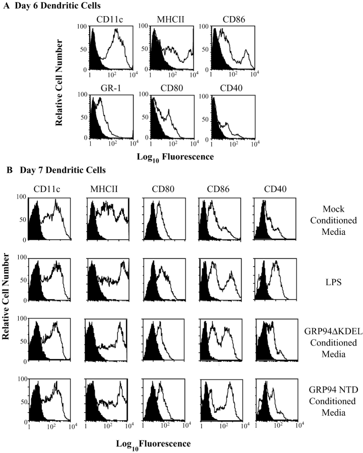
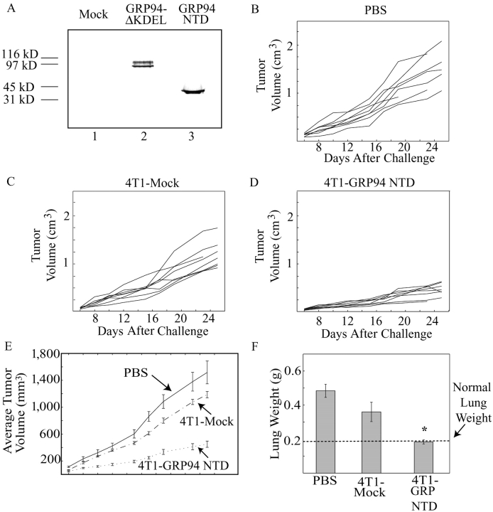
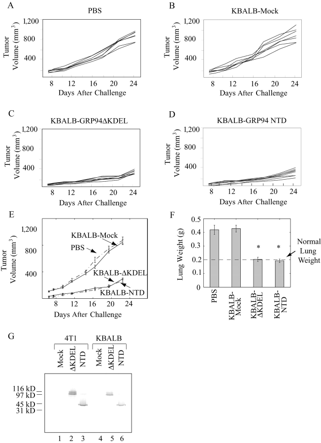
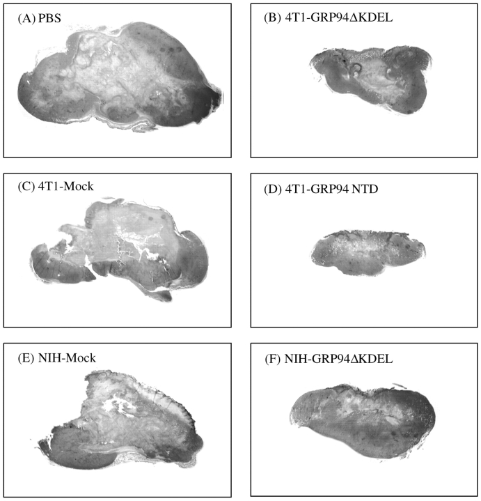
In chemical carcinogenesis models, GRP94 (gp96) elicits tumor-specific protective immunity. The tumor specificity of this response is thought to reflect immune responses to GRP94-bound peptide antigens, the cohort of which uniquely identifies the GRP94 tissue of origin. In this study, we examined the apparent tissue restriction of GRP94-elicited protective immunity in a 4T1 mammary carcinoma model. We report that the vaccination of BALB/c mice with irradiated fibroblasts expressing a secretory form of GRP94 markedly suppressed 4T1 tumor growth and metastasis. In addition, vaccination with irradiated cells secreting the GRP94 NH(2)-terminal geldanamycin-binding domain (NTD), a region lacking canonical peptide-binding motifs, yielded a similar suppression of tumor growth and metastatic progression. Conditioned media from cultures of GRP94 or GRP94 NTD-secreting fibroblasts elicited the up-regulation of major histocompatibility complex class II and CD86 in dendritic cell cultures, consistent with a natural adjuvant function for GRP94 and the GRP94 NTD. Based on these findings, we propose that GRP94-elicited tumor suppression can occur independent of the GRP94 tissue of origin and suggest a primary role for GRP4 natural adjuvant function in antitumor immune responses.


Figures

Figure 1.
Figure 1.
GRP94ΔKDEL is secreted by transfected 4T1 mammary carcinoma cells. (A) Immunofluorescence with anti-myc antibody reveals expression of GRP94ΔKDEL within transfected 4T1 cells. 4T1 cells were grown on glass coverslips and stained with anti-myc antibody (9E10) and TR-conjugated secondary antibody, and then visualized by confocal microscopy (100×). (B–C) Oligosaccharide processing of GRP94ΔKDEL. Media or cell lysates from mock-transfected (mock) or GRP94ΔKDEL-transfected (T) 4T1 cells were collected after metabolic labeling with [35S]Promix. GRP94 species were recovered by immunoprecipitation with either anti-GRP94 antibody or anti-myc antibody, as indicated. GRP94ΔKDEL appears as a doublet of bands distinct from endogenous, full-length GRP94 (*). (C) Glycosidase digestion of GRP94ΔKDEL. Media (M) or cell lysates (C) from GRP94ΔKDEL-transfected 4T1 cells were collected as described in B and then treated (+) or not treated (−) with Endo H or PNGase-F, as indicated. Endogenous GRP94 in the cell lysate samples (position “E”) and transfected forms of GRP94 are indicated (“T” positions). All samples were resolved by SDS-PAGE on 6% gels and visualized by PhosphorImager analysis.
Figure 2.
Figure 2.
Kinetics of GRP94ΔKDEL secretion from 4T1 breast carcinoma cells. (A) 4T1 cells were metabolically labeled with [35S]Promix and then media and cell lysates were collected and immunoprecipitated with anti-GRP94 antibody. All samples were treated with PNGase-F before resolution by SDS-PAGE on 6% gels for GRP94ΔKDEL or 10% gels for prolactin. Radiolabeled proteins were visualized by PhosphorImager analysis. (B) Bands from A were quantified using MacBAS-2.0 software and were used to determine percent total GRP94ΔKDEL or prolactin present in the media or cell lysate at each time point.
Figure 3.
Figure 3.
Vaccination with 4T1 mammary carcinoma cells or NIH-3T3 fibroblasts secreting GRP94ΔKDEL leads to delayed tumor growth rates and decreased tumor metastasis. (A) Irradiation does not alter the secretion of GRP94ΔKDEL. 4T1 cells were transfected with GRP94ΔKDEL (T and T/I) or mock-transfected (mock). 24 h after transfection, cells were irradiated with 10,000 rads (T/I) or left unirradiated (T). 72 h after transfection, cells were metabolically labeled with [35S]Promix and GRP94ΔKDEL was recovered from the media by immunoprecipitation. Proteins were resolved by SDS-PAGE on 6% gels and visualized by PhosphorImager analysis. (B–H) GRP94ΔKDEL secretion from either 4T1 cells or NIH-3T3 fibroblasts elicits delayed tumor growth. Female BALB/c mice were immunized with PBS or with irradiated, GRP94ΔKDEL-transfected or irradiated, mock-transfected 4T1 or NIH-3T3 cells, as indicated. Animals were then challenged with unirradiated 4T1 cells, as described in Materials and Methods, and tumor volumes were followed over time. Tumor growth curves for individual mice in each group are shown in B–F and average tumor volumes with standard error are shown in G and H. (I) GRP94ΔKDEL secretion from either 4T1 cells or NIH-3T3 fibroblasts elicits decreased metastatic tumor progression. After sacrifice, lungs were resected from mice in B–H and weighed to determine the extent of metastatic tumor burden. Average weights with standard error are shown with groups differing significantly from PBS control denoted by an asterisk (P ≤ 0.0012 for 4T1-ΔKDEL and P ≤ 0.025 for NIH-ΔKDEL). (J) Comparison of the relative levels of GRP94ΔKDEL secretion by 4T1 and NIH-3T3 cells. Equal numbers (106 cells) of 4T1 or NIH3T3 cells were transfected with GRP94ΔKDEL (ΔKDEL samples) or mock-transfected (mock samples). 24 h after transfection, cells were metabolically labeled with [35S]Promix and GRP94DKDEL was recovered from the media by immunoprecipitation. Proteins were resolved by SDS-PAGE on 6% gels and visualized by PhosphorImager analysis.
Figure 4.
Figure 4.
GRP94 NH2-terminal domain is cleared by APCs and colocalizes with full-length GRP94 upon internalization. (A) Cell type specificity of GRP94 NH2-terminal domain binding. Recombinantly expressed GRP94 NH2-terminal domain was fluorescently labeled and used in a standard binding assay with various cell types as described in Materials and Methods. The percentage of cells exhibiting mean fluorescence above control is indicated. (B) Clearance of GRP94 and GRP94 NH2-terminal regulatory domain by peritoneal macrophages. Elicited peritoneal macrophages were incubated with FITC-labeled GRP94 N-terminal domain (FITC-GRP94 NTD) and TR-labeled full-length GRP94 (Texas Red-GRP94). After washing at 4°C to remove unbound protein, cells were warmed to 37°C for the indicated times before fixation and visualization by confocal microscopy (100×). The overlay image displays dual channel imaging of both labeled proteins.
Figure 4.
Figure 4.
GRP94 NH2-terminal domain is cleared by APCs and colocalizes with full-length GRP94 upon internalization. (A) Cell type specificity of GRP94 NH2-terminal domain binding. Recombinantly expressed GRP94 NH2-terminal domain was fluorescently labeled and used in a standard binding assay with various cell types as described in Materials and Methods. The percentage of cells exhibiting mean fluorescence above control is indicated. (B) Clearance of GRP94 and GRP94 NH2-terminal regulatory domain by peritoneal macrophages. Elicited peritoneal macrophages were incubated with FITC-labeled GRP94 N-terminal domain (FITC-GRP94 NTD) and TR-labeled full-length GRP94 (Texas Red-GRP94). After washing at 4°C to remove unbound protein, cells were warmed to 37°C for the indicated times before fixation and visualization by confocal microscopy (100×). The overlay image displays dual channel imaging of both labeled proteins.
Figure 5.
Figure 5.
GRP94ΔKDEL and GRP94 N-terminal domain elicit DC maturation. Conditioned media from GRP94ΔKDEL-transfected, GRP94 NTD-transfected, or mock-transfected NIH-3T3 cells were collected for 72 h and incubated with day 6 DCs as described in Materials and Methods. (A) Phenotypic profile of day 6 DCs before incubation with conditioned media. (B) Phenotypic profile of day 7 DCs after incubation with conditioned media from mock-transfected, GRP94ΔKDEL-transfected, or GRP94 NTD-transfected NIH-3T3 cells. Incubation in media plus LPS was used as a positive control.
Figure 6.
Figure 6.
Vaccination with 4T1 mammary carcinoma cells secreting GRP94 NH2-terminal domain leads to delayed tumor growth and decreased tumor metastasis. (A) Expression and secretion of GRP94 NTD by transfected 4T1 cells. 4T1 cells were transfected with either GRP94ΔKDEL or GRP94 NTD constructs or were mock-transfected (mock) as indicated. Cells were metabolically labeled with [35S]Promix, and GRP94 domains were recovered from the media by immunoprecipitation before resolution by SDS-PAGE. Radiolabeled proteins were visualized by PhosphorImager analysis. (B–E) GRP94 NTD elicits delayed tumor growth rates. Female BALB/c mice were immunized with PBS or with irradiated, GRP94 NTD-transfected or irradiated, mock-transfected 4T1 cells as indicated. Animals were then challenged with unirradiated 4T1 cells and tumor volumes were followed over time, as described in Fig. 3. Tumor growth curves for individual mice are shown in B–D and average tumor volumes with standard error are shown in E. GRP94 NTD elicits decreased metastatic tumor progression. After animals were killed, lungs were resected from mice in B–D and weighed to determine the extent of metastatic tumor burden. Average lung weights with standard error are shown with groups differing significantly from PBS control denoted by an asterisk (P ≤ 0.00031).
Figure 7.
Figure 7.
GRP94ΔKDEL and GRP94 NH2-terminal domain secreted by syngeneic KBALB fibroblasts yield suppression of 4T1 tumor growth and metastasis. Female BALB/c mice were immunized with PBS or with irradiated, mock-transfected, GRP94ΔKDEL-transfected, or GRP94 NTD-transfected KBALB fibroblasts as indicated. Animals were then challenged with unirradiated 4T1 cells as described in Materials and Methods, and tumor volumes were followed over time. Tumor growth curves for individual mice in each group are shown in A–D and average tumor volumes with standard error are shown in E. (F) GRP94ΔKDEL or GRP94 NH2-terminal domain secretion from K-BALB fibroblasts yields decreased tumor metastasis. After animals were killed, lungs were resected from mice as shown in A–E and weighed. Average weights with standard error are shown, with groups differing significantly from PBS control denoted by an asterisk (P < 0.0003 for KBALB-ΔKDEL and P ≤ 0.0002 for KBALB-NTD). (G) Comparison of GRP94ΔKDEL and GRP94 NTD secretion by 4T1 and KBALB cells. Equal numbers (106 cells) of 4T1 KBALB cells were transfected with GRP94ΔKDEL (ΔKDEL samples), GRP94 NH2-terminal domain (NTD samples) or mock-transfected (mock samples). 24 h after transfection, cells were metabolically labeled with [35S]Promix and GRP94 species were recovered from the media by immunoprecipitation. Proteins were resolved by SDS-PAGE on 12.5% gels and visualized by PhosphorImager analysis.
Figure 8.
Figure 8.
Vaccination with GRP94ΔKDEL- or GRP94 NTD-secreting 4T1 or NIH-3T3 cells yields smaller 4T1 tumor size and decreased intratumoral necrosis. Female BALB/c mice were immunized with PBS or with irradiated, GRP94ΔKDEL- or GRP94 NTD-secreting 4T1 or NIH-3T3 cells as described in Figs. 3 and 4. Animals were then challenged with unirradiated 4T1 cells. When the animals were killed, tumors were formalin fixed and paraffin-embedded specimens were sectioned and stained with hematoxylin and eosin. Representative tumor sections from each vaccination group are shown (2.5×). Tumors are from mice immunized with (A) PBS, (B) irradiated, GRP94ΔKDEL-transfected 4T1 cells, (C) irradiated, mock-transfected 4T1 cells, (D) irradiated, GRP94 NTD-transfected 4T1 cells, (E) irradiated, mock-transfected NIH-3T3 cells, or (F) irradiated, GRP94ΔKDEL-transfected NIH-3T3 cells.

Similar articles

Cited by

References

    1. Srivastava, P.K. 2002. Interaction of heat shock proteins with peptides and antigen presenting cells: chaperoning of the innate and adaptive immune responses. Annu. Rev. Immunol. 20:395–425. - PubMed
    1. Srivastava, P.K., A.B. DeLeo, and L.J. Old. 1986. Tumor rejection antigens of chemically induced sarcomas of inbred mice. Proc. Natl. Acad. Sci. USA. 83:3407–3411. - PMC - PubMed
    1. Blachere, N.E., H. Udono, S. Janetzki, Z. Li, and P.K. Srivastava. 1993. Heat shock protein vaccines against cancer. J. Immunother. 14:352–356. - PubMed
    1. Tamura, Y., P. Peng, K. Liu, M. Daou, and P.K. Srivastava. 1997. Immunotherapy of tumors with autologous tumor-derived heat shock protein preparations. Science. 278:117–120. - PubMed
    1. Srivastava, P.K., and M. Heike. 1991. Stress-induced proteins in immune response to cancer. Curr. Top. Microbiol. Immunol. 167:109–123. - PubMed

Publication types

MeSH terms